- Physics Lab Equipment
- Chemistry Lab Equipment
- Biology Lab Equipment
- Anatomical Lab Equipment
- Learning
- Educational Lab Equipment
- Magnets
- Agriculture
- Astronomy
- Earth Science
- Geography Lab Equipment
- Safety Lab Coat
- Laboratory
- Meteorological
- Electricity & Magnetism
- Heat
- School Lab Equipment
- Lab Glassware
- Science Lab Equipment
- Tvet Lab Equipment
- Robotics Kits Supplier and Manufacturer for Schools India
- Math Lab Equipment Manufacturers and Exporters in Ambala
- STEM Kits Manufacturers and Supplier India
- Engineering Lab Equipment
- Hospital Lab Equipment
- Water, Sanitation & Hygiene Supplies
- Cold Chain, Solar & Communication Equipment
- Nutrition
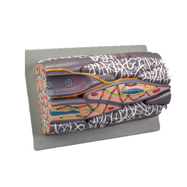
Microstructure of Smooth Muscle Model
The Microstructure of Smooth Muscle Model (Product Code: SLE/A/151) is a block model of a greatly enlarged bundle of smooth muscle fibers, revealing the structures inside and between the cells, from the nucleus, mitochondria and sarcoplasmic reticulum to the dense bodies and contractile myofilaments that produce contraction. Histology, physiology and zoology lab equipment buyers in India choose it to make an invisible tissue visible to a whole class. It is made by Jain Scientific Equipments Pvt Ltd (ScienceLab), Ambala.
Specifications
| Parameter | Detail |
|---|---|
| Type | Block model of a greatly enlarged bundle of smooth muscle fibers |
| Size | 28 × 16 × 11 cm |
| Base | Mounted on a durable polymer base |
| Structures shown | Significant extra- and intracellular structures, including sarcoplasmic reticulum, nucleus, mitochondria, dense bodies and contractile myofilaments |
Typical Use
Smooth muscle lines the walls of hollow organs and blood vessels, and its contraction works differently from the striated muscle students already know. On this block model, the nucleus, the mitochondria and the dense bodies that anchor the contractile myofilaments give the teacher a clear basis to explain why smooth muscle has no visible striations and how it can contract slowly and steadily.
Lecturers typically use it alongside prepared microscope slides of muscle tissue, so students first see the enlarged three-dimensional structure and then recognise the same tissue under the microscope. It suits undergraduate zoology, physiology, pharmacy and medical histology practicals.
Manufacturer
The model is manufactured by Jain Scientific Equipments Private Limited, an Indian company in Ambala, Haryana; its products for schools, colleges and laboratories are sold under the name ScienceLab. Details are on our company profile.
Related Human Models
Items for a complete muscle-tissue lesson:
- Skeletal Muscle Fiber Model — the striated counterpart, for a side-by-side comparison
- Microstructure of Artery and Vein Model — shows where smooth muscle layers sit in vessel walls
- Microslide Animal Tissue Set 2 Muscle, Bone, Connective — photographed microscope views of muscle and related tissues
- Micro Slide Viewer — for viewing the Microslide image strips without a microscope
For other microstructure models, see the Human Models range.
Frequently Asked Questions
Which cell structures can students identify?
The sarcoplasmic reticulum, nucleus, mitochondria, dense bodies and contractile myofilaments, plus significant structures between the fibers.
What magnification is the model?
It is described as greatly enlarged; an exact magnification is not stated in our documentation. Contact us if you need this figure.
How does it differ from a skeletal muscle model?
Smooth muscle has no sarcomere striations; its myofilaments are anchored to dense bodies, which this model shows clearly. Pair it with the Skeletal Muscle Fiber Model for comparison.
Where is smooth muscle found in the body?
In the walls of the digestive tract, blood vessels, airways, bladder and uterus, where it works involuntarily.
What size is it?
The block measures 28 × 16 × 11 cm and sits on a durable polymer base.
Can overseas universities order it?
Yes. We supply institutions and distributors in India and international markets; send the quantity and destination for a quotation.
Request a Quote
Ask for a quotation via our contact page, or place the Microstructure of Smooth Muscle Model in the enquiry cart with other histology items.
Last updated: 24 September 2026