- Physics Lab Equipment
- Chemistry Lab Equipment
- Biology Lab Equipment
- Anatomical Lab Equipment
- Learning
- Educational Lab Equipment
- Magnets
- Agriculture
- Astronomy
- Earth Science
- Geography Lab Equipment
- Safety Lab Coat
- Laboratory
- Meteorological
- Electricity & Magnetism
- Heat
- School Lab Equipment
- Lab Glassware
- Science Lab Equipment
- Tvet Lab Equipment
- Robotics Kits Supplier and Manufacturer for Schools India
- Math Lab Equipment Manufacturers and Exporters in Ambala
- STEM Kits Manufacturers and Supplier India
- Engineering Lab Equipment
- Hospital Lab Equipment
- Water, Sanitation & Hygiene Supplies
- Cold Chain, Solar & Communication Equipment
- Nutrition
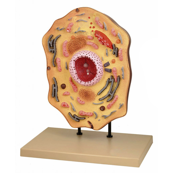
Animal Cell Model
The Animal Cell Model shows a typical animal cell enlarged 20,000 times, so a whole class can see organelles that a school microscope cannot resolve. It displays the nucleus, endoplasmic reticulum, mitochondria, ribosomes, Golgi apparatus, centrioles, lysosomes and fat vacuoles. At about 380 x 240 x 460 mm and 2350 g, it sits on a teacher's desk and moves easily between rooms.
Animal Cell Model specifications
| Parameter | Detail |
|---|---|
| Magnification | Enlarged 20,000 times |
| Structures shown | Nucleus, endoplasmic reticulum, mitochondria, ribosomes, Golgi apparatus, centrioles, lysosomes, fat vacuoles |
| Dimensions | 380 x 240 x 460 mm approx. |
| Weight | 2350 g approx. |
| Product code | SLE/BL/05 |
| Minimum order | 10 units |
| Dispatch | Usually within 7 to 10 working days |
| Manufacturer | ScienceLab (Jain Scientific Equipments Pvt Ltd), Ambala, India |
Cell biology lessons with the Animal Cell Model
From microscope to model. Students mount stained cheek cells and view them under a monocular microscope, drawing only what they can actually see. They then use the model to find the organelles that the light microscope could not show.
Plant and animal cells side by side. Stand the model next to a plant cell model and have students build a table of structures found in both cells or in only one. This prepares them for the familiar exam question on plant and animal cell differences.
Matching micrographs. Groups match each electron micrograph on the ultrastructure microslide of animal cells to the same organelle on the model, then write one line on what that organelle does.
Towards cell division. Point out the centrioles on the model, then move to the animal mitosis models to follow how the spindle forms and the chromosomes separate.
Frequently Asked Questions About the Animal Cell Model
How can students label the model by function as well as by name?
First name each structure, then add its job. The nucleus holds the DNA, ribosomes and the endoplasmic reticulum make proteins, the Golgi apparatus packages them, mitochondria release energy, lysosomes break down waste and centrioles take part in cell division.
Which structures does an animal cell share with a plant cell, and which does it lack?
Both have a nucleus, endoplasmic reticulum, ribosomes, mitochondria and a Golgi apparatus. The animal cell has no cell wall, no chloroplasts and no large central vacuole, and it has centrioles, which most plant cells do not. A plant cell model beside it makes the contrast easy to see.
Do only animal cells have mitochondria?
No. Plant cells also contain mitochondria and respire all the time, by day and by night. Many students assume respiration belongs to animals because plants photosynthesise, and the mitochondria on this model are a good place to correct that idea.
Why can't these organelles be seen under a school microscope?
Most of them are too small, or too faint without special stains, for a school light microscope. A stained cheek cell shows little more than the nucleus, cytoplasm and cell membrane. Ribosomes, the endoplasmic reticulum and lysosomes can be studied in detail only with an electron microscope, so the model shows the cell enlarged 20,000 times.
How can the lesson be adapted for younger and older classes?
Younger pupils can find and name the nucleus, cell membrane, cytoplasm and mitochondria. Older students can trace a protein from the ribosomes through the endoplasmic reticulum and Golgi apparatus to the cell surface, or explain why active muscle cells hold many mitochondria.
Cell biology models and slides
- Plant Cell Model
- Prokaryotic Cell Model
- Animal Mitosis Models
- Animal Meiosis Plaque
- Microslide Ultra Structure of Animal Cells
To request a quotation, send the product name, code and quantity through our contact page or add the item to your enquiry cart.
Last updated: 14 November 2026