- Physics Lab Equipment
- Chemistry Lab Equipment
- Biology Lab Equipment
- Anatomical Lab Equipment
- Learning
- Educational Lab Equipment
- Magnets
- Agriculture
- Astronomy
- Earth Science
- Geography Lab Equipment
- Safety Lab Coat
- Laboratory
- Meteorological
- Electricity & Magnetism
- Heat
- School Lab Equipment
- Lab Glassware
- Science Lab Equipment
- Tvet Lab Equipment
- Robotics Kits Supplier and Manufacturer for Schools India
- Math Lab Equipment Manufacturers and Exporters in Ambala
- STEM Kits Manufacturers and Supplier India
- Engineering Lab Equipment
- Hospital Lab Equipment
- Water, Sanitation & Hygiene Supplies
- Cold Chain, Solar & Communication Equipment
- Nutrition
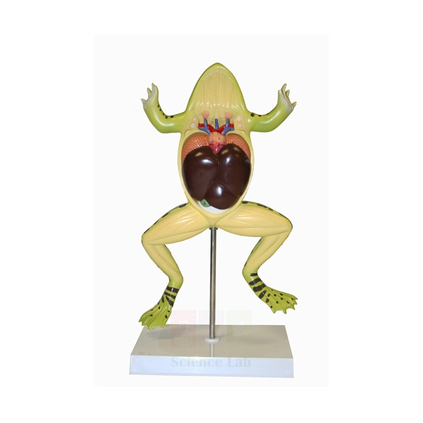
Frog Dissection Model
This frog model is opened from the belly side, the ventral view used in a classroom frog dissection. It is about two and a half times life size and shows the thoracic and abdominal structures. Four parts lift out and are held by magnets instead of pins or hooks, and the model comes off its stand for close study.
Frog dissection model specifications
| Parameter | Detail |
|---|---|
| Scale | About 2-1/2 x life size |
| Size without stand | 35 x 24 x 5 cm |
| Sectioning | Sectioned ventrally (opened from the belly side) |
| Removable parts | 4 |
| Fixing of parts | Magnets, no pins or hooks |
| Stand | Removable, so the model can be lifted off for examination |
| Structures shown | Thoracic and abdominal structures |
| Product code | SLE/A/119 |
| Minimum order | 10 units |
| Dispatch | Usually within 7 to 10 working days |
| Manufacturer | ScienceLab (Jain Scientific Equipments Pvt Ltd), Ambala, India |
Frog anatomy practicals with the dissection model
Organ checklist. Give each group a printed list of thoracic and abdominal organs. With all four parts in place, students point to each organ and tick it off. They then lift the magnetic pieces out one at a time, name what lies underneath and refit them before passing the model on.
Rehearsal before a specimen. Where a real frog is still dissected, the teacher first uses the model to show where the body wall is opened and how the organs sit. Students then work in a dissection pan with a student dissecting set, keeping the enlarged model beside the tray as their reference.
Organs inside the framework. Stand the model next to the Frog Skeleton so learners see how the backbone and the two limb girdles frame the body cavity. For individual bench work afterwards, each pair can take apart the life-size Mi-Own Frog Model.
From water to land. Pair the model with the Fish Dissection Model and ask students to list what changes when a vertebrate breathes air: gills give way to lungs, and the heart gains a second atrium.
Frequently Asked Questions About the Frog Dissection Model
Which organ should students find first in a frog dissection?
Start with the liver. It is the biggest organ in the body cavity and covers much of the gut, so it is the easiest landmark. From there, work up to the heart and lungs and down to the stomach and intestine, ticking each organ off a checklist as it is found.
What are the three lobes of the frog liver, and where is the gall bladder?
Most practical guides name a right lobe and two left lobes, the left anterior and the left posterior. The gall bladder is a small greenish sac tucked beneath the liver. It stores bile, which flows into the first part of the small intestine and helps break down fat.
What path does food take through the frog's digestive system?
Food passes from the mouth down the short oesophagus into the stomach. A ring of muscle, the pyloric sphincter, lets it into the duodenum, the straight first part of the small intestine. The coiled ileum follows, then the large intestine, which opens into the cloaca.
What are the yellow fat bodies, and why does their size change?
Fat bodies are finger-like yellow structures that sit near the kidneys and gonads. They are energy stores. A frog draws on them during winter hibernation, summer aestivation and the breeding season, so they are large before these periods and shrink afterwards.
What is the cloaca, and which systems open into it?
The cloaca is the shared chamber at the end of the gut. The large intestine, the ducts from the kidneys and the reproductive ducts all empty into it. Faeces, urine and eggs or sperm therefore leave the body through one opening.
How should the magnetic parts be handled and stored?
Lift each part straight out instead of prising it at an angle, and hold it by the edges. Refit all four pieces before the model goes back on its stand. Store it away from other strong magnets, which could pull the pieces out of their correct positions.
Frog and fish study models and dissection tools
- Frog Skeleton
- Mi-Own Frog Model
- Fish Dissection Model
- Dissection Pan
- Dissecting Set, Student
- Stereoscopic Dissection Microscopes
To request a quotation, send the product name, code and quantity through our contact page or add the item to your enquiry cart.
Last updated: 14 November 2026