- Physics Lab Equipment
- Chemistry Lab Equipment
- Biology Lab Equipment
- Anatomical Lab Equipment
- Learning
- Educational Lab Equipment
- Magnets
- Agriculture
- Astronomy
- Earth Science
- Geography Lab Equipment
- Safety Lab Coat
- Laboratory
- Meteorological
- Electricity & Magnetism
- Heat
- School Lab Equipment
- Lab Glassware
- Science Lab Equipment
- Tvet Lab Equipment
- Robotics Kits Supplier and Manufacturer for Schools India
- Math Lab Equipment Manufacturers and Exporters in Ambala
- STEM Kits Manufacturers and Supplier India
- Engineering Lab Equipment
- Hospital Lab Equipment
- Water, Sanitation & Hygiene Supplies
- Cold Chain, Solar & Communication Equipment
- Nutrition
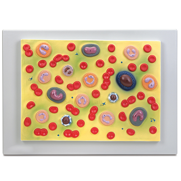
Human Blood Cell Types Model
A 54 x 38 cm plaque showing the formed elements of human blood: erythrocytes, platelets and five types of leukocytes. Each leukocyte is modelled with its characteristic nucleus, and every structure carries a number that matches the accompanying key. Teachers keep it beside the microscope bench while students learn to name cells on a stained smear.
Blood cell plaque specifications
| Parameter | Detail |
|---|---|
| Format | Plaque mount |
| Size | 54 x 38 cm |
| Red cells | Erythrocytes |
| White cells | 5 types of leukocytes, each with its characteristic nucleus |
| Other formed elements | Platelets |
| Key | Numbered structures referenced on the accompanying key |
| Product code | SLE/A/140 |
| Minimum order | 10 units |
| Dispatch | Usually within 7 to 10 working days |
| Manufacturer | ScienceLab (Jain Scientific Equipments Pvt Ltd), Ambala, India |
Haematology practicals with the blood cell plaque
Smear identification. Students scan a stained human blood smear under the oil-immersion objective of a 1000x LED monocular microscope. They sketch each white cell they meet, then confirm the name by matching its nucleus shape against the numbered cells on the plaque.
Differential count practice. Once the five types are familiar, students move along the thin end of the smear in a zigzag, tally every leukocyte they pass and turn the tallies into percentages. The plaque stays in view so that doubtful cells can be checked quickly.
Cell counting. Before a total count with the haemocytometer, use the plaque to compare red cells with white cells and platelets. Explain that red cells far outnumber white cells in blood, so the two counts need different dilutions and different squares of the grid.
Red cells and blood groups. After a grouping exercise with the simulated blood typing kit, return to the erythrocytes on the plaque and explain that the A, B and Rh antigens sit on the red cell surface.
Frequently Asked Questions About the Human Blood Cell Types Model
How do students tell the five white cell types apart?
Look first for granules, then at the shape of the nucleus. Neutrophils, eosinophils and basophils are granulocytes, separated by the colour of their granules and the lobing of the nucleus. Lymphocytes and monocytes have no obvious granules; the lymphocyte is small with a round nucleus, while the monocyte is larger with a folded one.
Which leukocytes belong to innate immunity and which to adaptive immunity?
Neutrophils, eosinophils, basophils and monocytes act in innate defence, responding fast to any invader without needing earlier contact. B and T lymphocytes provide adaptive immunity: they recognise particular antigens, make antibodies or kill infected cells, and leave memory cells behind after an infection.
What does a monocyte turn into once it leaves the blood?
In the tissues it develops into a macrophage or a dendritic cell. Both engulf microbes and debris and then display fragments of them to T lymphocytes, which is why they are called antigen-presenting cells.
Can B and T lymphocytes be told apart on the plaque or on a smear?
No. With routine blood stains both appear as small round cells with a large dark nucleus and a thin rim of cytoplasm. Separating them needs antibody labels against surface markers, read by flow cytometry or immunostaining, so the plaque shows a single lymphocyte type.
What are platelets, and why do they look so small?
Platelets are not complete cells. They are fragments pinched off large bone-marrow cells called megakaryocytes, they have no nucleus and they are far smaller than red cells. That is why they appear as small specks both on the plaque and on a stained smear.
Haematology teaching items
- Human Blood Smear
- Monocular Microscope 1000x, LED light
- Haemocytometer
- Blood Typing Kit, Simulated
- Stained Human Blood
To request a quotation, send the product name, code and quantity through our contact page or add the item to your enquiry cart.
Last updated: 14 November 2026