- Physics Lab Equipment
- Chemistry Lab Equipment
- Biology Lab Equipment
- Anatomical Lab Equipment
- Learning
- Educational Lab Equipment
- Magnets
- Agriculture
- Astronomy
- Earth Science
- Geography Lab Equipment
- Safety Lab Coat
- Laboratory
- Meteorological
- Electricity & Magnetism
- Heat
- School Lab Equipment
- Lab Glassware
- Science Lab Equipment
- Tvet Lab Equipment
- Robotics Kits Supplier and Manufacturer for Schools India
- Math Lab Equipment Manufacturers and Exporters in Ambala
- STEM Kits Manufacturers and Supplier India
- Engineering Lab Equipment
- Hospital Lab Equipment
- Water, Sanitation & Hygiene Supplies
- Cold Chain, Solar & Communication Equipment
- Nutrition
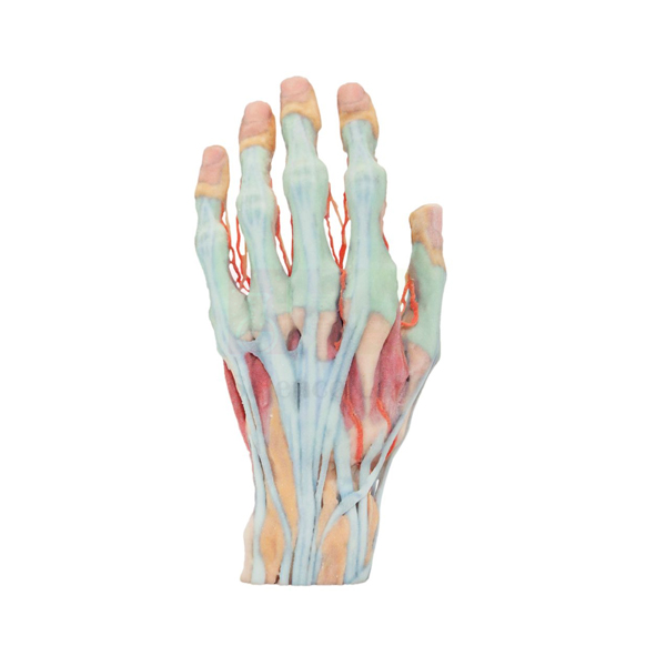
3-D Printed Human Superficial Hand Dissection Model
3-D Printed Human Superficial Hand Dissection Model (Product Code: SLE/A/102) reproduces a superficial dissection of the human hand at about life size, showing the vasculature, musculature and tendons in intricate detail. Because it is 3-D printed, students can study a specimen that is as realistic as possible without the need for human cadavers. It is used in anatomy, physiotherapy and occupational therapy teaching as biology lab equipment in India, and is one of the human anatomical models manufactured by Jain Scientific Equipments Pvt Ltd (ScienceLab), Ambala.
Specifications
| Parameter | Detail |
|---|---|
| Type | 3-D printed superficial dissection model of the human hand |
| Scale | About life size |
| Size | About 6-7/10" W x 5-9/10" H x 11-2/5" L |
| Structures shown | Vasculature, musculature and tendons of the hand, in intricate detail |
| Purpose | Realistic specimen study without the need for human cadavers |
Typical Use
The hand packs a great deal of anatomy into a small space, and this model lets learners examine the layer just beneath the skin: the blood vessels that supply the fingers and palm, the small muscles that give the hand its precision, and the tendons that transmit movement from the forearm. Instructors use it to explain grip, pinch and finger movement, and to show why injuries to a single tendon or vessel can affect function so noticeably.
For courses where cadaver access is limited, or where the biology dissection set is used only on animal specimens, the model gives a realistic human reference that can be handled in every session without preservation or special storage. Physiotherapy, occupational therapy, nursing and medical students use it for hand-anatomy practicals, and art and design departments use it to study the hand's form. Pairing it with a skeletal hand helps students relate each tendon to the bones it moves.
Manufacturer
The model is produced by Jain Scientific Equipments Private Limited, which trades as ScienceLab from its base in Ambala, Haryana, and serves teaching institutions in India and international markets. Buyers who need company registration details for procurement can request them with their quotation. The company profile has further background.
Related Human Models
Models that help build a complete picture of the hand and upper limb:
- Human Skeletal Hand Model — the bones beneath the muscles and tendons shown here
- Human Hand Joint Model with Ligaments — adds the joints and ligaments of the hand
- Human Arm and Hand model — follows the tendons back to the forearm muscles
- 3-D Printed Internal Heart Structures Model — another 3-D printed model for cadaver-free study
- 3D Printed Human Flexed Knee Joint — a 3-D printed joint for lower-limb lessons
See all Human Models for more anatomy teaching options.
Frequently Asked Questions
What does "superficial dissection" mean on this model?
It shows the structures that lie just beneath the skin of the hand: the vasculature, musculature and tendons.
How large is it?
About life size, measuring about 6-7/10" W x 5-9/10" H x 11-2/5" L.
Why choose a 3-D printed model over a cadaveric specimen?
It lets students study a realistic specimen without the need for human cadavers, so it can be used in ordinary classrooms and handled repeatedly.
Are the bones of the hand shown?
Our product details describe the vasculature, musculature and tendons. For bone anatomy, add the Human Skeletal Hand Model, or ask us about the model's deeper layers.
Does it come on a stand or base?
A stand is not listed for this model. Please contact us to confirm how it is supplied.
How should it be cleaned?
Dust it with a soft dry brush or cloth, and use only a lightly damp cloth for marks; avoid solvents and hot water.
What information speeds up a quotation?
The number of units, your delivery location and any registration documents your purchasing office requires. Send these via the contact page or the enquiry cart.
Request a Quote
To order the 3-D Printed Human Superficial Hand Dissection Model, contact us for pricing or add it to your enquiry cart.
Last updated: 24 September 2026