- Physics Lab Equipment
- Chemistry Lab Equipment
- Biology Lab Equipment
- Anatomical Lab Equipment
- Learning
- Educational Lab Equipment
- Magnets
- Agriculture
- Astronomy
- Earth Science
- Geography Lab Equipment
- Safety Lab Coat
- Laboratory
- Meteorological
- Electricity & Magnetism
- Heat
- School Lab Equipment
- Lab Glassware
- Science Lab Equipment
- Tvet Lab Equipment
- Robotics Kits Supplier and Manufacturer for Schools India
- Math Lab Equipment Manufacturers and Exporters in Ambala
- STEM Kits Manufacturers and Supplier India
- Engineering Lab Equipment
- Hospital Lab Equipment
- Water, Sanitation & Hygiene Supplies
- Cold Chain, Solar & Communication Equipment
- Nutrition
Human models are enlarged, dissectible and life-size replicas of the human body, its organs and systems, used as biology teaching aids in schools and for anatomy teaching in nursing, paramedical and medical colleges. Jain Scientific Equipments Pvt Ltd (ScienceLab), Ambala, is the OEM that makes this range at its own licensed factory in Ambala. Its 148 listed models, from torsos, sense organs and teeth to pathology examples and CPR trainers, form the largest part of the anatomical collection.
Human Models by Topic
| Topic | What it covers | Example products |
|---|---|---|
| Torsos and muscular figures | Trunk and whole-body models showing organ positions and muscle layers | Human Muscular Torso with Head and Open Back Model; Human Male and Female Torso Model, 20 Parts |
| Heart, circulation and breathing | Heart structure, blood flow and how the lungs fill and empty | Human Heart, Pumping Model; Human Circulatory System Model |
| Sense organs and skin | Eye, ear, nose, tongue and skin structure | Human Eye Model; Human Ear Model, 5 Parts |
| Nervous, endocrine and urinary systems | Nerve cells and pathways, hormone-producing glands and the kidneys | Human Nervous System Model; Human Endocrine Organs Model; Human Kidney with Adrenal Gland Model |
| Reproduction and development | Reproductive organs, pregnancy and foetal growth | Human Female Genital Organs Model; Human Embryo Development Set |
| Regional anatomy | Arm, hand, shoulder and neck structures studied region by region | Human Muscular Arm Model; Shoulder Girdle with Musculature Model |
| Teeth and pathology | Dentition, oral health and diseased tissue compared with healthy anatomy | Human Tooth Set; Dental Caries Model; Cataract Eye Model with 4 Forms of Cataract |
| Clinical-skills trainers | Hands-on practice for resuscitation, injections and ECG electrode placement | Advanced Child CPR Training Manikin; Adult Venipuncture and Injection Training Arm, Dark |
Featured Human Models
| Product | Product Code | Typical Use |
|---|---|---|
| Deluxe Human Male and Female Torso Model | SLE/A/22 | Life-size, 27-part torso with interchangeable male and female urogenital inserts |
| Human Muscular Figure Model | SLE/BL/71 | 32-part, 140 cm figure with detachable arm and leg and removable organs |
| Human Heart Model | SLE/A/12 | 3-piece heart at twice life size, showing coronary vessels and internal chambers |
| Human Kidney, Nephron, and Glomerulus Model Set | SLE/A/21 | Kidney, nephrons and glomerulus at three magnifications on one base |
| Human Teeth Model, Dentition Development | SLE/BL/85 | Upper and lower jaws at four stages from newborn to young adult |
| Acupuncture Model 84 cm, Deluxe | SLE/BL/01 | Figure mapping 14 primary channels and 361 acupoints, with a subcutaneous view on one side |
| Advanced Adult CPR Training Manikin | SLE/BL/02 | Compression and ventilation practice with light, audio and printed feedback |
OEM Manufacturing & Supply
ScienceLab, the trading name of Jain Scientific Equipments Private Limited, is the OEM for the Human Models range: the models are manufactured at the company's own licensed factory at 449, HSIIDC Industrial Area, Saha, Ambala, Haryana, and supplied factory-direct to schools, colleges, dealers, tender buyers and importers in India and abroad, with quotations straight from the manufacturer. A handful of third-party-branded models, such as the 3B, Somso and Denoyer-Geppert items, are supplied rather than made in Ambala. The company is incorporated under CIN U29309HR2020PTC087597, registered as an MSME (Udyam UDYAM-HR-01-0003714) and runs its factory under Factory Licence AMB-ONLINE-CHD-J-414.
Related Ranges
- Anatomical Lab Equipment: the parent hub linking all six anatomy sub-ranges.
- Bone Model: full skeletons, spinal columns, pelves and joints; a few skeletal pieces, such as the Human Skeletal Hand Model, are listed here instead.
- Brain Models: dissectible brains and sectioned heads, while nerve-cell and nervous-system models stay here.
- Digestive System: stomach, liver, gallbladder and pancreas organ models; whole-tract models such as the Human Digestive Tract Model are on this page.
- Skulls: adult, fetal and comparative skulls for osteology.
- Animal Model: animal and comparative anatomy models for zoology rather than human biology.
Frequently Asked Questions
Who manufactures these human anatomy models?
ScienceLab (Jain Scientific Equipments Private Limited) is the OEM for the Human Models range and makes the models at its licensed factory in Saha, Ambala. The only exceptions are a few third-party-branded models, such as 3B, Somso and Denoyer-Geppert items, which the company supplies.
Which human models should a school biology lab buy first?
For a school biology lab, a torso, a heart, an eye, an ear and a muscular figure from the Human Models range cover most units on the human body. Add a skeleton from the Bone Model sub-range to complete the core set.
Are human models suitable for nursing and paramedical courses?
Yes. Besides organ and system models, the Human Models range includes pathology models and clinical-skills trainers, such as adult and child CPR manikins, an injection training arm and a 15-lead ECG placement trainer, used in nursing and paramedical teaching.
Does the range include dental models?
Yes. The Human Models range includes teeth models, such as the Human Teeth Model, Dentition Development, the Human Tooth Set and single-tooth models, plus the Dental Caries Model, Odontopathies Model and Human Oral Hygiene Model for teaching dental health.
Can human models be quoted as a complete set for a tender?
Yes. Send your model list or tender schedule with product codes (for example SLE/A/12), quantities and the delivery city or country, and ScienceLab will quote the Human Models together with matching items from the other anatomy sub-ranges.
Request a Quote or Catalogue
To receive a quotation or our anatomy catalogue, write to us through the contact page, or add the models you need to the shopping cart and send them as one enquiry.
Last updated: 24 September 2026
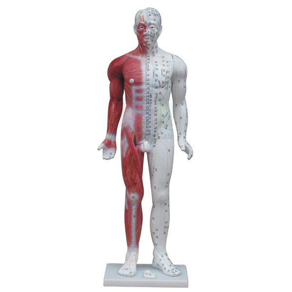
Acupuncture Model 84 cm, Deluxe
Acupuncture Model 84 cm, Deluxe (Product Code: SLE/BL/01) is a colourful, highly illustrated plastic figure for teaching the channel and point system used in Chinese medicine. Its right side maps 14 primary channels, 361 acupoints and 48 extraordinary acupoints with CUN markers for judging the distance between points, while the left side opens into a subcutaneous view of nerves, blood vessels and muscles. Colleges of acupuncture, massage and traditional medicine, physiotherapy trainers and wellness academies use it as a shared reference in class. It is made by Jain Scientific Equipments Pvt Ltd (ScienceLab), Ambala, an anatomical models manufacturer supplying teaching institutions.
Specifications
| Parameter | Detail |
|---|---|
| Height | 84 cm |
| Material | Colourful plastic, highly illustrated and detailed |
| Right side | 14 primary channels, 361 acupoints, 48 extraordinary acupoints, CUN markers |
| Left side | Subcutaneous view of neural distribution, artery, vein, blood vessel and muscular structures |
| Additional points (left side) | Upper and lower extremity Extra points not located on the primary channels |
| Suitable for | Teaching and learning of Chinese medicine, acupuncture and massage |
Typical Use
Instructors use the right side to walk students through each channel in turn, pointing out named acupoints and using the CUN markers to show how point locations are measured in proportional body units rather than centimetres. Once a pathway is learned on the surface, turning the model to the left side lets the class see what lies beneath — nerves, arteries, veins and muscles — which helps practitioners understand why some points need careful needle depth and angle.
The figure also works as a revision tool: students can quiz one another on point locations or trace the Extra points of the upper and lower limbs that sit off the primary channels. As one of the more specialised biology teaching aids India institutions stock, it lasts longest when kept upright away from direct sunlight and dusted with a soft, dry cloth rather than solvents that could fade the printed markings.
Manufacturer
Jain Scientific Equipments Private Limited produces anatomical and teaching models in Ambala, Haryana, and markets them under the ScienceLab brand. You can learn more about the company on our company profile.
Related Human Models
Teachers building an anatomy and therapy teaching set often add these models:
- Human Muscular Figure Model — for detailed study of the muscles that lie under many points.
- Human Nervous System Model — to follow nerve pathways referred to in point location.
- Human Circulatory System Model — shows the major vessels students must avoid when needling.
- Deluxe Human Male and Female Torso Model — internal organ anatomy to complement surface landmarks.
Explore the full range in our Human Models category.
Frequently Asked Questions
How many points and channels are shown?
The right side shows 14 primary channels, 361 acupoints and 48 extraordinary acupoints. The left side adds upper and lower extremity Extra points that are not on the primary channels.
How tall is the model?
It is 84 cm tall, large enough for a teacher to point out locations to a small group at the front of a class.
What are the CUN markers for?
They help students evaluate the distance between points using the proportional cun measurement used in acupuncture.
Does it show anatomy beneath the skin?
Yes. The left side is a subcutaneous view showing neural distribution, arteries, veins, blood vessels and muscular structures.
Is a key or point chart supplied?
Please ask us through the contact page about any key or printed guide supplied with the current model.
Can colleges order several models?
Yes, we supply institutions in India and international markets. Send the quantity, delivery address and billing details and we will send a quotation that includes warranty terms.
Request a Quote
Enquire about the Acupuncture Model 84 cm, Deluxe through our contact page or add it to the enquiry cart.
Last updated: 24 September 2026
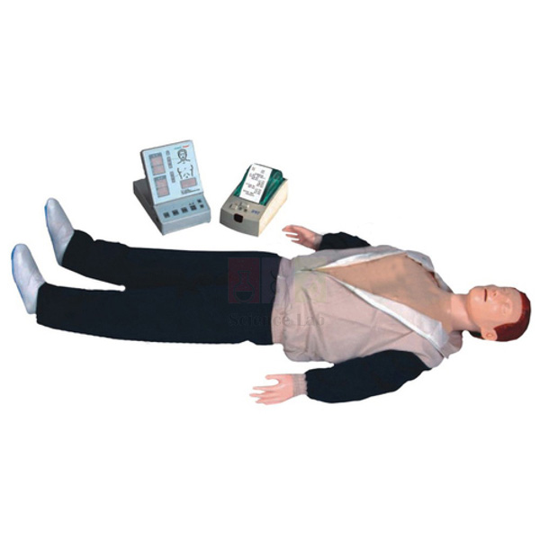
Advanced Adult CPR Training Manikin
Advanced Adult CPR Training Manikin (Product Code: SLE/BL/02) is an adult simulation model on which trainees practise reviving a patient with chest compressions (heart pressure) and mouth-to-mouth respiration. It tells the trainee whether each action is correct through light indicators, audio prompting and grade printing, and a digital counter is built into the feedback display. Hospitals, medical colleges and nursing training institutes use it for basic life support teaching, and it is a practical piece of lab equipment for college India skills labs. It is made by Jain Scientific Equipments Pvt Ltd (ScienceLab), Ambala.
Key Details
| Parameter | Detail |
|---|---|
| Type | Adult CPR simulation model |
| Design basis | Designed according to the International CPR standard |
| Skills practised | Heart pressure (chest compression) and mouth-to-mouth respiration |
| Ventilation feedback | Light indicator display for correct and incorrect artificial respiration (mouth-to-mouth) |
| Compression feedback | Light indicator display for correct compression position and wrong compression position |
| Other feedback | Audio prompting, grade printing, digital counter |
| Suitable for | Hospitals, medical colleges and nursing training institutes |
Typical Use
A typical session starts with the instructor demonstrating hand placement and head-tilt for rescue breaths, after which each trainee performs cycles of compressions and ventilations while the lights confirm correct or wrong hand position and correct or incorrect breaths. Audio prompts guide the trainee in real time, and the grade printout gives a record of the attempt that can be discussed afterwards or kept in the trainee's skills file — useful for assessment days in nursing schools and hospital in-service programmes.
Beyond clinical courses, the manikin can anchor a first aid training kit school or workplace safety teams use for awareness drills. Rotate trainees so everyone gets hands-on time, clean the face and mouth area between users according to your hygiene protocol, and store the manikin flat in its packing so the chest and indicator parts are not strained.
Manufacturer
This training manikin is offered by Jain Scientific Equipments Private Limited, an Ambala, Haryana manufacturer of medical education and science teaching equipment that trades as ScienceLab. Our company profile has more about the company.
Related Human Models
Training centres often build a full resuscitation and clinical skills set with:
- Advanced Child CPR Training Manikin — to teach the paediatric technique alongside the adult one.
- Advanced Infant CPR Training Manikin — completes an all-ages basic life support course.
- Adult Venipuncture and Injection Training Arm, Dark — for practising injections and blood draws in the same skills lab.
- Human Lung Model in Bell Jar — explains the breathing mechanics behind rescue ventilation.
See more in our Human Models category.
Frequently Asked Questions
What feedback does the manikin give?
Light indicators show correct and incorrect mouth-to-mouth respiration and correct or wrong compression position, backed by audio prompting, a digital counter and grade printing.
Which CPR guidelines does it follow?
It is designed according to the International CPR standard. If your course is tied to a particular guideline edition, please confirm the details with us before ordering.
What power supply and printing consumables does it need?
Please check the power source and printer paper requirements for the current model with us through the contact page.
How is hygiene managed between trainees?
Clean the face and mouth area between users following your institution’s protocol. Ask us which replacement hygiene parts are available for this model.
Who is this model suitable for?
It is designed for adult CPR training in hospitals, medical colleges and nursing training institutes, and is also used for first aid courses.
What should a tender or institutional enquiry include?
Quantity, delivery location, billing entity and your tender format. We supply India and international markets, and warranty terms are confirmed in the quotation.
Request a Quote
To price the Advanced Adult CPR Training Manikin, contact us via the contact page or add it to your enquiry cart.
Last updated: 24 September 2026
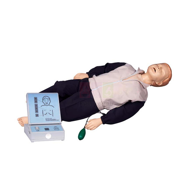
Advanced Child CPR Training Manikin
Advanced Child CPR Training Manikin (Product Code: SLE/BL/03) is a child-sized simulation model for practising paediatric resuscitation — chest compressions (heart pressure) and mouth-to-mouth respiration — with light indicators that show at once whether each step is done correctly. Designed according to the International CPR standard, it lets paediatric nurses, school health staff, paramedics and first aid instructors rehearse the adjustments needed when the patient is a child rather than an adult. It suits institutions assembling a health education kit school India programmes use for life-saving skills. It is made by Jain Scientific Equipments Pvt Ltd (ScienceLab), Ambala.
Key Details
| Parameter | Detail |
|---|---|
| Type | Child CPR simulation model |
| Design basis | Designed according to the International CPR standard |
| Skills practised | Patient revival through heart pressure and mouth-to-mouth respiration |
| Respiration indicator | Light indicator display for correct and incorrect artificial respiration (mouth-to-mouth) |
| Compression indicators | Correct compression position display and wrong compression position display |
| Suitable for | Paediatric CPR and first aid training |
Typical Use
Child CPR differs from adult technique in hand use, force and breath size, and trainees who have only practised on an adult model tend to over-press or over-inflate. On this manikin the instructor can let each trainee try, watch the compression-position lights, then correct placement straight away; the respiration lights do the same for rescue breaths, showing whether the airway was opened and the breath delivered properly. Short, repeated rounds work better than a single long attempt, so plan the class in rotations.
Schools, colleges and hospitals that source from a school lab equipment supplier India wide often pair this model with the adult and infant versions so that one course covers every age group. Between trainees, wipe the face and mouth area according to your hygiene procedure, and keep the manikin in a dry cupboard away from heat so the indicator components stay in good order.
Manufacturer
ScienceLab is the brand of Jain Scientific Equipments Private Limited, a company based in Ambala, Haryana that makes training models for medical and science education. It is registered as an MSME manufacturer under Udyam UDYAM-HR-01-0003714, and its international consignments move under IEC AAECJ8618A from the Directorate General of Foreign Trade. See our company profile for more.
Related Human Models
For a complete paediatric and general life support course, consider:
- Advanced Adult CPR Training Manikin — adds audio prompting and grade printing for adult technique.
- Advanced Infant CPR Training Manikin — for the youngest age group, where technique changes again.
- Human Respiratory System Model, 2-parts — to explain the airway that rescue breaths must reach.
- Human Heart Model — shows why chest compressions keep blood moving.
Browse our Human Models category for more.
Frequently Asked Questions
What does the child manikin indicate?
It has light displays for correct and incorrect mouth-to-mouth respiration, plus separate displays for correct and wrong compression position.
Does it include audio prompts or a printout like the adult model?
The features listed for this model are its light indicator displays. For audio or printing functions, please check the current specification with us through the contact page.
Which standard is it designed to?
It is designed according to the International CPR standard. Confirm with us if your training body requires a specific guideline edition.
Who typically buys the child version?
Nursing and medical colleges, paediatric wards, school health programmes and first aid trainers who need to teach child resuscitation specifically.
How do I keep it clean and working?
Disinfect the face and mouth area after each trainee as your protocol requires, avoid soaking the model, and ask us about any replacement parts you may need later.
Can you quote for a set of adult, child and infant manikins?
Yes. Tell us the quantity of each, your delivery location in India or abroad and your billing details; warranty terms are included in the quotation.
Request a Quote
Request pricing for the Advanced Child CPR Training Manikin through the contact page, or add it to your enquiry cart with the other manikins you need.
Last updated: 24 September 2026
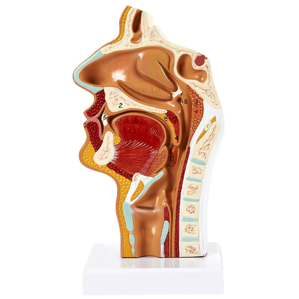
Anatomical Human Nasal Cavity Model
Anatomical Human Nasal Cavity Model (Product Code: SLE/BL/04) helps students understand the internal structure of the nose in three areas: the lateral nasal wall with its superior, middle and inferior nasal conchae, a section through the nasal bones and cartilages of the external nose, and the frontal, sphenoid and maxillary paranasal sinuses. Anatomy departments, ENT and physiology teachers, nursing schools and senior secondary biology classes use it to connect the nose with breathing, smell and sinus problems. As part of a wider range of biology lab equipment India schools and colleges order, it is made by Jain Scientific Equipments Pvt Ltd (ScienceLab), Ambala.
Key Details
| Parameter | Detail |
|---|---|
| Type | Anatomical model of the human nasal cavity |
| Nasal cavity | Lateral nasal wall showing the superior, middle and inferior nasal conchae projecting medially into the nasal cavity |
| External nose | Section of the nasal bones and cartilages |
| Paranasal sinuses | Frontal, sphenoid and maxillary sinuses |
| Suitable for | Anatomy, ENT, nursing and biology teaching |
Typical Use
Start with the external nose section to show how bone gives way to cartilage along the bridge, then move to the lateral wall, where the three conchae curl inward and create the narrow passages that warm, moisten and filter incoming air. Pointing out the frontal, sphenoid and maxillary sinuses around the cavity helps explain why a blocked nose often goes with sinus pressure, and why infections can spread between these spaces.
The model sits well within a human biology model set India classrooms use for the respiratory system and sense organs, and it is a handy prop for nursing students learning nasal procedures. Keep it covered when not in use, handle it by its base or edges rather than the fine structures, and clean it with a slightly damp cloth.
Manufacturer
Jain Scientific Equipments Private Limited makes human anatomy models for teaching at its operations in Ambala, Haryana, and sells them under the name ScienceLab. The company profile gives more information.
Related Human Models
Useful companions for teaching the nose, airway and senses:
- Human Nasal Cavity Model — an alternative nasal cavity model to compare.
- Human Nose Model on Base — another view of nasal anatomy for the same lesson.
- Human Respiratory System Model, 2-parts — follows the airway from the nose down to the lungs.
- Human Ear Model, 5 Parts — completes an ear, nose and throat teaching set.
Find more in our Human Models category.
Frequently Asked Questions
Which structures does the model show?
The nasal conchae on the lateral nasal wall, a section of the nasal bones and cartilages of the external nose, and the frontal, sphenoid and maxillary sinuses.
Is it suitable for school biology as well as medical teaching?
Yes. School classes use it for the respiratory system and sense of smell, while nursing and medical students use the same structures in more detail.
Does the model come apart?
The description does not list removable parts. Please ask us through the contact page for the current construction, size and base details.
Is a key to the structures included?
Please confirm with us whether a numbered key or guide is supplied with the model you order.
How should the model be cleaned?
Dust it regularly with a soft brush and wipe gently with a damp cloth. Avoid alcohol or solvent cleaners that could affect painted details.
Can you supply models for a new anatomy lab?
Yes, we supply institutions in India and international markets. Send the full list of models, quantities and delivery address, and our quotation will state warranty terms.
Request a Quote
To order the Anatomical Human Nasal Cavity Model, contact us through the contact page or add it to your enquiry cart.
Last updated: 24 September 2026
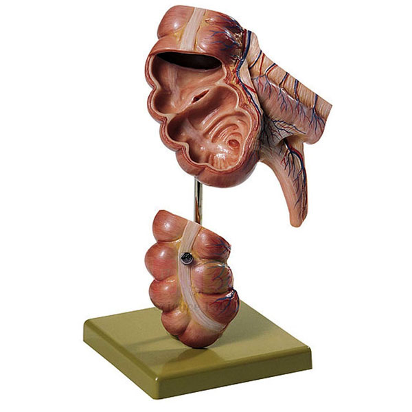
Appendix and Caecum Model
Appendix and Caecum Model (Product Code: SLE/BL/06) is a natural size PVC model of the junction between the small and large intestine, showing the wall of the caecum and the appendix and marking 17 positions for study. It separates into 2 parts and is mounted on a stand, so it can be taken apart during a lesson and reassembled afterwards. Medical and nursing colleges, surgical trainees and senior biology classes use it when teaching digestion and conditions such as appendicitis. Institutions looking for a biology lab equipment supplier India can order it from Jain Scientific Equipments Pvt Ltd (ScienceLab), Ambala, which makes it.
Specifications
| Parameter | Detail |
|---|---|
| Material | Advanced PVC |
| Size | Natural size |
| Structures shown | Wall of the caecum and appendix |
| Positions shown | 17 |
| Parts | Separates into 2 parts |
| Mounting | Mounted on a stand |
Typical Use
Because it is natural size, the model gives students a realistic sense of how small the appendix is compared with the caecum it hangs from — something diagrams rarely convey. In a lesson on the digestive tract, the teacher can show the model assembled first, then separate the two parts to look more closely at the caecum wall and the appendix. Medical and nursing students use the same model when learning about appendicitis, why the site of pain can vary, and what a surgeon looks for during an appendectomy.
For school biology lab equipment in India classrooms, it pairs well with a full digestive system model so learners can locate the caecum within the whole tract before looking at it in detail. Keep the stand on a flat surface, rejoin the two parts carefully after each lesson, and wipe the PVC with a mild soap solution when it needs cleaning.
Manufacturer
Jain Scientific Equipments Private Limited, trading as ScienceLab, produces anatomy teaching models in Ambala, Haryana for schools, colleges and hospitals. Tender and procurement teams needing vendor details can use GSTIN 06AAECJ8618A1ZT and Legal Entity Identifier 3358004HRD1JIVXKYX49. More about us is on the company profile.
Related Human Models
To teach the digestive system in context, buyers also choose:
- Human Digestive System Model — places the caecum and appendix within the whole digestive tract.
- Deluxe Human Male and Female Torso Model — shows abdominal organs in their body positions.
- Human Micro Sexless Torso Model — a compact torso for desk-level revision of organ layout.
See more in our Human Models category.
Frequently Asked Questions
Is the model life size?
Yes, it is natural size, which makes it realistic for demonstrating the proportions of the appendix and caecum.
What material is it made from?
It is made of advanced PVC, which can be wiped clean and handled regularly in class.
How many parts does it have?
It separates into 2 parts and is mounted on a stand.
What do the 17 positions refer to?
The model shows 17 positions. Please ask us through the contact page for the key that describes each one on the current model.
Which courses is it suitable for?
Senior secondary biology, nursing, medical and paramedical courses that cover the digestive system and abdominal conditions.
What do you need for an institutional quotation?
Quantity, delivery address, billing name and any tender or GST format required. We supply India and international markets, and warranty terms are included in the quotation.
Request a Quote
For a price on the Appendix and Caecum Model, write to us through the contact page or add it to your enquiry cart.
Last updated: 24 September 2026
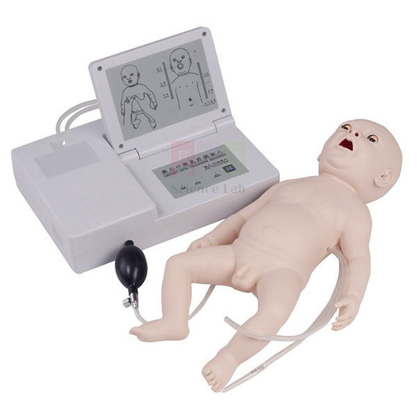
Advanced Infant CPR Training Manikin
Advanced Infant CPR Training Manikin (Product Code: SLE/BL/07) is an infant simulation model for learning how to revive a baby through heart pressure (chest compression) and mouth-to-mouth respiration. Designed according to the international CPR standard, it has light indicator displays that separate correct from wrong compression position and correct from incorrect artificial respiration, so learners see their mistakes as they make them. Neonatal and paediatric nurses, midwifery students, childcare workers and new-parent classes practise on it, and it fits well into the educational lab equipment India nursing schools keep for skills training. It is made by Jain Scientific Equipments Pvt Ltd (ScienceLab), Ambala.
Key Details
| Parameter | Detail |
|---|---|
| Type | Infant CPR simulation model |
| Design basis | Designed according to the international CPR standard |
| Skills practised | Heart pressure and mouth-to-mouth respiration for infant revival |
| Compression indicator | Light indicator display for correct and wrong compression position |
| Respiration indicator | Light indicator display for correct and incorrect artificial respiration (mouth-to-mouth) |
| Suitable for | Infant CPR and first aid courses |
Typical Use
Infant resuscitation calls for a very different touch: compressions are given with fingers rather than the heel of the hand, and rescue breaths are much smaller. Practising on an infant-sized model is the only realistic way to build that feel. Instructors usually demonstrate once, then let each learner try while the compression-position light confirms finger placement on the chest and the respiration light shows whether each breath was delivered correctly.
Hospitals running neonatal resuscitation refreshers, colleges teaching paediatric nursing and community programmes putting together a first aid training kit school staff and parents can use all find it useful. Clean the face and mouth after each learner, keep the manikin in a closed box when not in use, and check the indicator lights before each class so no session time is lost.
Manufacturer
Made in Ambala, Haryana, this manikin is part of the medical training range of Jain Scientific Equipments Private Limited, which does business under the ScienceLab name. Read about the company on our company profile.
Related Human Models
Items often purchased with the infant manikin for life support and physiology teaching:
- Advanced Adult CPR Training Manikin — for adult technique, with audio prompting and grade printing.
- Advanced Child CPR Training Manikin — bridges infant and adult technique for older children.
- Human Heart, Pumping Model — helps learners see what compressions are doing for circulation.
- Human Circulatory System Model — for background teaching on blood flow around the body.
More models are in our Human Models category.
Frequently Asked Questions
What skills can be practised on this manikin?
Infant chest compressions (heart pressure) and mouth-to-mouth respiration, with lights confirming correct or wrong compression position and correct or incorrect breaths.
Why buy an infant model when we already have an adult one?
Infant CPR uses different hand technique and much gentler breaths, which cannot be practised realistically on an adult-sized manikin.
Is it designed to a recognised CPR standard?
It is designed according to the international CPR standard. Please tell us if your programme follows a particular guideline version so we can advise.
How is the manikin powered?
Please check the power requirements for the indicator lights of the current model with us through the contact page.
How should it be cleaned between learners?
Wipe the face and mouth area with a suitable disinfectant wipe after each learner and let it dry. Ask us about replacement parts if your course has high usage.
Do you provide warranty and bulk pricing?
Warranty terms and pricing for multiple units are given in our quotation. We deliver to India and international markets.
Request a Quote
Ask for a quotation on the Advanced Infant CPR Training Manikin via the contact page, or place it in your enquiry cart.
Last updated: 24 September 2026
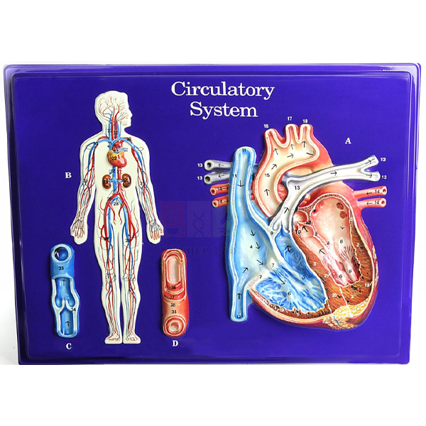
Circulatory System Model Activity Set
Circulatory System Model Activity Set (Product Code: SLE/BL/17) is a hands-on classroom set built around a full-colour, markable model of the circulatory system made from durable vinyl. Students trace blood vessels throughout the body, study a cutaway view of the heart’s interior and look at an enlarged cutaway section of a vein and an artery, while an activity guide and a three-overlay heart transparency reinforce how circulation works. Middle and secondary school biology teachers and teacher-training colleges use it as a ready-made human body model kit India classrooms can run as a guided activity. It is made by Jain Scientific Equipments Pvt Ltd (ScienceLab), Ambala.
Specifications
| Parameter | Detail |
|---|---|
| Model | Full-colour markable model of durable vinyl |
| Features shown | Vessels throughout the body, cutaway view of the interior of the heart, enlarged cutaway section of a vein and an artery |
| Transparency | Colourful three-overlay transparency of the heart |
| Activity guide | Background information, basic understandings, teacher-developed activities, glossary, key to model structures, colour transparencies and a blackline master |
Typical Use
Because the vinyl surface is markable, students can draw the path of blood directly on the model — for example, colouring the route from the body back to the heart, through the lungs and out again — and then compare their tracing with the key to model structures. The three-overlay heart transparency builds the heart up layer by layer on a projector, which helps a whole class follow the chambers and valves before pairs of students work with the cutaway heart on the model itself. Comparing the enlarged vein and artery sections leads naturally into a discussion of wall thickness, blood pressure and valves in veins.
The guide’s teacher-developed activities, glossary and blackline master save preparation time, making the set one of the more practical biology teaching aids India schools can use for a complete lesson sequence. Use the key to model structures for quick assessment, store transparencies flat in a folder, and ask us which markers are recommended for the vinyl surface.
Manufacturer
Jain Scientific Equipments Private Limited, which operates under the ScienceLab brand in Ambala, Haryana, supplies this activity set to schools and colleges. The company is incorporated as a private limited company (CIN U29309HR2020PTC087597) and manufactures at its licensed factory at 449, HSIIDC Industrial Area, Saha, Ambala (Factory Licence AMB-ONLINE-CHD-J-414). Our company profile has further details.
Related Human Models
Teachers extending a circulation unit often add:
- Human Circulatory System Model — a display model of the whole system for demonstration.
- Human Heart, Pumping Model — shows the heart’s pumping action to follow up the cutaway study.
- Human Cardiac Beat And Blood Circulation — demonstrates heartbeat and circulation together.
- Endocrine System Model Activity Set — a companion activity set for the next body-systems topic.
- Prepared Microscope Slide Set – Circulatory System — lets students see blood and vessel tissue under the microscope.
Browse all options in our Human Models category.
Frequently Asked Questions
What is included in the set?
A full-colour markable vinyl model and an activity guide containing background information, basic understandings, teacher-developed activities, a glossary, a key to model structures, colour transparencies and a blackline master, plus a three-overlay heart transparency.
Can students write on the model?
Yes, the model is markable, so students can trace vessels and label structures during activities. Please ask us which markers are best for cleaning off afterwards.
Which parts of the heart and vessels can be studied?
The model has a cutaway view of the interior of the heart and an enlarged cutaway section of a vein and an artery, alongside vessels traced throughout the body.
What is a blackline master used for?
It is a printable master sheet teachers can photocopy so each student has a worksheet to complete alongside the model.
What class levels is it suited to?
It suits school biology lessons on the circulatory system and introductory teacher-training or nursing courses. For curriculum questions, contact us through the contact page.
Can schools order several sets?
Yes, we supply schools in India and international markets. Share the number of sets and delivery details, and our quotation will include warranty terms.
Request a Quote
Request pricing for the Circulatory System Model Activity Set through our contact page, or add it to the enquiry cart with other biology teaching aids.
Last updated: 24 September 2026
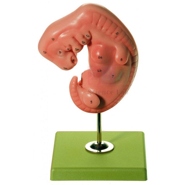
Embryo Model
The Embryo Model (Product Code: SLE/BL/32) is a stand-mounted teaching model of a human embryo at 4 weeks old, moulded in advanced PVC and marked with 12 positions for identification. It lets biology teachers, nursing tutors and embryology students show the very early body form in three dimensions, something that flat textbook drawings rarely convey well. The model is made by Jain Scientific Equipments Pvt Ltd (ScienceLab) in Ambala and sits comfortably among the biology teaching aids India's schools and colleges use to introduce development before birth.
Specifications
| Parameter | Detail |
|---|---|
| Type | Human embryo model |
| Stage represented | Embryo 4 weeks old |
| Positions shown | 12 |
| Material | Advanced PVC |
| Mounting | Mounted on stand |
Typical Use
In lessons on reproduction and human development, the Embryo Model gives students a fixed reference point for the first month after fertilisation. Teachers work through the 12 positions one at a time, explaining how the early body takes shape and how the foundations of the major organ systems are laid down long before the embryo resembles a baby. Because it stands upright, the model can be passed around a small group or placed on the front bench while the class sketches and labels it.
It is most effective when used as the first step in a sequence: start with this 4-week embryo, then move to later-stage fetus models so learners can see how dramatically form changes over the pregnancy. Schools assembling a human biology model set in India often place it next to placenta and uterus models for exactly this reason. For care, wipe the PVC surface with a soft damp cloth and mild soap, dry it, and keep it out of strong sunlight when stored.
Manufacturer
This embryo model comes from Jain Scientific Equipments Private Limited, the Ambala, Haryana company that trades under the ScienceLab name and produces anatomical models together with a wider range of laboratory teaching equipment. Background on the firm is available on our company profile page.
Related Human Models
To build a complete lesson on prenatal development, these models are commonly ordered together with the Embryo Model:
- Human Embryo Set, PVC — follows the fetus and uterus month by month through gestation, continuing where this 4-week stage leaves off
- Human Embryo Development Set — covers later fetal stages and fetal positions for the second half of the lesson
- Human Placenta Model — explains how the placenta and umbilical cord support the developing embryo
- Human Female Genital Organs Model — shows the organs where fertilisation and implantation take place
See every item in our Human Models range.
Frequently Asked Questions
What stage of development does the Embryo Model represent?
A human embryo at 4 weeks old. It is intended for teaching the earliest phase of development rather than the later fetal months.
How many features are identified on the model?
12 positions are shown. If you need a printed list of those positions for worksheets, please ask through our contact page when you enquire.
What is it made of, and how is it mounted?
It is made of advanced PVC and mounted on a stand, so it can be displayed upright on a bench or shelf and cleaned with a damp cloth.
What are the overall dimensions?
Dimensions are not listed in our current product details. Contact us and we will confirm the size before you order, which is useful when planning cabinet space.
Is it suitable for nursing and medical colleges as well as schools?
Yes. Senior school biology classes use it for the reproduction chapter, while nursing, midwifery and medical students use it when studying early embryology.
What should an institutional quotation request include?
Send the product code SLE/BL/32, the quantity, your delivery address and any paperwork your purchase department needs; we will reply with a formal quotation.
Do you supply outside India?
Yes. ScienceLab supplies schools, colleges and distributors in India and international markets; warranty and delivery terms are stated on each quotation.
Request a Quote
To order the Embryo Model, send your quantity and delivery location through our contact page, or add it to the enquiry cart along with other anatomy models so we can quote them together.
Last updated: 24 September 2026
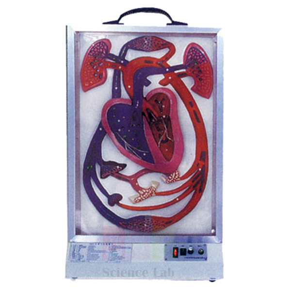
Human Cardiac Beat And Blood Circulation
The Human Cardiac Beat And Blood Circulation model (Product Code: SLE/BL/47) is a powered demonstration unit that pairs an enlarged, sectioned heart with a diagram of the body's blood circulation. Its flexible heart wall is moved mechanically from behind so the heart visibly beats, the valves open and close, and flashing lights trace the flow of blood — red for arteries and blue for veins. For institutions comparing heart model anatomy options in India, that movement is what separates it from static display models. It is manufactured by Jain Scientific Equipments Pvt Ltd (ScienceLab), Ambala, for schools, nursing colleges and medical teaching departments.
Specifications
| Parameter | Detail |
|---|---|
| Type | Working heart and blood circulation demonstration model |
| Size | 41 x 17 x 71 cm |
| Heart display | Enlarged heart shown in section, together with a blood circulation diagram |
| Heart wall | Flexible material, moved by a mechanism behind it to demonstrate the heartbeat |
| Valves | Shown opening and closing |
| Circulation display | Light flashes — red represents arteries, blue represents veins |
| Power | Requires a power supply |
Typical Use
Teachers switch the model on at the start of a cardiac cycle lesson and let the class watch several beats before explaining what is happening: the heart wall contracting and relaxing, the valves opening to let blood through and closing to stop it flowing backwards, and the lights showing oxygen-rich blood leaving through the arteries and returning through the veins. Having motion and colour at the same time helps students connect the pumping action of the heart with the route blood takes around the body.
As school biology lab equipment in India, it fits the circulation chapter in secondary classes and is equally useful for nursing and paramedical students revising cardiac physiology. The light flashes are easiest to follow in a dimmed room. Place the unit on a stable bench within reach of a socket, switch it off between lessons, and keep the flexible heart wall away from sharp objects and direct sunlight.
Manufacturer
Jain Scientific Equipments Private Limited produces this circulation model in Ambala, Haryana, and sells it under its trading name, ScienceLab, as part of a catalogue that spans anatomy models and general laboratory apparatus. Our company profile gives more detail about the business.
Related Laboratory Equipment
These items extend a heart and circulation lesson built around this model:
- Human Heart, Pumping Model — a hand-operated alternative in which a squeeze bulb drives simulated blood through clear chambers
- Human Heart Model — a static model for close study and labelling of heart anatomy after the demonstration
- Human Fetal Circulation Model — shows how circulation is arranged before birth, for comparison with the adult pattern
- Human Lungs with Heart and Larynx Model — links the heart to the lungs for the pulmonary part of the circuit
- Blood Pressure Monitor — lets students take real readings once they understand what drives blood pressure
More anatomy teaching models are listed in the Human Models category.
Frequently Asked Questions
Does the model need electricity?
Yes, it needs a power supply to run the heartbeat mechanism and the lights. For voltage and plug details, especially on export orders, please check with us through the contact page.
How much space does it take up?
The model measures 41 x 17 x 71 cm, so allow a bench or shelf position of at least that footprint and height.
How is the blood flow shown?
By flashing lights on the circulation diagram: red lights represent arteries and blue lights represent veins.
Can students actually see the valves working?
Yes. The valves open and close as the flexible heart wall moves, which makes the role of valves in one-way flow easy to explain.
Who should we contact if the mechanism or lights need attention?
Write to us with the product code SLE/BL/47 and a short description of the issue, and our team will advise on the next step. Warranty terms are stated on the quotation.
Can you supply several units or ship internationally?
Yes. We quote for single units, multiple-campus orders and distributor quantities for India and international markets.
Request a Quote
For pricing and delivery on the Human Cardiac Beat And Blood Circulation model, reach us via the contact page or place it in your enquiry cart with any related heart models you need.
Last updated: 24 September 2026
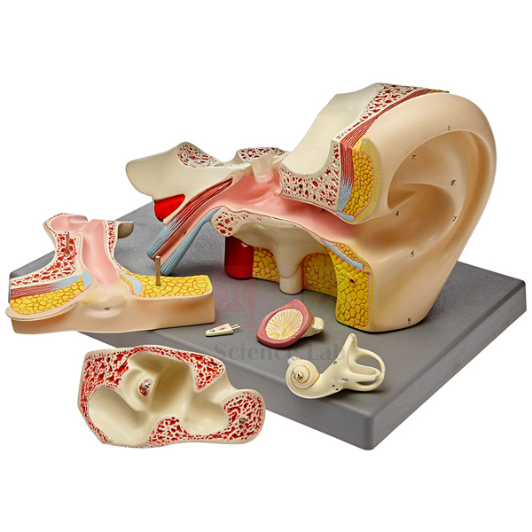
Human Ear Model, 5 Parts
The Human Ear Model, 5 Parts (Product Code: SLE/BL/50) is a dissectible model of the human ear, built 5–6 times larger than the natural ear so that small hearing structures can be pointed out clearly in front of a class. It separates into 5 parts, is coloured to distinguish the different structures, and is numbered with a key card. Schools, nursing colleges and audiology courses looking for an ear model for anatomy lab use in India choose it mainly for explaining how hearing works. It is manufactured by Jain Scientific Equipments Pvt Ltd (ScienceLab), Ambala, and supplied mounted on a base.
Specifications
| Parameter | Detail |
|---|---|
| Type | Dissectible human ear model |
| Number of parts | 5, dissectible |
| Enlargement | 5–6 times larger than the natural ear |
| Mounting | Mounted on a base |
| Finish | Appropriately coloured |
| Identification | Numbered, with key card |
| Teaching focus | Explaining the hearing mechanism |
Typical Use
The model is built for the lesson that asks, "How do we hear?" Teachers follow a sound wave from the outer ear, through the ear canal to the eardrum, across the tiny bones of the middle ear and into the inner ear, where vibrations are turned into nerve signals. Because the model is enlarged 5–6 times and comes apart into 5 pieces, students can see how the outer, middle and inner ear fit together and then examine each region in turn using the numbers and key card.
It works well in high school biology lab equipment sets for the sense organs chapter, and in nursing, speech therapy and audiology courses where ear anatomy underpins practical work. After each session, reassemble the 5 parts before storing so none go missing, keep the model covered to limit dust, and clean painted surfaces gently with a soft, slightly damp cloth.
Manufacturer
This ear model is made by Jain Scientific Equipments Private Limited, which operates in Ambala, Haryana, under the ScienceLab brand. The company is incorporated as a private limited company (CIN U29309HR2020PTC087597) and produces its range at a licensed factory at 449, HSIIDC Industrial Area, Saha, Ambala (Factory Licence AMB-ONLINE-CHD-J-414). You can read more on our company profile.
Related Laboratory Equipment
Items that pair with the Human Ear Model, 5 Parts in a hearing and balance lesson:
- Human Inner Ear Model, 18x — a far more magnified view of the inner ear alone for detailed study
- Human Inner Ear Model, 4x — a smaller inner-ear model suited to individual or small-group work
- Human Ear Model — an alternative ear model if you are equipping several benches
- Sight & Sound Health Lab — activity material on the senses of sight and hearing to follow the anatomy lesson
Browse our complete Human Models collection for other sense organ models.
Frequently Asked Questions
How many parts does the ear model separate into?
It is dissectible into 5 parts, which lets students look at individual regions of the ear and then put them back together.
How large is it compared with a real ear?
It is 5–6 times larger than the natural ear. Overall dimensions are not listed in our current details; contact us if you need them for cabinet planning.
How are the structures labelled?
Structures are numbered on the model and identified on the accompanying key card.
Is it suitable for explaining how hearing works?
Yes, that is its main purpose. The enlargement and removable parts make it straightforward to trace sound through the outer, middle and inner ear.
Can your company registration details be provided for vendor enrolment?
Yes. Our CIN and factory licence are listed above, and further registration details needed for tender or vendor forms can be requested through the contact page.
Do you supply ear models in quantity or for export?
Yes. We quote for classroom sets, college departments and distributors across India and international markets; warranty terms are confirmed on the quotation.
Request a Quote
Ask for pricing on the Human Ear Model, 5 Parts through our contact page, or add it to the enquiry cart together with the other sense organ models you need.
Last updated: 24 September 2026
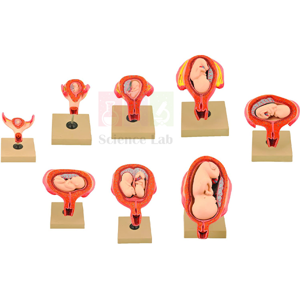
Human Embryo Development Set
The Human Embryo Development Set (Product Code: SLE/BL/51) is a series of models showing the fetus at different stages of the gestation period, from each of the first three months of pregnancy through to the fourth and fifth month, together with models of a breech presentation, a fetus lying on its back and twin five-month-old fetuses in the uterus. Each model is mounted on a base, coloured and numbered with a key card. It is produced by Jain Scientific Equipments Pvt Ltd (ScienceLab), an anatomical models manufacturer in Ambala, for biology departments, nursing and midwifery schools and medical colleges.
Specifications
| Parameter | Detail |
|---|---|
| Type | Set of fetal development models covering the gestation period |
| Models 1–3 | One model for each of the first three months of pregnancy |
| Model 4 | Fetus in the fourth to fifth month |
| Model 5 | Fetus in the breech position |
| Model 6 | Fetus lying on its back |
| Model 7 | Twin five-month-old fetuses in the uterus |
| Closer examination | Models 4–8 are described as allowing closer examination of the anatomical features that have developed |
| Mounting | Mounted on a base |
| Finish and identification | Appropriately coloured, numbered, with key card |
Typical Use
The set is laid out in order on the bench so students can walk along a timeline of pregnancy. The first three models show how quickly the embryo changes month by month early on; the fourth shows the more developed fetus of the fourth to fifth month. The remaining models move the discussion from growth to position: the breech model and the fetus on its back let tutors explain fetal presentation and why it matters as delivery approaches, while the twin model shows how two fetuses share the space of the uterus.
This makes the set especially useful in midwifery and obstetric nursing training, as well as in senior-school reproduction chapters. As part of a biology lab setup in schools in India, it is often shared between sections and kept in a closed cabinet; keep the key card with the set, dust the models with a soft brush and return each one to its place in the sequence after class.
Manufacturer
ScienceLab is the brand under which Jain Scientific Equipments Private Limited, based in Ambala, Haryana, makes and sells this development set and its other human anatomy teaching models. Visit the company profile to learn more about the company.
Related Laboratory Equipment
Models and aids that complete a pregnancy and birth lesson alongside this set:
- Human Embryo Set, PVC — a 10-part series showing the uterus and fetus for every month up to the ninth
- Embryo Model — the 4-week embryo, for the very start of the development timeline
- Human Placenta Model — explains how the fetus is supported through the placenta and umbilical cord
- Human Birth Demonstration Model — carries the lesson on from fetal position to the birth process
- Fetal Positions Before Birth — an additional teaching aid on presentations before delivery
Find further anatomy models in our Human Models category.
Frequently Asked Questions
Which stages of pregnancy does the set cover?
Models 1–3 each show one of the first three months, and model 4 shows the fetus in the fourth to fifth month. Further models show fetal positions and twins.
How many models are in the set?
Our product description details models 1 to 7 and also refers to models 4–8 for closer examination, so please confirm the exact piece count with us through the contact page before ordering.
Does the set show abnormal fetal positions?
Yes. Model 5 shows the breech position and model 6 shows the fetus lying on its back, alongside the standard developmental models.
Is a twin pregnancy included?
Yes. Model 7 shows twin five-month-old fetuses in the uterus.
How do students identify the structures?
The models are numbered and a key card is supplied, so students can check each numbered feature against its name.
What do you need to prepare a quotation?
Tell us the quantity of sets, the delivery address and your required timeline. We supply institutions and distributors in India and international markets, and warranty terms are confirmed in writing on the quotation.
Request a Quote
Request pricing for the Human Embryo Development Set on our contact page, or add it to the enquiry cart with any other development models you are considering.
Last updated: 24 September 2026
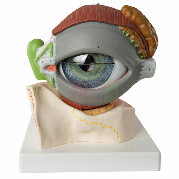
Human Eye Model with Eyelids
The Human Eye Model with Eyelids (Product Code: SLE/BL/52) is a 5x-enlarged model that shows the eyeball in its bony orbit together with the surrounding muscles, the eyelids, a tear duct and the lachrymal gland. The eyeball itself is horizontally sectioned, and the sclera with its clear cornea, as well as the muscles, can be removed. As an eye model for anatomy teaching in India, it goes a step beyond a plain eyeball by showing how the eye sits within and is served by the structures around it. It is manufactured by Jain Scientific Equipments Pvt Ltd (ScienceLab), Ambala, mounted on a base and numbered with a key card.
Specifications
| Parameter | Detail |
|---|---|
| Type | Eye-in-orbit model with eyelids |
| Enlargement | 5x |
| Structures included | Eyelids, bony orbit with muscles, tear duct, lachrymal gland |
| Eyeball | Horizontally sectioned |
| Removable parts | Sclera with clear cornea; muscles |
| Mounting and finish | Mounted on a base, appropriately coloured and nicely finished |
| Identification | Numbered, with key card |
Typical Use
The model is ideal for demonstrating the relationship between the eyeball and the musculature within the orbit. Tutors lift away the removable muscles to show where they attach and explain how they turn the eye in different directions, then remove the sclera and clear cornea to reveal the horizontally sectioned interior. The eyelids, lachrymal gland and tear duct add a second strand to the lesson: how the eye is protected and kept moist, and where tears are produced and drained.
Optometry, ophthalmic assistant and nursing students use it for these accessory structures, while senior school classes use it in the sense organs chapter. ScienceLab supplies it as part of the biology lab equipment range that institutions across India order for sense organ teaching. Handle the clear cornea by its edges to avoid scratches, clean it with a soft lint-free cloth, and keep the removable parts together on the base when the model is not in use.
Manufacturer
Jain Scientific Equipments Private Limited manufactures this eye model and markets it as ScienceLab from Ambala, Haryana, where the company designs and builds teaching models for biology and medical education. The company profile has the full background.
Related Human Models
Other eye models that suit the same vision and sense organ lessons:
- Human Eye in Orbit Model — another orbit-based design for comparing how the eye is held in the skull
- Human Eye Model — concentrates on the eyeball itself when the accessory structures are not needed
- Eye Microanatomy Model — takes the lesson down to the tissue-level structure of the eye
- Cataract Eye Model with 4 Forms of Cataract — links normal eye anatomy to a common clinical condition
The full Human Models range lists all our anatomy teaching models.
Frequently Asked Questions
How is this different from a standard eyeball model?
It includes the eyelids, bony orbit with muscles, tear duct and lachrymal gland, so it shows the eye in its surroundings rather than on its own.
How much is the eye enlarged?
The model is 5x enlarged. For overall dimensions, please contact us.
Which parts can be removed?
The sclera with clear cornea and the muscles are removable, and the eye itself is horizontally sectioned to show its interior.
Does it explain the tear system?
Yes. The lachrymal gland and tear duct are included, which lets tutors cover tear production and drainage in the same lesson.
Is a key to the numbered structures supplied?
Yes, the model is numbered and comes with a key card.
Can we order this for several labs or for export?
Yes. Quotations are prepared for single pieces, department sets and distributors in India and international markets, with warranty terms stated on the quotation.
Request a Quote
Send your requirement for the Human Eye Model with Eyelids through the contact page, or add it to the enquiry cart along with other eye models for a combined quotation.
Last updated: 24 September 2026
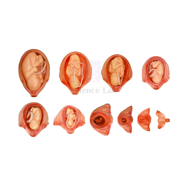
Human Embryo Set, PVC
The Human Embryo Set, PVC (Product Code: SLE/BL/53) is a 10-part model series that shows the relationship between the fetus and the uterus across the whole gestation period, beginning with a normal uterus and then covering each month of pregnancy up to the ninth. The parts are made of PVC and synthetic rubber. Made by Jain Scientific Equipments Pvt Ltd (ScienceLab) in Ambala, one of the human anatomical models manufacturers in India serving schools and colleges, the set is used in biology, nursing and midwifery teaching wherever a month-by-month picture of pregnancy is needed.
Specifications
| Parameter | Detail |
|---|---|
| Type | Embryo and fetus development series showing fetus and uterus |
| Number of parts | 10 |
| Material | PVC and synthetic rubber |
| Part 1 | Normal uterus, inner genitals |
| Part 2 (first month) | Placenta is shown |
| Part 3 (second month) | Embryo looks like a human |
| Part 4 (third month) | Fetus about 9 cm |
| Part 5 (fourth month) | Fetus about 16 cm |
| Part 6 (fifth month) | Fetus about 25 cm |
| Part 7 (sixth month) | Fetus about 30 cm |
| Part 8 (seventh month) | Fetus about 35 cm |
| Part 9 (eighth month) | Fetus about 40 cm |
| Part 10 (ninth month) | Fetus about 45 cm |
Typical Use
Because every month of pregnancy has its own part, the set is ideal for a growth-timeline activity. Students arrange the 10 parts in order, starting from the normal uterus, and note how the uterus changes as the fetus grows. The fetal heights listed for each stage — from about 9 cm in the third month to about 45 cm in the ninth — can be plotted on a simple graph, turning a descriptive topic into a data exercise. Tutors also use the second and third parts to discuss when the placenta appears and when the embryo takes on a recognisably human form.
In nursing and midwifery training the set supports antenatal teaching, where students must relate the month of pregnancy to what is happening inside the uterus. It also suits health education programmes in schools in India that cover pregnancy and prenatal care. Store the parts in order so the sequence is ready for the next class, and clean the PVC and rubber surfaces with a damp cloth only.
Manufacturer
The set is manufactured in Ambala, Haryana, by Jain Scientific Equipments Private Limited — the company behind the ScienceLab trading name — whose catalogue covers reproductive, developmental and other human anatomy models. See our company profile for more information.
Related Laboratory Equipment
These models round out a pregnancy and development module built around the 10-part set:
- Human Embryo Development Set — adds fetal positions such as breech and a twin pregnancy
- Embryo Model — a closer look at the 4-week embryo that precedes the later parts of this set
- Human Placenta Model — expands on the placenta introduced in part 2
- Human Birth Demonstration Model — continues the story after the ninth month
- Female Pelvis with Organs — places the uterus in the context of the surrounding pelvic organs
All our anatomy teaching models are grouped in the Human Models category.
Frequently Asked Questions
How many parts are in the Human Embryo Set?
There are 10 parts: a normal uterus followed by one part for each month of gestation from the first to the ninth.
What material is it made from?
The set is made of PVC and synthetic rubber, which can be wiped clean with a damp cloth.
Are fetal sizes given for each stage?
Yes. The reference heights run from about 9 cm (third month) to about 45 cm (ninth month). For the physical dimensions of the model parts themselves, please contact us.
At which stage is the placenta shown?
The placenta is shown in part 2, which represents the first month of gestation.
Is it suitable for midwifery and nursing courses?
Yes. Relating each month of pregnancy to the fetus and uterus is central to antenatal teaching, and the set is organised exactly that way.
Can we place a biology model kit bulk order that includes this set?
Yes. We quote combined orders for institutions and distributors in India and international markets. Include quantities, delivery address and any documentation required; warranty terms are confirmed on the quotation.
Request a Quote
Ask for a quotation on the Human Embryo Set, PVC through the contact page, or add it to the enquiry cart with the other development models on your list.
Last updated: 24 September 2026
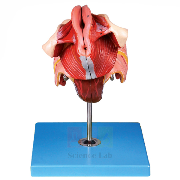
Human Female Genital Organs Model
The Human Female Genital Organs Model (Product Code: SLE/BL/54) is a dissected model showing both the internal and the external female genital organs, with 40 positions marked for identification. It is made of advanced PVC and mounted on a stand. Manufactured by Jain Scientific Equipments Pvt Ltd (ScienceLab), Ambala, it serves as reproductive-system biology lab equipment in India for senior school classes, nursing and midwifery programmes, and medical and paramedical colleges.
Specifications
| Parameter | Detail |
|---|---|
| Type | Dissected female genital organs model |
| Coverage | Internal and external female genital organs |
| Positions shown | 40 |
| Material | Advanced PVC |
| Mounting | Mounted on a stand |
Typical Use
Because it shows the external and internal organs in one dissected view, the model lets a tutor move from the outer anatomy inward in a single, continuous explanation, rather than switching between separate diagrams. With 40 positions to work through, it supports both an introductory overview for school students and the more detailed identification expected in nursing, midwifery and medical anatomy practicals. Typical topics include the structure of the reproductive tract, the menstrual cycle, fertilisation and the anatomical basis of common gynaecological examinations.
It works well in small-group sessions where students take turns naming the numbered positions, and it is also a sensible, matter-of-fact aid for reproductive health education. Keep the model on its stand when not in use, wipe it with a damp cloth and mild detergent, and avoid alcohol-based cleaners that can dull painted PVC surfaces.
Manufacturer
This model is made by Jain Scientific Equipments Private Limited in Ambala, Haryana, the company that trades as ScienceLab. It is a registered MSME enterprise with manufacturing as its major activity (Udyam UDYAM-HR-01-0003714), and its export consignments are handled under Importer-Exporter Code AAECJ8618A issued by the DGFT. Further details are on our company profile.
Related Human Models
Models that are often taught together with the Human Female Genital Organs Model:
- Human Male Genital Organs Model — the male counterpart for side-by-side comparison in the same lesson
- Human Female Ovary Model — a focused model for studying the ovary in more detail
- Female Pelvis with Organs — shows the reproductive organs in their position within the pelvis
- Human Female Urogenital Model — connects the reproductive organs with the kidneys, ureters and bladder
View the complete Human Models range for other anatomy teaching models.
Frequently Asked Questions
Does the model show both internal and external organs?
Yes. It shows the internal and external female genital organs in dissected form.
How many structures are marked?
40 positions are shown. If you need the identification list in advance for course material, please request it through our contact page.
What is it made of?
Advanced PVC, mounted on a stand for display and demonstration.
What size is the model?
Dimensions are not listed in our current product details. Contact us and we will confirm them before you order.
Can this model be included in a biology model kit bulk order?
Yes. We regularly combine anatomy models into a single quotation for colleges and distributors; send us the list of items and quantities.
Can you export to institutions outside India?
Yes. We supply India and international markets and can share our IEC and other registration details for your vendor documentation. Warranty terms are stated on the quotation.
Request a Quote
For a price on the Human Female Genital Organs Model, write to us through the contact page or add the model to your enquiry cart with any related reproductive-system models.
Last updated: 24 September 2026
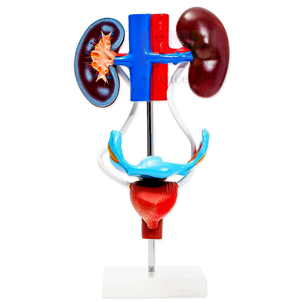
Human Female Urogenital Model
The Human Female Urogenital Model (Product Code: SLE/BL/55) brings the female urinary and reproductive systems together in a single PVC plastic model measuring 20 x 20 x 50 cm. It shows the kidney, ureters and urinary bladder alongside the uterus and its accessories, the vagina, the ovary membrane and the supporting ligaments, including the uterus ligament and its artery. The model is manufactured by Jain Scientific Equipments Pvt Ltd (ScienceLab), Ambala, and is used as a combined excretory and reproductive teaching aid in schools, nursing colleges and medical institutions.
Specifications
| Parameter | Detail |
|---|---|
| Type | Female urogenital system model |
| Material | PVC plastic |
| Size | 20 x 20 x 50 cm |
| Urinary structures | Kidney, ureters, urinary bladder |
| Reproductive structures | Uterus and accessories of the uterus, vagina, ovary membrane |
| Supporting structures | Ligaments, including the uterus ligament and its artery |
Typical Use
Textbooks usually present the urinary and reproductive systems in separate chapters, yet in the body they lie close together in the pelvis. This model is useful precisely because it shows them in one piece: students can follow urine from the kidney down the ureters to the bladder, and see at the same time how the bladder sits in relation to the uterus and vagina. Tutors also use the ligaments and the uterine artery to explain how the uterus is held in position and supplied with blood.
For nursing and midwifery students, understanding this close relationship is useful background for clinical procedures and for recognising how pregnancy affects the urinary tract. In high school biology lab equipment collections it supports both the excretion and the reproduction chapters with one purchase. At 20 x 20 x 50 cm it needs a clear shelf; clean it with a slightly damp cloth and keep it away from heat sources.
Manufacturer
ScienceLab is the trading name used by Jain Scientific Equipments Private Limited of Ambala, Haryana, which manufactures this urogenital model among its anatomy and life-science teaching products. Our company profile describes the company in more detail.
Related Laboratory Equipment
Models that extend a urinary and reproductive system lesson:
- Human Male and Female Urogenital Systems Model — for comparing male and female arrangements side by side
- Human Kidney Model — an enlarged look at kidney structure beyond what this combined model shows
- Human Kidney, Nephron, and Glomerulus Model Set — explains how the kidney filters blood at microscopic level
- Female Half Pelvis Model — a sectioned view of the female pelvis and its organs
- Human Female Genital Organs Model — more detail on the internal and external genital organs
Explore more anatomy models in our Human Models category.
Frequently Asked Questions
Which organs does the model show?
The kidney, ureters, urinary bladder, uterus with its accessories, vagina, ovary membrane and ligaments, including the uterus ligament and its artery.
What are its dimensions?
The model measures 20 x 20 x 50 cm.
What material is used?
It is made of PVC plastic, which is durable in regular classroom handling and easy to wipe clean.
Does it show the blood supply?
It includes the uterus ligament and its artery. Other vessels are not listed in our product details, so please contact us if a specific vessel is important for your syllabus.
Are the structures numbered or supplied with a key?
Our current listing does not state this. Ask us when you enquire and we will confirm what identification material is included.
What information should a purchase enquiry contain?
The product code SLE/BL/55, quantity, delivery address and any tender format you must follow. We supply institutions in India and international markets, with warranty terms given on the quotation.
Request a Quote
To receive a quotation for the Human Female Urogenital Model, use our contact page or add it to the enquiry cart together with any kidney or pelvis models you need.
Last updated: 24 September 2026
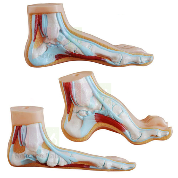
Human Foot Set Model
The Human Foot Set Model (Product Code: SLE/BL/56) is a set of three life-size, finely detailed foot models that compare a flattened foot, an arching foot and a normal foot. Each foot carries 23 numbered parts for identification, and the models are one-piece (not dissectible), so there are no loose pieces to lose between classes. Physiotherapy, podiatry, sports science and nursing tutors use the set to explain the arches of the foot and what happens when they are lost or exaggerated. It is manufactured by Jain Scientific Equipments Pvt Ltd (ScienceLab), Ambala, as one of its biology teaching aids for Indian and overseas institutions.
Specifications
| Parameter | Detail |
|---|---|
| Type | Set of foot models for comparing arch forms |
| Feet in the set | Flattened foot, arching foot, normal foot |
| Scale | Life size |
| Numbered parts | 23 on each foot |
| Construction | Not dissectible |
| Finish | Finely detailed |
Typical Use
The strength of this set is comparison. Placed side by side, the three feet let students see at once how the normal arch distributes body weight, how a flattened foot has lost that arch, and how an arching foot sits higher off the ground. Because every foot uses 23 numbered parts, learners can find the same structure on all three and describe how its position changes, which is exactly the kind of reasoning expected in physiotherapy and podiatry assessments.
The set is also useful for posture and footwear discussions in physical education and sports science, and for patient education in clinics where a practitioner needs to show a patient why an arch problem causes discomfort. It fits neatly into a human biology model set in India's colleges that already includes skeletal models of the leg and foot. Since the models do not come apart, care is simple: dust them regularly and wipe with a damp cloth.
Manufacturer
Jain Scientific Equipments Private Limited, trading as ScienceLab, makes this foot set in Ambala, Haryana, where it produces musculoskeletal and other anatomical models for teaching. Read about the company on our company profile.
Related Laboratory Equipment
Useful companions for a lesson on the foot and lower limb:
- Human Skeletal Foot Model — shows the bones that form the arches seen on the surface models
- Human Foot Model Set — an alternative foot set for institutions comparing options
- Human Leg and Portion of Pelvis Model — connects the foot to the leg and hip in one view
- Human Skeleton Model, Disarticulated — lets students handle individual foot bones
For more anatomy models, see the Human Models category.
Frequently Asked Questions
Which foot types are included?
Three: a flattened foot, an arching foot and a normal foot.
Are the models life size?
Yes, each foot is life size, which makes it easy to relate the models to real feet and footwear.
Can the feet be taken apart?
No. The models are not dissectible; they are designed for external comparison of arch form.
How many parts are numbered?
Each foot has 23 numbered parts. If you need the list of numbered parts ahead of delivery, ask us through the contact page.
Who typically buys this set?
Physiotherapy and podiatry departments, sports science and physical education faculties, nursing colleges and biology departments teaching the skeletal and muscular systems.
Do you accept institutional and export orders?
Yes. Send the quantity and delivery details for a formal quotation; we supply India and international markets, with warranty terms confirmed on the quotation.
Request a Quote
Enquire about the Human Foot Set Model through our contact page, or add it to the enquiry cart with any skeletal models you plan to order.
Last updated: 24 September 2026
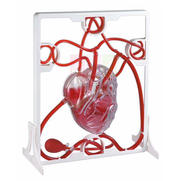
Human Heart, Pumping Model
The Human Heart, Pumping Model (Product Code: SLE/BL/61) is a 3-D working plastic heart with clear chambers, in which a squeeze bulb pumps simulated blood through the chambers in the same way a real heart moves blood. It is easy to assemble, comes with a complete instruction manual and a display stand, and includes acrylic paints and a brush so students can colour the finished heart themselves. Manufactured by Jain Scientific Equipments Pvt Ltd (ScienceLab), Ambala, it works as a science model kit for school projects as well as a hands-on aid for circulation lessons.
Specifications
| Parameter | Detail |
|---|---|
| Type | 3-D working heart model |
| Material | Plastic |
| Chambers | Clear, so the movement of simulated blood is visible |
| Pumping action | Squeeze bulb pumps simulated blood through the heart chambers |
| Assembly | Easy to assemble, with complete instruction manual |
| Supplied with | Display stand, acrylic paints and brush, instruction manual |
Typical Use
This model turns the heart lesson into a make-and-test activity. Students first assemble the heart following the instruction manual, then paint it with the acrylic paints and brush — teachers can ask them to use colour to separate the sides of the heart that carry oxygen-rich and oxygen-poor blood. Once it is on its display stand, squeezing the bulb sends simulated blood through the clear chambers, so the class can watch the direction of flow and discuss why the chambers and valves are arranged as they are.
It suits science fairs, holiday projects and classroom demonstrations alike, and heart model anatomy lessons in India's middle and secondary schools benefit from a model students have built themselves. Younger learners should assemble and paint it under teacher supervision, allow the paint to dry fully before pumping, and rinse and dry the chambers after use according to the manual.
Manufacturer
Behind the ScienceLab brand is Jain Scientific Equipments Private Limited, an Ambala, Haryana manufacturer whose range stretches from student project models like this pumping heart to full-size anatomy figures. Learn more on the company profile.
Related Human Models
These models take the circulation topic further once students have built the pumping heart:
- Human Cardiac Beat And Blood Circulation — a powered demonstration with a beating wall, moving valves and lights for arteries and veins
- Human Heart Model — a detailed anatomical model for labelling after the practical activity
- Heart 3 times Natural Size Model — an enlarged heart that the whole class can see from a distance
- Circulatory System Model Activity Set — extends the activity from the heart to the whole circulatory system
Our full range is in the Human Models category.
Frequently Asked Questions
How does the pumping action work?
A squeeze bulb pushes simulated blood through the clear heart chambers, imitating the way a real heart moves blood.
Does it arrive assembled?
It is designed to be easy to assemble and comes with a complete instruction manual, so students can put it together as part of the activity.
What is included?
The heart model parts, a display stand, acrylic paints, a brush and the instruction manual.
Is the simulated blood supplied?
The model is designed to pump simulated blood; please confirm through our contact page exactly what fluid is included with your order.
Is it suitable for school science projects?
Yes. Assembling, painting and then operating the heart makes it well suited to projects and exhibitions, with teacher supervision for younger students.
Can we buy class sets or order for export?
Yes. We quote class quantities for schools and distributors in India and international markets; warranty terms are confirmed on the quotation.
Request a Quote
For class-set pricing on the Human Heart, Pumping Model, contact us through the contact page or add it to the enquiry cart with other circulation models.
Last updated: 24 September 2026
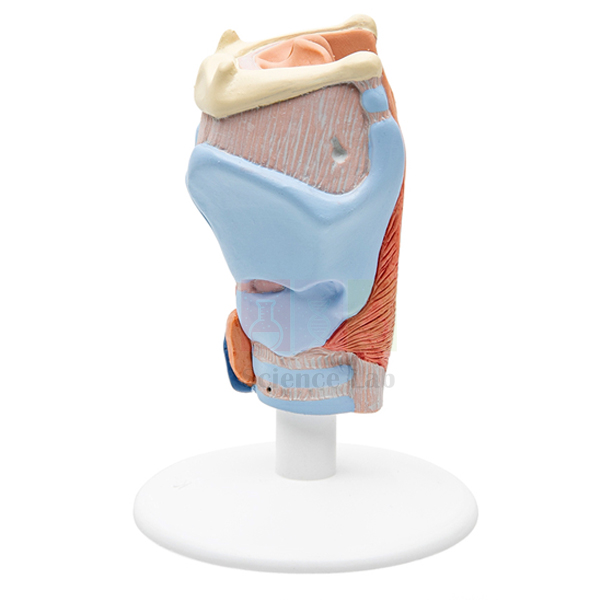
Human Larynx Model
Human Larynx Model (Product Code: SLE/BL/63) is a full-size, two-part anatomical model of the voice box used to teach the larynx and the muscles, thyroid gland, hyoid bone, windpipe, ligaments, vessels and nerves around it in medical, nursing, speech therapy and school biology classes. Every structure is numbered and identified on a key card, and the model is mounted on a base. It is made by Jain Scientific Equipments Pvt Ltd (ScienceLab), an OEM anatomical models manufacturer, at its own factory in Ambala, India.
Specifications
| Parameter | Detail |
|---|---|
| Structures shown | Muscles, thyroid gland, larynx, hyoid bone, windpipe (trachea), ligaments, vessels, nerves |
| Size | Full size |
| Parts | Divided into 2 parts |
| Identification | Numbered, with key card |
| Mounting | Mounted on a base |
| Finish | Appropriately coloured and nicely finished |
| Product code | SLE/BL/63 |
| Manufacturer | Jain Scientific Equipments Pvt Ltd (ScienceLab) — OEM |
| Country of origin | India (made in Ambala, Haryana) |
What Is the Larynx Model Used For?
Opening the model into its two parts reveals structures that are hidden in the intact neck, so a lecturer can move from the outer muscles and thyroid gland to the larynx itself and the windpipe below it. The numbered key card makes it suitable for self-study and spot tests: students name each numbered structure, then check their answers against the card. Speech and language therapy students relate the cartilages, ligaments and muscles to voice production, while nursing students use it to understand the airway.
In schools, the larynx often joins a respiratory-system display next to lung and nasal cavity models, forming part of a human biology model set for the topic. Keep the key card with the model, reassemble the two halves after each class, and store it covered to prevent dust collecting in the detail.
OEM Manufacturer
ScienceLab is the trading name of Jain Scientific Equipments Private Limited, the OEM (original equipment manufacturer) that makes this larynx model at its own licensed factory at Saha, Ambala, Haryana, India. Because the model is produced by the manufacturer rather than sourced from a trader, schools, medical and nursing colleges, dealers and importers buy it factory-direct.
A replacement key card, a repeat order for another laboratory or a matching respiratory model is handled by the same manufacturer — just ask us. More about the company is on the profile page.
Related Human Models
Models that continue the airway and respiratory lesson:
- Human Larynx, Trachea, and Bronchi Model — follows the airway from the larynx down into the bronchi
- Human Lungs with Heart and Larynx Model — shows the larynx in relation to the lungs and heart
- Anatomical Human Nasal Cavity Model — covers the upper airway above the larynx
- Human Respiratory System Model, 2-parts — an overview of the whole respiratory system
- Pathology of the Human Thyroid Model — extends the thyroid gland seen on this model
This model is part of the Human Models range of anatomical teaching models.
Frequently Asked Questions
Which structures can students identify on the Human Larynx Model?
Students can identify the muscles, thyroid gland, larynx, hyoid bone, windpipe, ligaments, vessels and nerves on the Human Larynx Model. Each structure is numbered and listed on the key card, which makes the model suitable for self-study and practical spot tests.
Does the Human Larynx Model come apart?
Yes. The Human Larynx Model divides into 2 parts, so a teacher can move from the outer muscles and thyroid gland to the inner larynx and windpipe. Reassemble the halves after each class so that the parts stay together.
Is the Human Larynx Model full size?
Yes. The Human Larynx Model is described as full size, which helps students relate it to their own neck anatomy. Overall dimensions of the model and base are not listed, so ask us through the contact page if you need them for display planning.
How should the Human Larynx Model be cleaned?
Dust the Human Larynx Model with a soft brush and wipe it with a lightly damp cloth. Avoid solvents, which can damage the coloured finish, keep the key card dry, and store the model covered to stop dust settling in the detail.
Who manufactures the Human Larynx Model?
The Human Larynx Model is manufactured by Jain Scientific Equipments Pvt Ltd (ScienceLab), the OEM, at its own factory at Saha, Ambala, Haryana, India. Institutions and dealers buy it factory-direct, with quotations coming straight from the manufacturer.
What should an institution include in an enquiry for the Human Larynx Model?
An enquiry for the Human Larynx Model should give product code SLE/BL/63, the quantity, the delivery address and any vendor documents your purchasing office needs, so ScienceLab can reply with one complete quotation.
Can the Human Larynx Model be supplied outside India?
Yes. ScienceLab supplies the Human Larynx Model to institutions, dealers and importers in India and international markets. Include the destination and quantity in your enquiry so that packing and shipping for the mounted model can be quoted with it.
Request a Quote
Quotations for the Human Larynx Model come direct from the manufacturer. Send your requirement on the contact page, or add the larynx model to the enquiry cart along with other respiratory models for one combined quotation.
Last updated: 24 September 2026
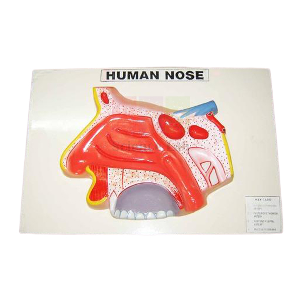
Human Nose Model on Base
The Human Nose Model on Base (Product Code: SLE/BL/64) is an enlarged model that shows the structure of the human nose, coloured to separate its parts and numbered with a key card. Mounted on a flat base, it sits steadily on a desk or lab bench where students can study it at close range. It is made by Jain Scientific Equipments Pvt Ltd (ScienceLab), Ambala, and is one of the biology teaching aids India's schools, nursing colleges and ENT training departments use for the sense of smell and the upper airway.
Key Details
| Parameter | Detail |
|---|---|
| Type | Enlarged human nose model |
| Shows | Structure of the nose |
| Mounting | Mounted on a base |
| Finish | Appropriately coloured |
| Identification | Numbered, with key card |
| Suitable for | Sense organ, respiratory system and ENT anatomy lessons |
Typical Use
The nose has two jobs that students often find hard to picture: it conditions the air we breathe, and it houses the sense of smell. With this enlarged model on the bench, teachers can show how the nose is built and explain how incoming air is filtered, warmed and moistened on its way to the lungs, then move on to how odours are detected. Small groups can gather round the base and use the numbers and key card to identify each structure for themselves, which works well as a short practical or a revision exercise.
It is a practical addition to school biology lab equipment in India for the sense organs and breathing chapters, and nursing and ENT trainees use it to revise nasal anatomy before clinical rotations. Keep the key card with the model, dust it with a soft brush, and wipe the coloured surfaces with a lightly damp cloth.
Manufacturer
The model is produced by Jain Scientific Equipments Private Limited, a company in Ambala, Haryana, that trades as ScienceLab and makes human anatomy models for classrooms and training institutes. Visit our company profile to find out more.
Related Human Models
Models for extending the lesson from the nose into the rest of the airway:
- Human Nose Model on Stand — the same subject on a stand, if you prefer a raised display
- Human Nose Model — another nose model option for equipping additional benches
- Anatomical Human Nasal Cavity Model — focuses on the internal nasal cavity behind the external nose
- Human Respiratory System Model, 2-parts — follows the airway down to the lungs
See the complete Human Models range for other sense organ models.
Frequently Asked Questions
What does the model show?
The structure of the human nose, enlarged, coloured and numbered, with a key card to identify each numbered feature.
By how much is it enlarged?
The model is enlarged, but the scale factor is not stated in our current product details. Please contact us and we will confirm it along with the dimensions.
What is the difference between this and the Human Nose Model on Stand?
According to our listings, the two differ in how they are mounted: this version stands on a flat base for desk use, while the other is held on a stand.
Can I see which structures are numbered before ordering?
Yes. Ask us for the key card details when you send your enquiry.
How should the model be cleaned?
Dust with a soft brush and wipe gently with a lightly damp cloth; avoid solvents that could affect the colours.
Do you supply schools outside India?
Yes. We serve institutions and distributors in India and international markets, and warranty terms are confirmed on each quotation.
Request a Quote
Get a quotation for the Human Nose Model on Base via the contact page, or add it to your enquiry cart along with any other respiratory or sense organ models.
Last updated: 24 September 2026
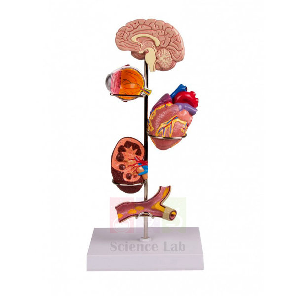
Human Hypertension Model
Human Hypertension Model (Product Code: SLE/BL/69) is a life-size teaching model on a stand with base used to explain high blood pressure and its effects in nursing, medical, pharmacy and health-education classes. Measuring 40 × 12 × 12 cm, it gives presenters something concrete to point to during lessons and awareness sessions on a condition that often has no symptoms. It is made by Jain Scientific Equipments Pvt Ltd (ScienceLab), an OEM manufacturer, at its own factory in Ambala, India.
Specifications
| Parameter | Detail |
|---|---|
| Type | Human anatomical teaching model |
| Topic | Hypertension (high blood pressure) |
| Size | 40 × 12 × 12 cm |
| Scale | Life size |
| Mounting | On stand with base |
| Structures shown | Not itemised in our description — photographs available on request |
| Product code | SLE/BL/69 |
| Manufacturer | Jain Scientific Equipments Pvt Ltd (ScienceLab) — OEM |
| Country of origin | India (made in Ambala, Haryana) |
What Is the Hypertension Model Used For?
In health education sessions the model gives the presenter something to point to while explaining what blood pressure is, how it is measured and why persistently raised pressure strains the heart and blood vessels over the years. Pairing it with a live reading taken on a blood pressure monitor turns an abstract number into a practical lesson that students and patients remember.
Nursing and pharmacy tutors use it when teaching lifestyle advice and the role of medication, and it sits naturally beside a heart model in anatomy displays on the circulatory system. Because it stands on its own base, it can stay on a reception counter or health-camp table all day. Dust it regularly and keep it out of strong sunlight.
OEM Manufacturer
Jain Scientific Equipments Private Limited, trading as ScienceLab, is the OEM (original equipment manufacturer) of the Human Hypertension Model and makes it at its own licensed factory at Saha, Ambala, Haryana, India. As an anatomical models manufacturer selling factory-direct, it deals with colleges, health programmes, dealers and importers without a trading intermediary: quotations, photographs of the current model and repeat orders all come from the maker. Company information is on the profile page.
Related Human Models
Items that support a blood pressure and circulation lesson:
- Human Heart Model — shows the organ that works harder when blood pressure is high
- Human Circulatory System Model — explains the network of vessels that carries blood around the body
- Microstructure of Artery and Vein Model — shows the vessel walls that raised pressure acts on
- Human Kidney Model — useful when discussing the kidney's link with blood pressure
- Blood Pressure Monitor — for taking real readings during the same session
The model belongs to the Human Models range of anatomical teaching models.
Frequently Asked Questions
What size is the Human Hypertension Model?
The Human Hypertension Model measures 40 × 12 × 12 cm. It is described as life size and comes on its own stand with base, so it can be placed on a demonstration bench, reception counter or health-camp table without extra support.
Which organs or changes does the Human Hypertension Model show?
The individual structures shown on the Human Hypertension Model are not listed in our current description. Request photographs and a detailed description through our contact page before ordering, so you can confirm the model matches your syllabus or awareness programme.
Who is the Human Hypertension Model suitable for?
The Human Hypertension Model suits nursing, medical and pharmacy students, health educators and clinics that run blood pressure awareness sessions. It gives presenters a concrete visual when explaining how persistently raised pressure strains the heart and blood vessels.
Does the Human Hypertension Model include a blood pressure measuring device?
No. A measuring device is not part of the Human Hypertension Model. A Blood Pressure Monitor is listed separately in the ScienceLab range if you want to pair the model with live readings during the same session.
How should the Human Hypertension Model be maintained?
Wipe the Human Hypertension Model with a dry or slightly damp cloth, dust it regularly and keep it on its base when not in use. Store it out of strong sunlight so that its surface finish lasts longer on display.
Who manufactures the Human Hypertension Model?
The Human Hypertension Model is manufactured by Jain Scientific Equipments Pvt Ltd (ScienceLab), the OEM, at its own factory at Saha, Ambala, Haryana, India, and is supplied factory-direct to colleges, health programmes, dealers and importers.
Can the Human Hypertension Model be supplied in quantity for health programmes?
Yes. ScienceLab quotes the Human Hypertension Model for single units and larger orders for institutions and health programmes in India and international markets. State the quantity and delivery locations so the manufacturer can quote in one step.
Request a Quote
For price, photographs or availability, write to the manufacturer through the contact page or add the model to the enquiry cart; quotations come direct from ScienceLab.
Last updated: 24 September 2026
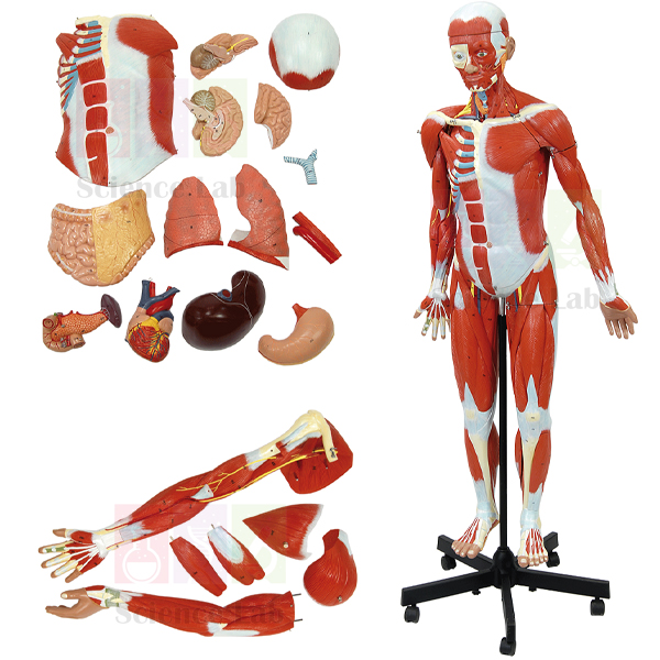
Human Muscular Figure Model
The Human Muscular Figure Model (Product Code: SLE/BL/71) is a 32-part, 140 cm tall figure that shows the musculature of the body together with removable internal organs. A 6-part muscular arm and an 8-part muscular leg detach for close study, the skullcap lifts off to reveal a bisected brain, and the heart, lungs and main abdominal organs can all be taken out. Every structure is numbered and referenced on the accompanying key card. Built by Jain Scientific Equipments Pvt Ltd (ScienceLab), one of the human anatomical models manufacturers in India based in Ambala, it is rod-mounted on a base with casters for use in medical, nursing, physiotherapy and sports science teaching.
Specifications
| Parameter | Detail |
|---|---|
| Type | Full muscular figure with removable organs |
| Total parts | 32 |
| Arm | Removable 6-part muscular arm |
| Leg | Removable 8-part muscular leg |
| Head and brain | Removable skullcap; brain bisected along the longitudinal fissure, showing internal structures and vasculature |
| Heart | Removable 2-part heart representing the atria, ventricles, valves and sections of the heart wall |
| Lungs | Bisected and removable, showing the bronchial tree and vascular system |
| Abdominal organs (removable) | Liver with gallbladder, 2-part stomach, duodenum with pancreas and spleen, large intestine |
| Identification | Structures numbered and referenced on the accompanying key card |
| Mounting | Rod-mounted on base with casters |
| Model height | 140 cm |
| Total size | 150 x 50 x 40 cm |
| Weight | Approximately 12 kg |
Typical Use
A figure of this size lets a whole class study the muscular system in its true proportions. Tutors usually begin with the surface muscles of the trunk and limbs, then detach the 6-part arm and the 8-part leg so students can examine the limb muscles piece by piece. With the chest and abdomen opened up, the removable heart, lungs, liver, stomach and intestine let the same model double as a lesson on the organs of the torso — the bisected brain and lungs show internal structure, not just outer surfaces.
Medical and nursing colleges use it for anatomy practicals, physiotherapy and sports science departments for muscle function and injury discussions, and fine-arts faculties for figure study. It suits institutions building a human biology model set in India around one central figure. The casters make it easy to wheel between lecture hall and lab; make sure the figure is standing steady before removing parts, return each piece to its matching position, and cover it when not in use.
Manufacturer
Jain Scientific Equipments Private Limited, known to customers as ScienceLab, manufactures this full-size muscular figure in Ambala, Haryana, alongside smaller anatomy models and laboratory apparatus. The company profile explains more about the organisation.
Related Laboratory Equipment
Items that complement a full muscular figure in anatomy teaching:
- Human Sexless Muscular Figure — an alternative muscular figure for comparison when equipping several rooms
- Miniature Human Muscular Figure — a desk-sized figure for individual student revision
- Human Muscular Arm Model — more detail on the muscles of the upper limb
- Human Muscular and Ligamentous Skeleton — shows how muscles and ligaments attach to the skeleton
- Skeletal Muscle Fiber Model — explains muscle structure at the level of the fibre
Browse more figures in the Human Models category.
Frequently Asked Questions
How many parts does the figure have?
32 parts in total, including a 6-part arm, an 8-part leg, a 2-part heart and a 2-part stomach.
How tall is it and how much space does it need?
The model is 140 cm tall; the total size is 150 x 50 x 40 cm and it weighs approximately 12 kg.
Which internal organs can be removed?
The 2-part heart, the bisected lungs, the liver with gallbladder, the 2-part stomach, the duodenum with pancreas and spleen, and the large intestine. The skullcap also lifts off to show the bisected brain.
Can it be moved between rooms?
Yes. It is rod-mounted on a base with casters, so it can be wheeled between the lecture hall and the lab.
How are the structures identified?
Anatomical structures are numbered on the model and referenced on the accompanying key card.
What are the packed dimensions for shipping?
Packed size and gross weight depend on the packing method chosen for the destination. Contact us and we will provide them with the quotation.
Do you export this figure?
Yes. We supply colleges, hospitals' teaching departments and distributors in India and international markets; warranty terms are set out on the quotation.
Request a Quote
Request a formal quotation for the Human Muscular Figure Model through our contact page, or add it to the enquiry cart with any supporting muscle and skeleton models.
Last updated: 24 September 2026
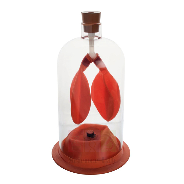
Human Lung Model in Bell Jar
The Human Lung Model in Bell Jar (Product Code: SLE/BL/72) is a simple working model of the breathing mechanism. Two balloons attached to a Y-tube act as the lungs, the bell jar with its open top represents the chest cavity, and a stout rubber sheet across the bottom acts as the diaphragm. Pull the rubber sheet down and the balloons inflate; let it go and they deflate. Made by Jain Scientific Equipments Pvt Ltd (ScienceLab), Ambala, it serves as a direct, hands-on biology experiment kit for students learning how breathing works, and is used from middle school through to nursing and physiology courses.
Specifications
| Parameter | Detail |
|---|---|
| Type | Bell jar breathing mechanism model |
| Lungs | Two balloons attached to a Y-tube |
| Chest cavity | Bell jar with open top |
| Diaphragm | Stout rubber sheet |
| Principle shown | Pulling the diaphragm down lowers the pressure inside the chamber, so the lungs expand and take in air; releasing it lets the lungs deflate |
| Teaching purpose | Shows how the lungs are filled and emptied by the contraction and relaxation of the diaphragm muscle |
Typical Use
This is a demonstration students can operate themselves. When the rubber diaphragm is pulled downward, the space inside the bell jar increases and the pressure there falls below atmospheric pressure, so air rushes in through the Y-tube and the balloon lungs expand — just as the lungs fill when the diaphragm contracts and flattens. When the sheet is released, it returns to its resting position and the balloons empty, modelling exhalation as the diaphragm relaxes. Asking students to describe the pressure change at each step links the biology lesson to basic gas laws from physics.
The model is a natural fit for health education programmes in schools in India that cover breathing, exercise and the effects of smoking, and for first-year nursing physiology. Pull the diaphragm gently and evenly rather than snapping it, check that the Y-tube and balloons are seated firmly so air does not leak, and store the model away from heat and sunlight so the rubber parts last longer.
Manufacturer
This breathing model is made in Ambala, Haryana, by Jain Scientific Equipments Private Limited, which trades as ScienceLab and supplies both simple demonstration apparatus and detailed anatomical models to education. Our company profile has more background.
Related Laboratory Equipment
Equipment for a complete breathing and respiration practical alongside the bell jar model:
- Lung Demonstration Model — another demonstration of lung function for comparison or a second bench
- Lung Capacity Gauge — lets students measure their own lung capacity after seeing the mechanism
- Lung Volume Bags — for collecting and comparing breath volumes in class
- Human Respiratory System Model, 2-parts — shows the real anatomy of the airway and lungs that the balloons represent
- Breathing Fitness Health Lab — activities connecting breathing with fitness and health
For anatomical models, visit our Human Models category.
Frequently Asked Questions
How does the bell jar lung model work?
Pulling the rubber diaphragm down lowers the pressure inside the jar, so the balloon lungs fill with air; releasing it lets them deflate.
What represents each part of the body?
The two balloons represent the lungs, the Y-tube the airways, the bell jar the chest cavity, and the rubber sheet the diaphragm.
Can students operate it themselves?
Yes. It is a simple hands-on model, and operating the diaphragm themselves helps students remember the sequence of inhalation and exhalation.
Is the bell jar made of glass or plastic?
The material of the jar is not specified in our current product details. Please contact us to confirm before ordering, especially for younger classes.
Can the balloons or rubber sheet be replaced?
Rubber parts naturally wear with repeated use. Ask us about replacement balloons and diaphragm sheets when you order.
Do you supply this model in quantity?
Yes. We quote for class sets, school chains and distributors in India and international markets, with warranty terms stated on the quotation.
Request a Quote
To price the Human Lung Model in Bell Jar for your school or college, use our contact page or add it to the enquiry cart together with other respiration equipment.
Last updated: 24 September 2026
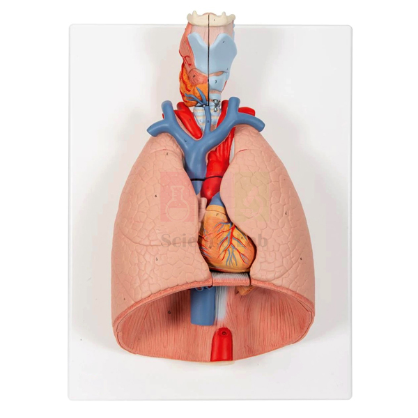
Human Lungs with Heart and Larynx Model
Human Lungs with Heart and Larynx Model (Product Code: SLE/BL/73) is a dissectible anatomy model used to teach the respiratory tract and heart–lung circulation in school, nursing and college classes. Schools assembling a human biology model set in India choose it because it combines a removable 2-part larynx, 2-part heart and 2-part lungs with the trachea, great vessels, diaphragm and oesophagus, numbered against a key card on a base. It is made by Jain Scientific Equipments Pvt Ltd (ScienceLab), an OEM manufacturer, at its own factory in Ambala, India.
Key Details
| Parameter | Detail |
|---|---|
| Type | Dissectible model of lungs, heart and larynx |
| Removable parts (as listed) | 2-part larynx, trachea with bronchial tree, vena cava, aorta, 2-part heart, subclavian artery and vein, pulmonary artery, 2-part lungs, diaphragm, oesophagus |
| Finish | Appropriately coloured |
| Identification | Numbered, with key card |
| Mounting | Mounted on a base |
| Product code | SLE/BL/73 |
| Manufacturer | Jain Scientific Equipments Pvt Ltd (ScienceLab) — OEM |
| Country of origin | India (made in Ambala, Haryana) |
What Is the Lungs, Heart and Larynx Model Used For?
A lesson on respiration can start at the larynx, where opening the two halves reveals the internal cavity, then move down the trachea into the branching bronchial tree and out into each lung. Lifting away the lung parts exposes the heart, and separating the heart halves lets the teacher show how deoxygenated blood returning through the vena cava is sent to the lungs along the pulmonary artery, while oxygenated blood leaves through the aorta. With the diaphragm at the base and the oesophagus running behind the airway, students also see why swallowing and breathing share the same space in the neck and chest.
Because it combines several organ systems, the model works well as the centrepiece of a human body model kit for India's secondary schools, nursing institutes and physiotherapy courses. Hand out the numbered key card and ask groups to reassemble the parts in order after each session; this reinforces anatomy and ensures nothing goes missing. Keep the removable parts together in a closed cabinet and wipe them with a soft, dry cloth only.
OEM Manufacturer
Jain Scientific Equipments Private Limited, which trades as ScienceLab, is the OEM (original equipment manufacturer) of this lungs, heart and larynx model. It is made and assembled at the company's own licensed factory at Saha, Ambala, Haryana, India, rather than bought in from a trader, and goes factory-direct to schools, colleges, nursing institutes, dealers and importers. If a removable part is lost or damaged, ask us about a replacement from the manufacturer. More background is on our company profile.
Related Human Models
Add these models to teach the chest organs in more depth:
- Human Thoracic Organs Model — a similar set of chest organs with removable front lung halves
- Human Segmented Lungs Model — shows the internal segments of the lungs
- Human Heart Model — a larger view of the heart chambers and valves
- Human Larynx, Trachea, and Bronchi Model — focuses on the upper and middle airway
- Pathology of the Human Lung Model — for comparing healthy lungs with common diseases
This model is listed in our Human Models category with other anatomy models.
Frequently Asked Questions
Which parts of the Human Lungs with Heart and Larynx Model can be removed?
The Human Lungs with Heart and Larynx Model lists its larynx, heart and lungs as 2-part sections, alongside the trachea with bronchial tree, vena cava, aorta, subclavian vessels, pulmonary artery, diaphragm and oesophagus, all numbered on the key card.
Why does the lungs, heart and larynx model include the oesophagus and diaphragm?
The lungs, heart and larynx model includes the oesophagus and diaphragm to show real relationships in the chest: the oesophagus runs behind the trachea, and the diaphragm forms the floor on which the lungs and heart rest during breathing.
Is a key card provided with the model?
Yes. Every structure on the Human Lungs with Heart and Larynx Model is appropriately coloured and numbered, and a key card identifies each number so students can name the parts during practicals and revision.
What are the size and material of the model?
The size and material of the Human Lungs with Heart and Larynx Model are not stated in its listing. Ask us through the contact page and we will confirm the dimensions and material in your quotation.
Who manufactures the Human Lungs with Heart and Larynx Model?
Jain Scientific Equipments Private Limited (ScienceLab) is the OEM that manufactures the Human Lungs with Heart and Larynx Model at its own factory at Saha, Ambala, Haryana, India, supplying schools, colleges, dealers and importers directly.
How does it compare with the Human Thoracic Organs Model?
Both the Human Lungs with Heart and Larynx Model and the Human Thoracic Organs Model show the heart, lungs and airway; the thoracic model is built around removable front lung halves. Tell us your syllabus focus and we will suggest the better fit.
Can colleges or distributors order several units?
Yes. The Human Lungs with Heart and Larynx Model can be quoted for colleges, schools and distributors in India and international markets; send product code SLE/BL/73, the quantity and the delivery location, and the quotation comes directly from the manufacturer.
Request a Quote
Quotations for the Human Lungs with Heart and Larynx Model come straight from the manufacturer. Send your requirement through our contact page, or add the model to the enquiry cart.
Last updated: 24 September 2026
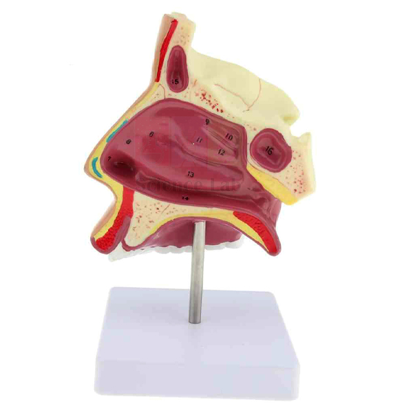
Human Nose Model on Stand
The Human Nose Model on Stand (Product Code: SLE/BL/74) is an enlarged, colour-finished model of the structure of the human nose, held on a stand and numbered with a key card. Presenting the nose on a stand makes it well suited to front-of-class demonstration, anatomy room displays and ENT teaching corners where the model is viewed as much as it is handled. It is manufactured by Jain Scientific Equipments Pvt Ltd (ScienceLab), Ambala, and is ordered by colleges and schools that need a school lab equipment supplier in India for sense organ and airway models.
Key Details
| Parameter | Detail |
|---|---|
| Type | Enlarged model of the human nose |
| Shows | Structure of the nose |
| Mounting | Mounted on a stand |
| Finish | Appropriately coloured |
| Identification | Numbered, with key card |
| Suitable for | ENT training, nursing anatomy, sense organ lessons and anatomy displays |
Typical Use
In ENT and nursing teaching, the nose is the entry point to the respiratory tract and the site of many everyday complaints, from blocked passages to nosebleeds. Tutors use this model to walk trainees through the structure of the nose in order, pointing to each numbered feature and checking recall with the key card. Because the stand presents the model upright, it can be positioned on the lecturer's desk and turned to face the class while the lesson moves from external shape to the passages that carry air inward.
In school classes it supports the chapter on the sense organs, where students learn how smell is detected and how the nose prepares air before it reaches the lungs. Between sessions it can stay on display in a glass-fronted cabinet; dust it regularly, keep the key card filed with it, and avoid placing it where the stand could be knocked from a shelf edge.
Manufacturer
ScienceLab models such as this one are manufactured by Jain Scientific Equipments Private Limited in Ambala, Haryana. For vendor registration, buyers can note the company's Corporate Identity Number U29309HR2020PTC087597 and its Factory Licence AMB-ONLINE-CHD-J-414, which covers the production premises at 449, HSIIDC Industrial Area, Saha, Ambala. More about the company is on our company profile.
Related Human Models
Models often chosen with the Human Nose Model on Stand for ENT and sense organ teaching:
- Human Nose Model on Base — the base-mounted version for close desk study by small groups
- Human Nasal Cavity Model — continues inward to the nasal cavity
- Anatomical Human Nasal Cavity Model — a further option for studying the internal nasal passages
- Human Tongue Model — pairs smell with taste in the chemical senses lesson
- Human Ear Model, 5 Parts — completes an ear, nose and throat teaching set
All our anatomy models are listed in the Human Models range.
Frequently Asked Questions
Why choose the stand-mounted version?
The stand presents the model upright, which suits demonstrations to a whole class and display in anatomy rooms. If the model will mainly be handled on a desk by small groups, the base-mounted version may suit better.
Is a key card included?
Yes. The model is numbered and comes with a key card for identifying its structures.
What scale is the model?
It is an enlarged model. The exact enlargement and dimensions are not in our current listing, so please contact us for them.
Is it suitable for ENT and nursing courses?
Yes. It is used to teach the structure of the nose as part of ENT, nursing and paramedical anatomy, as well as school sense organ lessons.
Can you provide registration details for our tender or vendor file?
Yes. Our CIN and factory licence number are given above; other registration details can be requested through the contact page.
Do you ship to institutions abroad?
Yes. ScienceLab supplies India and international markets. Warranty and delivery terms are confirmed on the quotation.
Request a Quote
Send your enquiry for the Human Nose Model on Stand through our contact page, or add it to the enquiry cart with other ENT models to receive one combined quotation.
Last updated: 24 September 2026
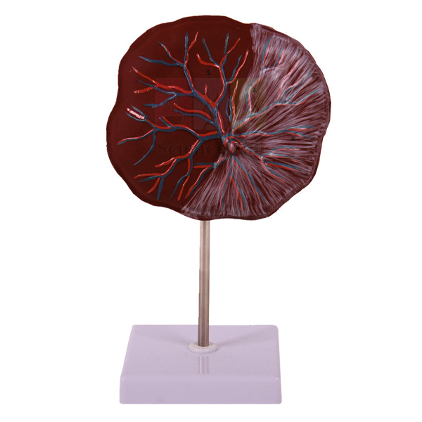
Human Placenta Model
The Human Placenta Model (Product Code: SLE/BL/79) shows the structure of the placenta and the relationship between the placenta and the umbilical cord, with 7 positions displayed for identification. It is made of advanced PVC and mounted on a stand. Midwifery and obstetric nursing programmes, medical colleges and senior school biology classes use it to explain how a developing baby is nourished during pregnancy. The model is manufactured by Jain Scientific Equipments Pvt Ltd (ScienceLab), Ambala, a biology lab equipment supplier in India for schools and higher education.
Specifications
| Parameter | Detail |
|---|---|
| Type | Placenta and umbilical cord model |
| Shows | Structure of the placenta and its relation to the umbilical cord |
| Positions displayed | 7 |
| Material | Advanced PVC |
| Mounting | Mounted on stand |
Typical Use
The placenta is the organ through which the fetus receives oxygen and nutrients and passes waste back to the mother's blood, and the umbilical cord is the connection that carries that exchange. This model lets tutors show both in one piece: students see the overall structure of the placenta, then follow how the cord joins it. Working through the 7 displayed positions gives a clear order for the explanation and a ready-made set of labelling questions for tests and practical records.
In midwifery training it supports teaching on the delivery of the placenta and its examination after birth, while in school biology it answers the common question of how a baby "breathes" and "eats" before it is born. Institutions building a human biology model set often pair it with fetal development models to show the placenta in context. Wipe the PVC with a soft damp cloth and keep the model on its stand when stored.
Manufacturer
Jain Scientific Equipments Private Limited makes the Human Placenta Model at Ambala in Haryana and sells it under the ScienceLab name, together with its range of reproductive and developmental anatomy models. For more about us, see the company profile.
Related Human Models
Models that place the placenta in the wider story of pregnancy:
- Human Embryo Set, PVC — shows the placenta early in gestation and the fetus in every month that follows
- Human Embryo Development Set — fetal stages and positions within the uterus
- Embryo Model — the 4-week embryo, before the placenta is fully formed
- Human Fetal Circulation Model — how blood from the umbilical cord circulates through the fetus
The full Human Models category lists our other anatomy teaching models.
Frequently Asked Questions
What does the Human Placenta Model show?
The structure of the placenta and the relation between the placenta and the umbilical cord.
How many positions are displayed?
7 positions are displayed. For the list of positions to prepare worksheets, please ask through our contact page.
What is it made from?
Advanced PVC, mounted on a stand.
What are its dimensions?
Dimensions are not listed in our current product details. Contact us and we will confirm them before dispatch.
Is it useful for midwifery training?
Yes. It helps trainees understand placental structure and the cord attachment, which underpins teaching on the third stage of labour and placental examination.
How do we request an institutional or export quotation?
Send the product code SLE/BL/79, quantity and delivery location. We supply India and international markets and confirm warranty terms on the quotation.
Request a Quote
For pricing on the Human Placenta Model, contact us via the contact page or add it to the enquiry cart with any embryo or fetal development models you need.
Last updated: 24 September 2026
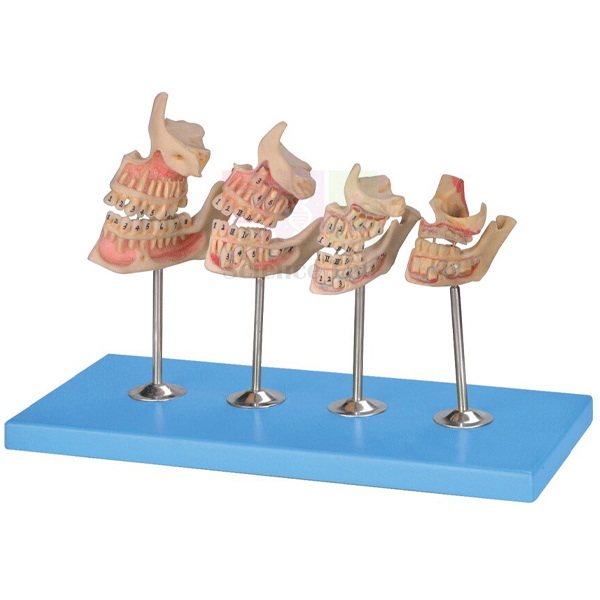
Human Teeth Model, Dentition Development
Human Teeth Model, Dentition Development (Product Code: SLE/BL/85) is a stand-mounted anatomy model used to teach how human teeth develop from birth to adulthood in biology classrooms and health science teaching. It displays the upper and lower jaws at four stages (a newborn, a 5-year-old child, a 9-year-old child and a young adult), coloured, numbered and keyed with a key card. It is made by Jain Scientific Equipments Pvt Ltd (ScienceLab), an OEM anatomical models manufacturer, at its own factory in Ambala, India.
Specifications
| Parameter | Detail |
|---|---|
| Stages shown | Upper and lower jaw of a newborn child, a 5-year-old child, a 9-year-old child and a young adult |
| Mounting | On a stand |
| Identification | Numbered, with key card |
| Finish | Appropriately coloured and neatly finished |
| Individual parts | Each part also available individually on demand |
| Product code | SLE/BL/85 |
| Manufacturer | Jain Scientific Equipments Pvt Ltd (ScienceLab) — OEM |
| Country of origin | India (made in Ambala, Haryana) |
What Is the Dentition Development Model Used For?
Placing four jaws side by side lets students see that the dentition is not fixed but changes through childhood. Teachers use the stages to explain the milk (deciduous) teeth of early childhood, the mixed period when permanent teeth begin to replace them, and the complete permanent set of the young adult, linking the model to lessons on growth, nutrition and dental care.
Because every stage is numbered and keyed, the model works well for identification practice and practical tests: students name the numbered teeth, then check against the key card. It is a clear addition to biology teaching aids in India's school, nursing and paramedical classrooms. Dust it with a soft, dry cloth and keep it away from direct sunlight and solvents so the colours last.
What to Specify When Ordering
- Complete four-stage model on its stand, or individual stages only (each part is available individually on demand)
- If ordering individually: which stage you need (newborn, 5-year-old, 9-year-old or young adult)
- Number of models per laboratory or classroom
- Any other teeth models you want quoted together, such as the lower jaw or incisor models
OEM Manufacturer
The Human Teeth Model, Dentition Development is produced by Jain Scientific Equipments Private Limited, which trades as ScienceLab and is the original equipment manufacturer (OEM) of this model. It is made and finished at the company's own licensed factory at Saha, Ambala, Haryana, rather than bought in from a trader, so schools, colleges, dealers and importers receive it factory-direct. Requests for single stages, replacement parts or repeat orders go straight to the manufacturer. Read more on our company profile.
Related Human Models
Other dental and skull models that support the same lessons on teeth and jaw growth:
- Human Teeth Model, Lower Jaw — shows the position of every tooth in the lower jaw in detail
- Human Teeth Model, Incisor — an enlarged incisor that divides into two parts to show inside the tooth
- Human Teeth Model — an enlarged teeth model with a tooth brush for oral hygiene lessons
- Human Skull Model, Fetal Child — for comparing the skull and jaws before birth
Browse all anatomy models in our Human Models category.
Frequently Asked Questions
Which stages does the Human Teeth Model, Dentition Development show?
The Human Teeth Model, Dentition Development shows the upper and lower jaws of a newborn child, a 5-year-old child, a 9-year-old child and a young adult, so students can compare how the teeth change from birth to adulthood in a single display.
Is the dentition model labelled?
Yes, the Human Teeth Model, Dentition Development is numbered and comes with a key card identifying the numbered features. This helps students revise on their own and lets teachers use the model for identification tests.
Can one stage of the model be bought on its own?
Yes, each part of the Human Teeth Model, Dentition Development is also available individually on demand. State which stage you need (newborn, 5-year-old, 9-year-old or young adult) when you request a quotation.
How is the Human Teeth Model, Dentition Development mounted?
The Human Teeth Model, Dentition Development is mounted on a stand and is appropriately coloured and neatly finished, so all four stages stay together as one display on a bench, shelf or teacher's desk.
What material and size is the model?
The material and overall dimensions of the Human Teeth Model, Dentition Development are not stated in this listing. Please ask through our contact page and we will confirm them with your quotation.
Who manufactures the Human Teeth Model, Dentition Development?
The Human Teeth Model, Dentition Development is manufactured by Jain Scientific Equipments Pvt Ltd (ScienceLab), the OEM, at its own licensed factory at Saha, Ambala, Haryana, India, and is sent factory-direct to schools, colleges, dealers and importers.
Request a Quote
Quotations for the Human Teeth Model, Dentition Development come directly from the manufacturer. Send your quantity and whether you need the full set or single stages through our contact page, or add the model to the enquiry cart.
Last updated: 24 September 2026
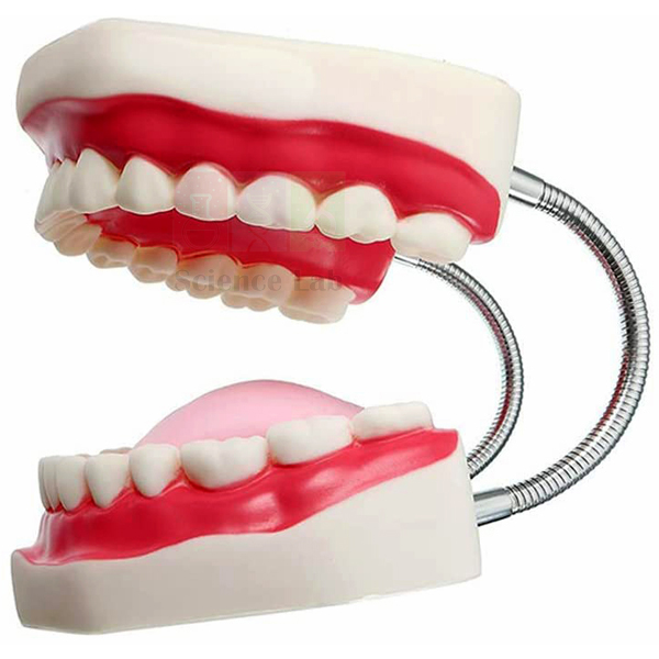
Human Teeth Model
Human Teeth Model (Product Code: SLE/BL/87) is an enlarged teeth model with a tooth brush, used to demonstrate tooth brushing and oral hygiene to a whole group in schools, health education sessions and dental awareness programmes. Its enlarged size lets everyone in the room follow each brushing step, and it is appropriately coloured and neatly finished. It is made by Jain Scientific Equipments Pvt Ltd (ScienceLab), an OEM manufacturer, at its own factory in Ambala, India, and fits well into a health education kit for schools.
Key Details
| Parameter | Detail |
|---|---|
| Type | Human teeth model, enlarged for demonstration to a group |
| Accessory | Tooth brush |
| Finish | Appropriately coloured and neatly finished |
| Product code | SLE/BL/87 |
| Manufacturer | Jain Scientific Equipments Pvt Ltd (ScienceLab) — OEM |
| Country of origin | India (made in Ambala, Haryana) |
What Is the Human Teeth Model Used For?
Teaching children to brush is far easier with a large model the whole class can see than with a mirror or a drawing. The teacher or health worker holds up the enlarged teeth and uses the tooth brush to show each part of the routine: outer surfaces, inner surfaces and chewing surfaces, reaching the back teeth, and gentle strokes along the gum line. Pupils then copy the movements, and volunteers can try the brush on the model themselves.
The same model supports lessons on why teeth decay, how plaque collects where brushing is missed and why sugary snacks matter. Schools building a human biology model set in India often start with a teeth model, because dental care is taught from the early grades onward. After each session, wipe the model with a soft damp cloth, dry it before storage and keep the brush clean and separate.
OEM Manufacturer
ScienceLab is the trading name of Jain Scientific Equipments Private Limited, the original equipment manufacturer (OEM) behind this Human Teeth Model. The model is produced at the company's own licensed factory at Saha, Ambala, Haryana, and sold directly by the maker to schools, health programmes, dealers and importers, so questions about the model, the tooth brush or repeat orders are answered by the people who make it. Company background is on our profile page.
Related Human Models
Teeth models that extend an oral hygiene lesson into tooth structure and growth:
- Human Teeth Model, Dentition Development — shows how the teeth change from birth to adulthood
- Human Teeth Model, Lower Jaw — for naming each tooth by its position in the jaw
- Human Teeth Model, Lower Molar with One Root — shows the root, nerves and vessels inside a molar
- Human Teeth Model, Incisor — an enlarged front tooth that divides into two parts
See the full range in our Human Models category.
Frequently Asked Questions
What is included with the Human Teeth Model?
The Human Teeth Model comes with a tooth brush, so a teacher or health worker can demonstrate correct brushing directly on the enlarged teeth. Dimensions and material are not specified in this listing; ask us and we will confirm them with your quotation.
Why is the Human Teeth Model enlarged?
The Human Teeth Model is enlarged for demonstration to a group, so everyone in a classroom or health session can see the brush movements and tooth surfaces clearly instead of crowding round a small, life-size model.
Who is the Human Teeth Model intended for?
The Human Teeth Model suits primary and secondary schools, nursing and health-worker training, and dental awareness programmes that teach oral hygiene to groups. Its main role is demonstrating tooth brushing, supported by simple lessons on tooth structure and decay.
How should the model be cleaned after use?
After each demonstration, wipe the Human Teeth Model with a soft damp cloth and let it dry before storing. Rinse and dry the tooth brush separately, and avoid solvents or abrasive cleaners that could damage the coloured finish.
Who manufactures the Human Teeth Model?
The Human Teeth Model is manufactured by Jain Scientific Equipments Pvt Ltd (ScienceLab), the OEM, at its own licensed factory at Saha, Ambala, Haryana, India. Quotations and repeat orders are handled directly by the manufacturer rather than a trader.
Can the Human Teeth Model be ordered in quantity or for export?
Yes, the Human Teeth Model can be quoted in quantity for schools, health programmes, dealers and importers in India and international markets. Share the number of models and the delivery destination on our contact page.
Request a Quote
Your quotation for the Human Teeth Model comes straight from the manufacturer. Send the quantity and destination via our contact page, or add the model to the enquiry cart with other anatomy models.
Last updated: 24 September 2026
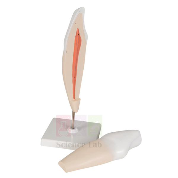
Human Teeth Model, Incisor
Human Teeth Model, Incisor (Product Code: SLE/BL/88) is an enlarged model of a human incisor used to teach the shape and structure of the front cutting teeth in biology, dental and health science classes. The model is divided into two parts, numbered with a key card and mounted on a stand, appropriately coloured and neatly finished. It is made by Jain Scientific Equipments Pvt Ltd (ScienceLab), an OEM among human anatomical models manufacturers in India, at its own factory in Ambala.
Key Details
| Parameter | Detail |
|---|---|
| Tooth represented | Human incisor |
| Scale | Enlarged |
| Parts | Divided into two parts |
| Identification | Numbered, with key card |
| Mounting | On a stand |
| Finish | Appropriately coloured and neatly finished |
| Product code | SLE/BL/88 |
| Manufacturer | Jain Scientific Equipments Pvt Ltd (ScienceLab) — OEM |
| Country of origin | India (made in Ambala, Haryana) |
What Is the Incisor Tooth Model Used For?
Incisors are the chisel-shaped front teeth that bite into and cut food, and an enlarged model makes their form easy for a whole class to see. Teachers use it to explain the crown with its thin cutting edge, the neck and the root, and to introduce the layers of a tooth when discussing how decay progresses from the surface inwards.
Because the model separates into two parts, students can examine each piece and match the numbered features with the key card, which turns a demonstration into a hands-on identification exercise. Placed beside a molar model, it shows clearly how cutting and grinding teeth differ. It is a practical piece of high school biology lab equipment for units on nutrition and the digestive system, where the mouth is the first stage; handle the separated parts over a table so they are not dropped.
OEM Manufacturer
Jain Scientific Equipments Private Limited, trading as ScienceLab, is the OEM of the Human Teeth Model, Incisor and makes it at its own licensed factory at Saha, Ambala, Haryana. Buying from the manufacturer means the model reaches schools, colleges, dealers and importers without a trading intermediary, and quotations, replacement parts or key cards and repeat batches are handled by the same team that makes it; just ask us. Learn more about the company on our profile.
Related Human Models
Teeth models that pair with the incisor when comparing tooth types:
- Human Teeth Model, Lower Molar with One Root — a grinding tooth to contrast with the cutting incisor
- Human Teeth Model, Triple Root Molar — for comparing single and multiple root forms
- Human Teeth Model, Lower Jaw — shows where the incisors sit among the other teeth
- Human Teeth Model, Dentition Development — follows the teeth from birth to adulthood
Explore more anatomy teaching models in the Human Models category.
Frequently Asked Questions
What does the Human Teeth Model, Incisor show?
The Human Teeth Model, Incisor shows an enlarged human incisor, the chisel-shaped front tooth used for cutting food. It is divided into two parts and numbered with a key card so students can identify its features during class study.
How many parts does the incisor model have?
The Human Teeth Model, Incisor is divided into two parts, which students can examine separately and match against the numbered key card. Handling the separated parts over a table helps prevent them being dropped or lost.
Is the Human Teeth Model, Incisor mounted?
Yes, the Human Teeth Model, Incisor is mounted on a stand and is appropriately coloured and neatly finished, so it can stand on a bench, teacher's desk or display shelf between lessons.
What size and material is the incisor model?
The exact enlargement, dimensions and material of the Human Teeth Model, Incisor are not stated in this listing. Contact us and we will confirm them in your quotation so you can plan storage and display space.
Who manufactures the Human Teeth Model, Incisor?
The Human Teeth Model, Incisor is manufactured by Jain Scientific Equipments Pvt Ltd (ScienceLab), the original equipment manufacturer, at its own licensed factory at Saha, Ambala, Haryana, India, and is sold factory-direct to institutions, dealers and importers.
Can the incisor model be ordered with other teeth models?
Yes, the Human Teeth Model, Incisor can be quoted together with the molar, lower jaw and dentition development teeth models, so a school or college can assemble a complete dental teaching set in one order from the manufacturer.
Request a Quote
Request a manufacturer's quotation for the Human Teeth Model, Incisor through our contact page, or add it to the enquiry cart together with other teeth and anatomy models.
Last updated: 24 September 2026
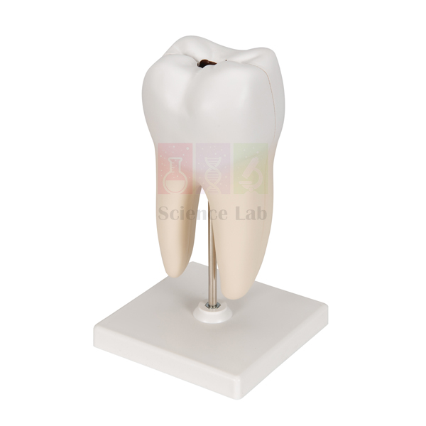
Human Teeth Model, Lower Molar with One Root
Human Teeth Model, Lower Molar with One Root (Product Code: SLE/BL/89) is a stand-mounted model of a lower molar used to show the nerves and vessels inside a tooth in biology, dental and nursing classes. It presents the molar with one root together with its nerves and vessels, appropriately coloured and numbered with a key card. It is made by Jain Scientific Equipments Pvt Ltd (ScienceLab), an OEM biology lab equipment manufacturer in India, at its own factory in Ambala.
Key Details
| Parameter | Detail |
|---|---|
| Tooth represented | Lower molar with one root |
| Structures shown | Nerves and vessels |
| Identification | Numbered, with key card |
| Mounting | On a stand |
| Finish | Appropriately coloured and neatly finished |
| Product code | SLE/BL/89 |
| Manufacturer | Jain Scientific Equipments Pvt Ltd (ScienceLab) — OEM |
| Country of origin | India (made in Ambala, Haryana) |
What Is the Lower Molar Model Used For?
Molars are the broad grinding teeth at the back of the mouth, and toothache usually means that decay or infection has reached the nerves and blood vessels inside the tooth. By showing those nerves and vessels, the model helps students see that a tooth is living tissue with its own blood and nerve supply, and why deep decay becomes painful and needs treatment.
The numbered features and key card make it suitable for identification practice and practical assessments in dental assistant, nursing and biology courses. As school biology lab equipment in India, it fits lessons on teeth, nutrition and oral health from middle school upward, and it pairs well with an incisor or multi-rooted molar model when comparing tooth types. Keep it on its stand and dust it with a soft, dry cloth.
OEM Manufacturer
ScienceLab, the trade name of Jain Scientific Equipments Private Limited, is the original equipment manufacturer of the Human Teeth Model, Lower Molar with One Root. The model is produced at the company's own licensed factory in the HSIIDC Industrial Area at Saha, Ambala, Haryana, and goes factory-direct to schools, colleges, dealers and importers in India and international markets. Buyers deal with the maker itself for quotations, spare key cards and repeat orders. More on our company profile.
Related Human Models
Teeth models that complement the lower molar in lessons on tooth structure:
- Human Teeth Model, Triple Root Molar — for comparing a single-rooted and a three-rooted molar
- Human Teeth Model, Incisor — a cutting tooth to contrast with the grinding molar
- Human Teeth Model, Lower Jaw — shows where the molars sit in the lower jaw
- Human Teeth Model — for brushing demonstrations that help prevent deep decay
View all anatomy teaching models in the Human Models category.
Frequently Asked Questions
What does the Human Teeth Model, Lower Molar with One Root show?
The Human Teeth Model, Lower Molar with One Root shows a lower molar with one root together with its nerves and vessels. It is numbered with a key card, so students can identify each structure and link it to lessons on tooth anatomy and decay.
Why does the molar model show nerves and vessels?
The Human Teeth Model, Lower Molar with One Root shows nerves and vessels to explain that a tooth is living tissue with its own blood and nerve supply, which helps students understand sensitivity, toothache and why deep decay needs treatment.
Is the Human Teeth Model, Lower Molar with One Root mounted?
Yes, the Human Teeth Model, Lower Molar with One Root is mounted on a stand and is appropriately coloured and neatly finished, so it can be displayed on a bench or shelf and passed between groups without handling loose parts.
How does it differ from the triple root molar model?
The Human Teeth Model, Lower Molar with One Root represents a molar with a single root and shows its nerves and vessels, while the Human Teeth Model, Triple Root Molar is a separate item for studying a three-rooted molar. Teachers often use both to compare root forms.
What size and material is the molar model?
The dimensions and material of the Human Teeth Model, Lower Molar with One Root are not stated in this listing. Please ask through our contact page and we will confirm them along with your quotation.
Who manufactures the Human Teeth Model, Lower Molar with One Root?
The Human Teeth Model, Lower Molar with One Root is manufactured by Jain Scientific Equipments Pvt Ltd (ScienceLab), the OEM, at its own licensed factory at Saha, Ambala, Haryana, India, and quotations come straight from the manufacturer.
Request a Quote
For a quotation direct from the manufacturer, send your quantity for the Human Teeth Model, Lower Molar with One Root through the contact page, or add it to our enquiry cart.
Last updated: 24 September 2026
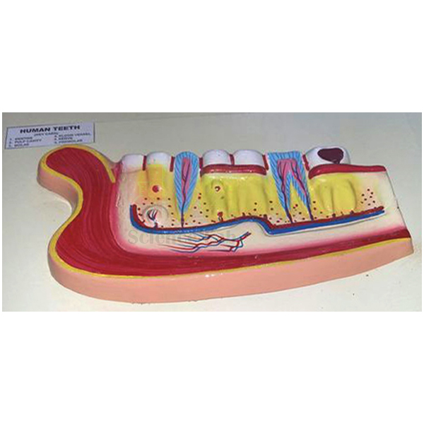
Human Teeth Model, Lower Jaw
Human Teeth Model, Lower Jaw (Product Code: SLE/BL/90) is a base-mounted model of the human lower jaw used to show the position of every tooth in the lower jaw for biology, dental and nursing teaching. Each tooth position is numbered and identified on a key card, and the model is appropriately coloured and neatly finished. It is made by Jain Scientific Equipments Pvt Ltd (ScienceLab), an OEM and school laboratory equipment manufacturer, at its own factory in Ambala, India.
Key Details
| Parameter | Detail |
|---|---|
| Shows | Position of all the teeth in the lower jaw |
| Identification | Numbered, with key card |
| Mounting | On a base |
| Finish | Appropriately coloured and neatly finished |
| Product code | SLE/BL/90 |
| Manufacturer | Jain Scientific Equipments Pvt Ltd (ScienceLab) — OEM |
| Country of origin | India (made in Ambala, Haryana) |
What Is the Lower Jaw Teeth Model Used For?
Learning the names and order of the teeth, from the incisors at the front through the canines and premolars to the molars at the back, is a basic step in human biology, dental and nursing courses. This model lays out the teeth of the lower jaw in their natural positions, so students can count them, name each type from the midline outward and relate each tooth's shape to its job in biting, tearing or grinding food.
Because every position is numbered, a teacher can cover the key card and ask students to identify the teeth, which makes the model a quick tool for revision and practical tests. The base keeps it steady on a bench during close group work. Schools completing a biology lab setup in India often place it on the anatomy shelf beside the skull and dentition models.
What to Specify When Ordering
- Presentation: this base-mounted model, or the separately listed lower jaw model on a stand
- Number of models per laboratory or classroom
- Any other teeth models to be quoted together, such as the incisor, molar or dentition development models
OEM Manufacturer
Jain Scientific Equipments Private Limited, which trades as ScienceLab, is the OEM of the Human Teeth Model, Lower Jaw and manufactures it at its own licensed factory at Saha, Ambala, Haryana. The company is a registered MSME (Udyam UDYAM-HR-01-0003714), and its export shipments are handled under Importer-Exporter Code AAECJ8618A (DGFT), so institutions, dealers and importers abroad can buy directly from the maker. Quotations, spare key cards and repeat orders are dealt with by the manufacturer. Read our company profile.
Related Human Models
Models that build on the lower jaw in lessons about teeth and the skull:
- Human Teeth Model, Lower Jaw, on Stand — the lower jaw presented on a stand instead of a base
- Human Teeth Model, Dentition Development — upper and lower jaws at four ages from newborn to young adult
- Human Teeth Model, Lower Molar with One Root — a close look inside one of the back teeth
- Human Skull Model, Life Size Colored — places the jaw in the context of the whole skull
See every anatomy teaching model in our Human Models category.
Frequently Asked Questions
What does the Human Teeth Model, Lower Jaw show?
The Human Teeth Model, Lower Jaw shows the position of all the teeth in the human lower jaw. Each position is numbered and identified on a key card, which helps students learn the order and names of the teeth.
How is the Human Teeth Model, Lower Jaw mounted?
The Human Teeth Model, Lower Jaw is mounted on a base, so it sits steadily on a bench or table for close group study. A separately listed lower jaw model on a stand is also available if you prefer that presentation.
Does the model include the upper jaw?
No, the Human Teeth Model, Lower Jaw represents the lower jaw only. For both jaws at different ages, the Human Teeth Model, Dentition Development shows upper and lower jaws from a newborn to a young adult.
How can the lower jaw model be used for assessment?
The Human Teeth Model, Lower Jaw is numbered and keyed, so teachers can hide the key card and ask students to name each numbered tooth, then check answers against the card. This suits practical tests in biology, nursing and dental courses.
What size and material is the lower jaw model?
The dimensions and material of the Human Teeth Model, Lower Jaw are not stated in this listing. Ask us through the contact page and we will confirm them together with your quotation.
Who manufactures the Human Teeth Model, Lower Jaw?
The Human Teeth Model, Lower Jaw is manufactured by Jain Scientific Equipments Pvt Ltd (ScienceLab), the OEM, at its own licensed factory at Saha, Ambala, Haryana, India, and is sent factory-direct to schools, colleges, dealers and importers.
Request a Quote
Quotations for the Human Teeth Model, Lower Jaw come direct from the manufacturer: send your quantity through the contact page or add the model to the enquiry cart with other teeth models.
Last updated: 24 September 2026
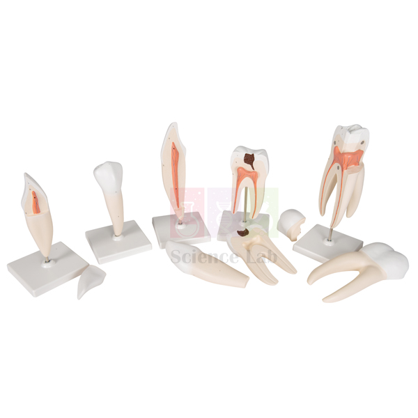
Human Tooth Set
Human Tooth Set (Product Code: SLE/BL/91) is a human biology model set of five enlarged teeth used to teach tooth types and dental decay in school and college biology classrooms. Each tooth stands on its own base at about 8 times life size, and a lower molar with caries lets teachers show decay beside healthy teeth. The set is made by Jain Scientific Equipments Pvt Ltd (ScienceLab), an OEM manufacturer, at its own factory in Ambala, India.
Specifications
| Parameter | Detail |
|---|---|
| Teeth in the set | Five: lower incisor, lower canine, lower premolar, lower molar with caries, upper molar |
| Lower incisor tooth | 2 parts |
| Lower canine tooth | 2 parts |
| Lower premolar tooth | 1 part |
| Lower molar tooth with caries | 2 parts |
| Upper molar tooth | 3 parts |
| Mounting | Each tooth on its own stand |
| Enlargement | About 8 times life size |
| Product code | SLE/BL/91 |
| Manufacturer | Jain Scientific Equipments Pvt Ltd (ScienceLab) — OEM |
| Country of origin | India (made in Ambala, Haryana) |
What Is the Human Tooth Set Used For?
In biology and health lessons the Human Tooth Set lets a teacher hold up each tooth type in turn and link its shape to its job: incisors cut, canines tear, and premolars and molars crush and grind food. Because every model is about 8 times life size, a whole class can follow the comparison from their desks, something a real tooth or a wall chart cannot offer.
As biology teaching aids for oral-health topics, the lower molar with caries is the natural talking point: place it next to the upper molar so students see the difference between a decayed tooth and a sound one, then discuss diet and brushing. Four of the five teeth are made in two or three parts, so check that every piece is back on its stand before the set is put away.
OEM Manufacturer
The Human Tooth Set is produced by Jain Scientific Equipments Private Limited, which trades under the brand ScienceLab. As the OEM (original equipment manufacturer), the company makes and assembles its anatomy models at its own licensed factory at Saha, Ambala, Haryana, India, rather than buying them in from a trader.
Schools, colleges, institutions, dealers and importers therefore buy this dental model set factory-direct, and questions about replacement stands, missing parts or repeat orders go to the maker itself — just ask us. Company background is on the ScienceLab company profile page.
Related Human Models
A tooth set is usually taught together with jaw and dentition models. These ScienceLab models extend the same dental lesson:
- Human Teeth Model, Triple Root Molar — a single enlarged molar that separates into six numbered parts for closer study.
- Human Teeth Model, Lower Jaw, on Stand — shows teeth in position in the jaw with roots, nerves and vessels exposed.
- Human Teeth Model, Dentition Development — follows the jaws from a newborn to a young adult to explain tooth eruption.
- Human Teeth Model, Incisor — an enlarged two-part front tooth for a focused look at one incisor.
See the complete range of Human Models for organ and body-system models used in the same anatomy course.
Frequently Asked Questions
What is included in the Human Tooth Set?
The Human Tooth Set includes five model teeth: a lower incisor (2 parts), a lower canine (2 parts), a lower premolar (1 part), a lower molar with caries (2 parts) and an upper molar (3 parts). Each tooth is supplied mounted on its own stand.
How large are the teeth in the Human Tooth Set?
Each tooth in the Human Tooth Set is about 8 times life size. The enlargement makes the outline of every tooth and the decayed area on the lower molar easy to see from the back of a classroom during a group demonstration.
Why does the Human Tooth Set include a tooth with caries?
The Human Tooth Set includes a lower molar with caries so teachers can show what tooth decay looks like next to healthy teeth. It gives a concrete starting point for lessons on oral hygiene, diet and dental care in biology and health education classes.
Who manufactures the Human Tooth Set?
The Human Tooth Set is manufactured by Jain Scientific Equipments Pvt Ltd (ScienceLab), the OEM, at its own licensed factory in Saha, Ambala, Haryana, India. Buyers deal directly with the manufacturer for quotations, repeat orders and any questions about replacement parts.
How should the Human Tooth Set be looked after?
Keep the Human Tooth Set on its stands away from direct sunlight and heat, and wipe the teeth with a soft, dry cloth. Reassemble the multi-part teeth after every lesson so that no pieces are lost between classes.
What information is needed to quote for the Human Tooth Set?
To quote for the Human Tooth Set, tell us how many sets you need, the delivery location and any other dental models for the same order. ScienceLab supplies India and international markets; send the details through the contact page.
Request a Quote
For a price on the Human Tooth Set, write to us through the ScienceLab contact page or add the set to the enquiry cart. Quotations come direct from the manufacturer, together with supply details for your order.
Last updated: 24 September 2026
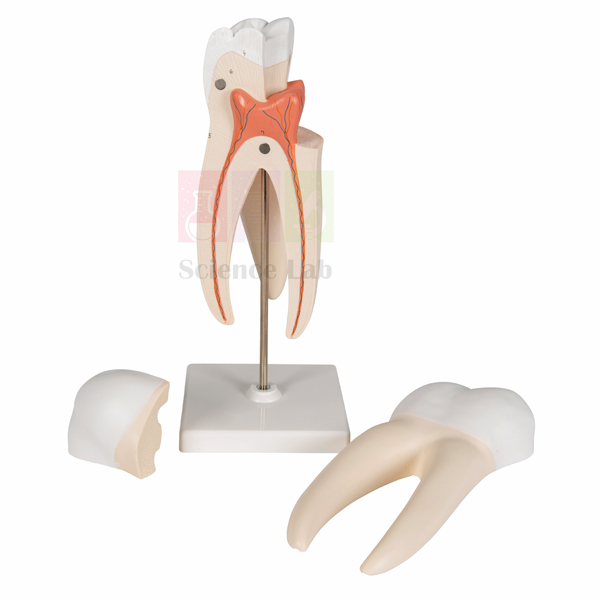
Human Teeth Model, Triple Root Molar
Human Teeth Model, Triple Root Molar (Product Code: SLE/BL/92) is an enlarged human anatomical model of a three-rooted molar used to study tooth structure and root anatomy in biology, nursing and dental-science classrooms. It separates into six parts and is numbered with a key card, so students can take the tooth apart and name each feature. It is made by Jain Scientific Equipments Pvt Ltd (ScienceLab), an OEM manufacturer, at its own factory in Ambala, India.
Specifications
| Parameter | Detail |
|---|---|
| Model | Human molar tooth with three roots, enlarged |
| Parts | Divided into six parts |
| Identification | Numbered, with key card |
| Finish | Appropriately coloured and neatly finished |
| Mounting | On a stand |
| Product code | SLE/BL/92 |
| Manufacturer | Jain Scientific Equipments Pvt Ltd (ScienceLab) — OEM |
| Country of origin | India (made in Ambala, Haryana) |
What Is the Triple Root Molar Model Used For?
In the human mouth, molars with three roots are normally found in the upper jaw, with two roots angled towards the cheek and one towards the palate, giving the tooth firm anchorage for heavy chewing. The Triple Root Molar model makes this root arrangement visible at an enlarged scale, which is difficult to show with an extracted tooth or a textbook diagram.
During a practical, students can lift off the six parts, match each numbered feature with the key card and then rebuild the tooth — a quick self-test before examinations. As a piece of school biology lab equipment it also works well beside a single-rooted molar model, prompting a discussion of why root number differs from tooth to tooth.
OEM Manufacturer
ScienceLab is the trading name of Jain Scientific Equipments Private Limited, the OEM (original equipment manufacturer) of this molar model. The company manufactures its anatomy models at its own licensed factory at Saha, Ambala, Haryana, India, and supplies them factory-direct to schools, colleges, institutions, dealers and importers.
Since the maker quotes directly, questions about quantities, a replacement key card or spare parts reach the source instead of passing through a trader — ask us. Read more about the company on the ScienceLab profile page.
Related Human Models
Root anatomy is easiest to teach by comparison. These models from the same range sit well alongside the Triple Root Molar:
- Human Teeth Model, Lower Molar with One Root — a single-rooted molar showing nerves and vessels, for contrasting root forms.
- Human Tooth Set — five enlarged teeth on stands for comparing incisors, canines, premolars and molars.
- Human Teeth Model, Incisor — a two-part front tooth to set against the molar’s broader crown.
- Human Teeth Model, Lower Jaw, on Stand — places the teeth in the jaw with roots, nerves and vessels exposed.
Explore all Human Models for more anatomy teaching models.
Frequently Asked Questions
How many parts does the Human Teeth Model, Triple Root Molar have?
The Human Teeth Model, Triple Root Molar is divided into six parts. The parts can be separated during a lesson and fitted back together, and the numbered features on the model are identified using its key card.
Is the Triple Root Molar model life size?
No, the Human Teeth Model, Triple Root Molar is enlarged so that its shape and three roots can be seen by a group. The exact enlargement factor is not listed, so please ask us through the contact page if you need dimensions.
What is the key card for?
The key card supplied with the Human Teeth Model, Triple Root Molar explains what each number on the model refers to. Students can use it to identify structures on their own, which makes the model useful for revision and practical tests as well as demonstrations.
Who manufactures the Human Teeth Model, Triple Root Molar?
Jain Scientific Equipments Pvt Ltd (ScienceLab) manufactures the Human Teeth Model, Triple Root Molar as the OEM at its own licensed factory in Saha, Ambala, Haryana, India. Quotations, repeat orders and spare-part questions are handled directly by the manufacturer.
How should the Triple Root Molar model be stored?
The Human Teeth Model, Triple Root Molar is mounted on a stand, so it can stay on a bench or display shelf between lessons. Keep it away from heat and direct sun, wipe it with a soft dry cloth, and keep all six parts with the key card.
What do I need to send for a quotation?
For a quotation on the Human Teeth Model, Triple Root Molar, send the quantity, delivery location and any other anatomy models you need through the contact page. ScienceLab supplies institutions and dealers in India and international markets.
Request a Quote
To price the Human Teeth Model, Triple Root Molar for your lab, use the ScienceLab contact form or place it in the enquiry cart. You receive the quotation direct from the manufacturer.
Last updated: 24 September 2026
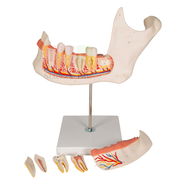
Human Teeth Model, Lower Jaw, on Stand
Human Teeth Model, Lower Jaw, on Stand (Product Code: SLE/BL/93) is a dissected model of the lower left human jaw used to show how teeth are rooted in the jaw and served by nerves and blood vessels, in biology, anatomy and dental-science teaching. Enlarged about 3.5 times, it is coloured and identified with a numbered key card. It is made by Jain Scientific Equipments Pvt Ltd (ScienceLab), an OEM manufacturer, at its own factory in Ambala, India.
Specifications
| Parameter | Detail |
|---|---|
| Model | Lower left human jaw with teeth |
| Enlargement | About 3.5 times |
| Dissection | Jaw dissected to show roots, nerves and vessels |
| Colouring | Appropriately coloured |
| Identification | Numbered key card |
| Mounting | Mounted on a base |
| Product code | SLE/BL/93 |
| Manufacturer | Jain Scientific Equipments Pvt Ltd (ScienceLab) — OEM |
| Country of origin | India (made in Ambala, Haryana) |
What Is the Lower Jaw Teeth Model Used For?
From the outside a tooth looks small and simple, yet much of it lies hidden inside the jaw. By opening up the lower left jaw, this model shows how the roots sit in the bone and how nerves and vessels reach them — the background students need when a lesson turns to toothache, dental anaesthesia or root-canal treatment.
Teachers can point to each numbered structure and ask learners to name it from the key card, turning the model into a quick oral quiz. As school biology lab equipment it pairs well with a whole-jaw model: one shows where every tooth stands, the other shows what lies beneath.
OEM Manufacturer
Jain Scientific Equipments Private Limited, known to customers by its brand ScienceLab, is the OEM (original equipment manufacturer) behind this jaw model. As an anatomical models manufacturer it produces at its own licensed factory at Saha, Ambala, Haryana, India, so buyers deal with the maker rather than a reseller.
Schools, medical and dental colleges, dealers and importers receive quotations straight from the manufacturer, and a replacement key card or a repeat order for matching lab sets can be requested from the same team — ask us. Find out more on the ScienceLab company profile.
Related Human Models
These dental models help complete a lesson that starts with the dissected lower jaw:
- Human Teeth Model, Lower Jaw — shows the position of all the teeth in the lower jaw, the surface view to set beside this dissection.
- Human Teeth Model, Lower Molar with One Root — a single molar with its nerves and vessels for a close-up of one tooth.
- Human Teeth Model, Dentition Development — upper and lower jaws from a newborn to a young adult for lessons on tooth eruption.
- Human Teeth Model, Triple Root Molar — a six-part molar for comparing a three-rooted tooth with the roots seen here.
The full Human Models range covers organs and body systems for the rest of the anatomy syllabus.
Frequently Asked Questions
How much is the Human Teeth Model, Lower Jaw, on Stand enlarged?
The Human Teeth Model, Lower Jaw, on Stand is enlarged about 3.5 times. At this scale the roots, nerves and vessels exposed by the dissection are large enough for a class to follow during a teacher-led demonstration.
What does the dissection on this lower jaw model show?
The Human Teeth Model, Lower Jaw, on Stand is dissected to show the roots of the teeth together with nerves and vessels inside the lower left jaw. The structures are coloured and numbered, and a key card explains what each number marks.
Which part of the jaw does the model represent?
The Human Teeth Model, Lower Jaw, on Stand represents the lower left jaw. For a view of every tooth position in the lower jaw, use it together with the Human Teeth Model, Lower Jaw, which is listed among the related models on this page.
Who manufactures the Human Teeth Model, Lower Jaw, on Stand?
The Human Teeth Model, Lower Jaw, on Stand is manufactured by Jain Scientific Equipments Pvt Ltd (ScienceLab), the OEM, at its own licensed factory in Saha, Ambala, Haryana, India. Institutions and dealers buy it directly from the manufacturer.
How should the lower jaw model be kept?
The Human Teeth Model, Lower Jaw, on Stand is mounted on a base, so it stands upright on a bench or shelf. Clean it with a soft dry cloth, keep it out of direct sunlight, and store the key card with the model for the next class.
What should a quotation request include?
A quotation request for the Human Teeth Model, Lower Jaw, on Stand should give the quantity, the delivery location and any other dental or anatomy models needed. ScienceLab serves buyers in India and international markets; send the details via the contact page.
Request a Quote
Ask for a price on the Human Teeth Model, Lower Jaw, on Stand through the ScienceLab contact page, or collect it with other models in the enquiry cart. The quotation is issued directly by the manufacturer.
Last updated: 24 September 2026
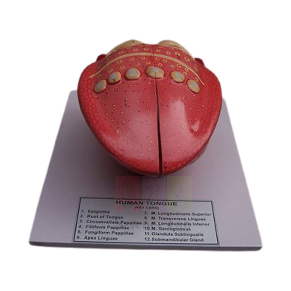
Human Tongue Model
Human Tongue Model (Product Code: SLE/BL/94) is a coloured, numbered teaching model of the human tongue that lets students trace its surface papillae, its muscle layers and the neighbouring salivary glands in a single view. It suits school and college biology classes, nursing and dental foundation courses, and any department assembling biology teaching aids in India for lessons on taste and digestion. The model is made by Jain Scientific Equipments Pvt Ltd (ScienceLab) in Ambala, India, and arrives mounted on a base with its key card.
Specifications
| Parameter | Detail |
|---|---|
| Model | Human tongue with associated muscles and glands |
| Mounting | Mounted on a base |
| Finish | Appropriately coloured and numbered |
| Key | Key card included |
| Surface structures shown | Epiglottis, root of tongue, circumvallate papillae, filiform papillae, fungiform papillae, apex linguae (tip of the tongue) |
| Muscles shown | M. longitudinalis superior, M. transversus linguae, M. longitudinalis inferior, M. genioglossus |
| Glands shown | Glandula sublingualis (sublingual gland), submandibular gland |
Typical Use
Teachers bring out the tongue model when covering the special senses and the mouth as the entry point of the digestive system. The three kinds of papillae on the upper surface lead naturally into a discussion of taste, while the longitudinal, transverse and genioglossus muscles show how the tongue changes shape during chewing, swallowing and speech. Because every structure carries a number, students can work through the key card in pairs, point to each part, and then quiz one another with the card covered.
The epiglottis and root of the tongue help explain why food passes into the oesophagus rather than the airway, and the sublingual and submandibular glands tie the lesson to saliva and the first stage of digestion. As part of school biology lab equipment in India, the model works well beside a prepared slide of tongue tissue so learners can compare the whole-organ view with what they see under the microscope. Store it covered and away from direct sunlight so the colouring stays clear.
Manufacturer
This tongue model is produced by Jain Scientific Equipments Private Limited, the company behind the ScienceLab name, which operates from Ambala, Haryana and makes anatomy models alongside a wider range of educational laboratory equipment. Schools and procurement offices that need vendor registration details can use its GSTIN 06AAECJ8618A1ZT and LEI 3358004HRD1JIVXKYX49. Background on the company is available on the ScienceLab company profile.
Related Human Models
These models and slides extend a lesson on the mouth, taste and the upper digestive and respiratory tracts:
- Microstructure of the Human Tongue Model — a magnified look at the tissue behind the papillae seen on this model.
- Prepared Microscope Slide Set – Digestion (Liver, Pancreas and Tongue) — real tongue sections for microscope work after the model demonstration.
- Larynx Human Model — continues the story below the epiglottis into the voice box.
- Human Nasal Cavity Model — covers smell, which works together with taste in flavour perception.
- Human Digestive System Model — places the tongue at the start of the complete digestive tract.
Browse every anatomy teaching model in the Human Models category.
Frequently Asked Questions
Which parts of the tongue can students identify on this model?
The epiglottis, root of the tongue, circumvallate, filiform and fungiform papillae, the apex, four named muscles (superior and inferior longitudinal, transverse and genioglossus) and the sublingual and submandibular glands. Each is numbered and listed on the key card.
Is the model supplied with a key and a base?
Yes. It comes mounted on a base, and a key card matching the numbered structures is included, so it is ready for classroom use on arrival.
What are the dimensions of the Human Tongue Model?
Overall dimensions are not listed on this page. Please ask through our contact page and we will confirm the size before you order.
How should the model be cleaned?
Wipe it with a soft cloth, lightly dampened if needed, and let it dry before storing. Avoid solvents or abrasive cleaners, which can dull the painted colours and numbers.
Does the model come with a warranty?
Warranty terms are not stated here. Ask us through the contact page and we will set them out in writing with your quotation.
Can you supply several units for a school network or an overseas buyer?
Yes. ScienceLab supplies schools, colleges and distributors across India and international markets; share the quantity and destination so we can quote packing and delivery.
Request a Quote
For a quotation on the Human Tongue Model, send the product code SLE/BL/94, the quantity and your delivery location through our contact page, or add it to the enquiry cart along with any other models you need.
Last updated: 24 September 2026
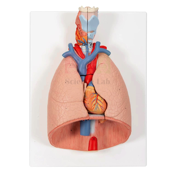
Human Thoracic Organs Model
Human Thoracic Organs Model (Product Code: SLE/BL/98) is an anatomy model used to teach the layout of the chest, with two removable front halves of the lungs that reveal the heart and great vessels in their natural position. It also shows a two-part larynx and heart, the trachea and bronchial tree, subclavian vessels, vena cava, aorta, pulmonary artery, oesophagus and diaphragm, numbered on a key card. It is made by Jain Scientific Equipments Pvt Ltd (ScienceLab), an OEM anatomical models manufacturer, at its own factory in Ambala, India.
Key Details
| Parameter | Detail |
|---|---|
| Type | Thoracic organs anatomy model |
| Removable parts | Two front halves of the lungs |
| Two-part organs | Larynx, heart |
| Airway and vessels | Trachea and bronchial tree, subclavian artery and vein, vena cava, aorta, pulmonary artery |
| Other structures | Oesophagus, diaphragm |
| Finish and key | Appropriately coloured, numbered with key card |
| Mounting | Mounted on a base |
| Custom builds | Custom-made anatomical models available for bulk quantity |
| Product code | SLE/BL/98 |
| Manufacturer | Jain Scientific Equipments Pvt Ltd (ScienceLab) — OEM |
| Country of origin | India (made in Ambala, Haryana) |
What Is the Human Thoracic Organs Model Used For?
With the front lung halves in position, the model shows what an anatomist first sees on opening the chest: the lungs filling most of the cavity and the diaphragm below. Removing them exposes the mediastinum, the central region where the heart, aorta, vena cava, pulmonary artery, trachea and oesophagus run close together. This makes it a clear way to explain relations between organs, such as why the oesophagus lies behind the heart, how the subclavian vessels arch towards the arms and how the pulmonary artery links the right side of the heart to the lungs.
As high school biology lab equipment it supports chapters on breathing and circulation, and in nursing and medical courses it serves as a revision aid before cadaver or imaging sessions. Ask students to predict which structure lies beneath each lung half before lifting it, then confirm with the numbered key card. Store the loose lung halves with the base and clean all surfaces with a soft, dry cloth.
What to Specify When Ordering
- Whether you need the standard model or a custom-made version, which is offered for bulk quantity
- The number of units for each lab, department or distributor order
- Any companion chest models, such as the Human Lungs with Heart and Larynx Model, to be quoted together
OEM Manufacturer
This thoracic model is manufactured by Jain Scientific Equipments Private Limited (CIN U29309HR2020PTC087597), the OEM (original equipment manufacturer) behind the ScienceLab brand. Production takes place at the company's own licensed factory at 449, HSIIDC Industrial Area, Saha, Ambala, Haryana (Factory Licence AMB-ONLINE-CHD-J-414), where custom-made anatomical models can also be discussed for bulk quantities. Colleges, schools, dealers and importers buy factory-direct, and replacement lung halves or repeat orders are handled by the manufacturer itself, so just ask us. Learn more on our profile page.
Related Human Models
Pair this model with the following for a complete chest and trunk lesson:
- Human Lungs with Heart and Larynx Model — a comparable set of organs with 2-part lungs
- Human Heart on Diaphragm Model — a closer look at the heart resting on the diaphragm
- Segmented Lung — shows the internal segments that the front lung halves cover
- Human Sexless Torso Model — places the thoracic organs within the whole trunk
- Human Skeleton with Internal Organs & Muscles — relates the organs to the rib cage and spine
The model is listed in our Human Models category.
Frequently Asked Questions
Which parts of the Human Thoracic Organs Model come off?
On the Human Thoracic Organs Model, the two front halves of the lungs are removable, and the larynx and heart are each made in two parts, so students can open the chest layer by layer during a lesson.
What is visible once the lung halves are removed?
Removing the lung halves of the Human Thoracic Organs Model exposes the heart, aorta, vena cava, pulmonary artery, subclavian artery and vein, trachea with bronchial tree and oesophagus, with the diaphragm forming the floor of the chest below.
Can a customised version of the thoracic organs model be made?
Custom-made anatomical models are listed as available for bulk quantity alongside the Human Thoracic Organs Model. Share your requirement and quantity through the contact page so we can discuss what is feasible before quoting.
How does it differ from the Human Lungs with Heart and Larynx Model?
The Human Thoracic Organs Model and the Human Lungs with Heart and Larynx Model cover the same organ group; the thoracic model is built around removable front lung halves that reveal the organs in their natural position. We can help you choose.
What size is the Human Thoracic Organs Model?
Dimensions and material of the Human Thoracic Organs Model are not stated in its listing. Ask us through the contact page and we will confirm them, along with packing details, in the quotation.
Who manufactures the Human Thoracic Organs Model?
Jain Scientific Equipments Private Limited (ScienceLab) is the OEM that manufactures the Human Thoracic Organs Model at its own licensed factory at Saha, Ambala, Haryana, India, and supplies it directly to schools, colleges, dealers and importers.
What should a tender or institutional enquiry include?
A tender or institutional enquiry for the Human Thoracic Organs Model should give product code SLE/BL/98, the quantity, the delivery location in India or abroad and any customisation needed; quotations come directly from the manufacturer.
Request a Quote
For pricing on the Human Thoracic Organs Model, contact the manufacturer through our contact page or add it to the enquiry cart.
Last updated: 24 September 2026
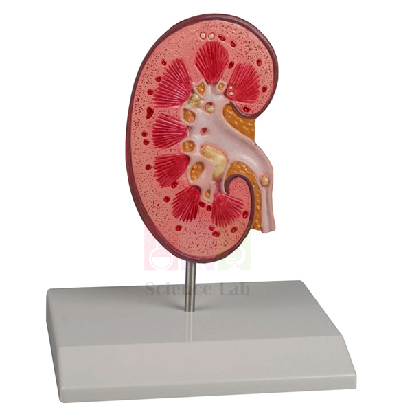
Kidney Stone Model
Kidney Stone Model (Product Code: SLE/BL/99) is a pathology teaching model used to explain kidney stones (nephrolithiasis) and stones in the urinary tract (urolithiasis) to students and patients. Its renal calices, renal pelvis and ureter are shown opened so stones can be located from the renal pyramids down to the ureter, which suits nursing institutes, urology clinics and pharmacy college lab equipment lists. It is made by Jain Scientific Equipments Pvt Ltd (ScienceLab), an OEM manufacturer, at its own factory in Ambala, India.
Key Details
| Parameter | Detail |
|---|---|
| Type | Pathology model of kidney and urinary stones |
| Conditions illustrated | Nephrolithiasis (kidney stones) and urolithiasis (urinary stones) |
| Opened structures | Renal calices, renal pelvis, ureter |
| Stone positions shown | Area of the renal pyramids; origin of the upper calix group; renal cortex; connecting tubules of the lower calix group, causing congestion of the minor calices (partially closed, partially opened); ureter |
| Finish and key | Appropriately coloured, numbered with key card |
| Mounting | Mounted on a base |
| Product code | SLE/BL/99 |
| Manufacturer | Jain Scientific Equipments Pvt Ltd (ScienceLab) — OEM |
| Country of origin | India (made in Ambala, Haryana) |
What Is the Kidney Stone Model Used For?
In the classroom, the opened collecting system lets students follow urine from the renal pyramids through the minor and major calices into the renal pelvis and down the ureter, and then see how a stone at each point changes the picture. The lower calix group, with its partly blocked minor calices, shows how an obstruction backs up urine inside the kidney, while the stone in the ureter explains the sudden, severe pain of ureteric colic. These comparisons support lectures on renal physiology, urinary tract pathology and the diet advice given for stone prevention.
Doctors and nurses also use the model in outpatient clinics to show patients where a stone sits and why a particular treatment is being discussed, and it adds a practical element to a health education kit for schools in India covering hydration and kidney health. Point to each numbered site while reading from the key card, and keep the model covered and dust-free between sessions.
OEM Manufacturer
ScienceLab is the trading name of Jain Scientific Equipments Private Limited, the OEM (original equipment manufacturer) of this kidney stone model. The company makes its anatomy and pathology models at its own licensed factory at Saha, Ambala, Haryana, India, and sells them factory-direct to medical and nursing colleges, hospitals, schools, dealers and importers in India and international markets. Repeat orders and quotations are handled by the manufacturer itself. Visit the company profile for background.
Related Human Models
For a complete lesson on the kidney and urinary tract, consider:
- Human Kidney Model — normal kidney anatomy to compare with the stone sites
- Human Kidney, Nephron, and Glomerulus Model Set — explains the tubules and filtration units involved
- Human Kidney, Nephron and Renal Corpuscle Model — enlarges the nephron and renal corpuscle for detailed study
- Human Kidney with Adrenal Gland Model — shows the kidney with its adrenal gland and vessels
- Gallstone Model — contrasts urinary stones with stones in the biliary system
This model is listed in our Human Models category.
Frequently Asked Questions
What conditions does the Kidney Stone Model explain?
The Kidney Stone Model explains kidney stones (nephrolithiasis) and stones elsewhere in the urinary tract (urolithiasis), showing where stones form or lodge in the opened calices, renal pelvis and ureter of a coloured, numbered kidney.
Where are the stones shown on the Kidney Stone Model?
On the Kidney Stone Model, stones are shown in the area of the renal pyramids, at the origin of the upper calix group, in the renal cortex, in the connecting tubules of the lower calix group and in the ureter.
Is the kidney stone model suitable for patient education?
Yes. The opened calices, renal pelvis and ureter of the Kidney Stone Model make it straightforward for doctors and nurses to show a patient where a stone sits and why it can cause pain or block the flow of urine.
Does the Kidney Stone Model come with a key?
Yes. The Kidney Stone Model is appropriately coloured and numbered, and the numbered features are identified on an accompanying key card, which makes it easy to use for self-study, practical examinations and counselling.
Is the Kidney Stone Model life size?
Scale, size and material of the Kidney Stone Model are not stated in its listing. Ask us through the contact page and we will confirm these details, together with packing information, in your quotation.
Who manufactures the Kidney Stone Model?
Jain Scientific Equipments Private Limited (ScienceLab) is the OEM that manufactures the Kidney Stone Model at its own factory at Saha, Ambala, Haryana, India, and supplies it directly to medical and nursing colleges, hospitals, dealers and importers.
Can hospitals or distributors order the model in quantity?
Yes. Hospitals, colleges and distributors in India and international markets can order the Kidney Stone Model in quantity; state product code SLE/BL/99 and the number of units, and the quotation comes straight from the manufacturer.
Request a Quote
Ask the manufacturer for a quotation on the Kidney Stone Model through our contact page, or add it to the enquiry cart with related kidney models.
Last updated: 24 September 2026
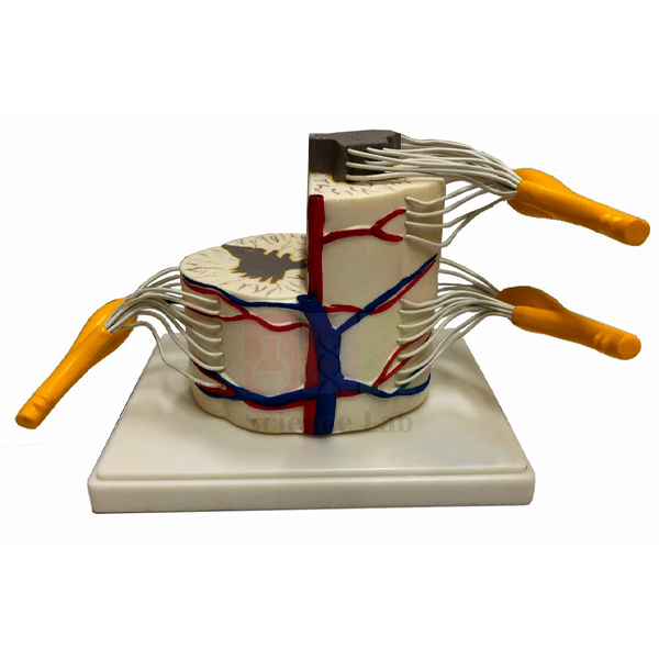
Human Vertebral And Partial Spinal Cord Model
Human Vertebral And Partial Spinal Cord Model (Product Code: SLE/BL/100) reproduces a segment of the upper thoracic spinal cord at six times life size, with the anterior and posterior nerve roots exposed and colour coded so the direction of nerve signals is easy to follow. Physiology teachers, nursing and physiotherapy tutors and senior school biology departments use it to explain how the cord connects to the body through spinal nerves. It is manufactured by Jain Scientific Equipments Pvt Ltd (ScienceLab), Ambala, and fits naturally into a human biology model set for the nervous system.
Specifications
| Parameter | Detail |
|---|---|
| Region represented | Segment of the upper thoracic spinal cord |
| Scale | Six times life size |
| Material | Vinyl plastic, chosen for durability |
| Nerve roots | Anterior and posterior roots exposed and colour coded |
| Size | 30 x 15 x 15 cm |
Typical Use
The enlargement makes the difference between the two root types visible from the back of a classroom. A tutor can trace the posterior (sensory) root into the cord and the anterior (motor) root out of it, then use the model to walk through a simple reflex arc or to explain why an injury at one level affects a particular band of the body. Because the roots are colour coded, students copying labelled diagrams into notebooks can match colours to function without constant prompting.
In high school biology lab equipment collections the model sits well between a whole nervous-system chart and prepared slides of spinal cord sections: it bridges the gross and microscopic views. Its vinyl construction tolerates repeated handling during practical sessions and viva preparation; keep it on a flat shelf and wipe it with a damp cloth after use.
Manufacturer
Jain Scientific Equipments Private Limited makes this spinal cord model under its trading name ScienceLab at Ambala in Haryana, a long-standing centre for scientific instrument making in India. The company designs and produces anatomy models for teaching institutions and dispatches them within India and abroad; you can read more on the company profile page.
Related Human Models
Pair this segment with models that show the whole spine or the wider nervous system:
- Human Spinal Cord Section Model — a sectional view for comparing grey and white matter.
- Human Thoracic Spinal Column Model — shows the thoracic vertebrae that surround this region of the cord.
- Human Reflex Pathway Model — follows the sensory and motor roots through a complete reflex arc.
- Human Nervous System Model — puts the spinal nerves in the context of the whole body.
All anatomy teaching models are listed in the Human Models category.
Frequently Asked Questions
Which part of the spinal cord does the model represent?
A segment from the upper thoracic region, enlarged six times so the nerve roots and their attachments are clear.
How large is the model?
It measures 30 x 15 x 15 cm, a comfortable size for bench demonstrations and small-group work.
Will it stand up to regular student handling?
It is moulded in vinyl plastic for durability. Normal classroom handling is fine; avoid dropping it or leaving it near heat sources.
Is a printed key supplied?
A key is not listed in the product details. If you need a labelled key or a structure list for your syllabus, ask us through the contact page before ordering.
What should an institutional enquiry include?
Give the product code SLE/BL/100, quantity, delivery address and any tender or purchase-order format you must follow, and we will prepare the quotation accordingly.
Do you ship outside India?
Yes. ScienceLab serves buyers in India and international markets; share the destination country and quantity for a packed and delivered quote.
Request a Quote
Ask for a price on the Human Vertebral And Partial Spinal Cord Model through our contact page, or place it in the enquiry cart with the other anatomy items on your list.
Last updated: 24 September 2026
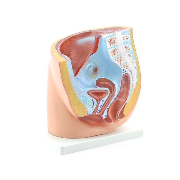
Female Half Pelvis Model
Female Half Pelvis Model (Product Code: SLE/A/04) is an approximately life-size sectional model that lets students look straight into the female pelvis and see how the bladder, uterus and colon sit in relation to one another and to the lower spine. It is used in secondary and senior secondary biology, nursing, midwifery and paramedical classrooms whenever reproductive or pelvic anatomy is taught. As an anatomical models manufacturer, Jain Scientific Equipments Pvt Ltd (ScienceLab) of Ambala supplies it mounted on a blue base with a laminated key.
Specifications
| Parameter | Detail |
|---|---|
| Scale | About life size |
| View | Cross section of a female pelvis |
| Organs in section | Bladder, uterus and colon |
| Skeletal and nervous structures | Structure of the spine and the spinal cord present in the pelvis |
| Base | Mounted on a blue base |
| Key | Laminated key included |
Typical Use
Sectional models answer a question that flat textbook diagrams leave vague: what lies in front of, behind and above what. With this half pelvis, a teacher can show that the bladder sits at the front, the uterus behind it and the colon further back against the spine, then explain how a filling bladder or a growing uterus changes that arrangement. Nursing and midwifery tutors use the same view when discussing catheterisation, pelvic examination or the changes of pregnancy in general terms.
The laminated key survives repeated handling in practical rooms, so students can use it for self-testing without damaging a paper sheet. When building a biology lab equipment India classroom set for reproduction and excretion, many schools place this model next to a skeletal female pelvis so learners can compare the bony framework with the soft organs it protects.
Manufacturer
ScienceLab is the brand under which Jain Scientific Equipments Private Limited, based in Ambala, Haryana, makes anatomy and biology teaching models for schools, colleges and training institutes. Details of the company and its product range are on the ScienceLab profile page.
Related Human Models
Useful companions for a lesson on the pelvis and reproductive system:
- Female Pelvis with Organs — a further view of pelvic organs for side-by-side comparison.
- Female Skeletal Pelvis — the bony pelvis alone, for teaching landmarks and pelvic shape.
- Male Pelvis Section Model — lets students contrast male and female pelvic anatomy.
- Human Female Genital Organs Model — detail of the reproductive organs seen in section here.
See the complete range in the Human Models category.
Frequently Asked Questions
Is the model life size?
It is about life size, which helps students relate what they see to the real body.
Which organs can be seen in the section?
The bladder, uterus and colon are shown in cross section, together with the spine and the spinal cord where they pass through the pelvis.
Does it come with labels?
A laminated key is included, and the model is mounted on a blue base for display.
Is it suitable for school students as well as nursing trainees?
Yes. The sectional view is simple enough for secondary school reproduction topics and detailed enough for introductory nursing and midwifery anatomy.
Can you provide exact dimensions or weight for shipping?
These are not listed on this page. Contact us through the contact page and we will confirm them with your quotation.
Do you supply colleges and distributors outside India?
Yes, ScienceLab supplies institutions in India and international markets; tell us the quantity and destination.
Request a Quote
To order the Female Half Pelvis Model (SLE/A/04), write to us via the contact page or add it to the enquiry cart and submit your list in one go.
Last updated: 24 September 2026
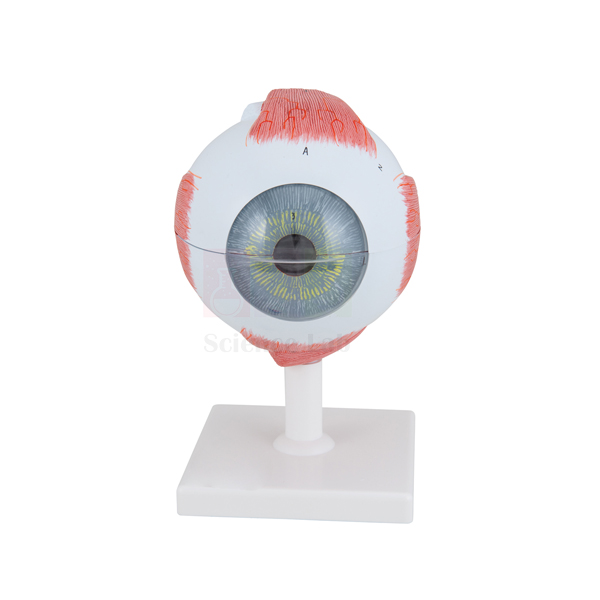
Human Eye Model
Human Eye Model (Product Code: SLE/A/10) is a five-times-enlarged, bisected eyeball that opens up the inside of the eye for teaching sight, focusing and eye defects. Its lens, iris, aqueous humor and cornea lift out, so students can handle the parts that bend light and see where each one sits. For schools and colleges searching for an eye model for anatomy teaching in India, it offers labelled structures and eye muscles on a compact blue base. It is manufactured by Jain Scientific Equipments Pvt Ltd (ScienceLab), Ambala.
Specifications
| Parameter | Detail |
|---|---|
| Scale | 5x life size |
| Size (with base) | 21 x 14 x 12 cm |
| Base | Blue base |
| Construction | Eye bisected to show internal structure |
| Removable parts | Lens, iris, aqueous humor and cornea |
| Labelling | Eye structures and musculature labelled; key included |
Typical Use
In a lesson on image formation, the teacher lifts out the cornea and lens to show the two structures that refract incoming light, then replaces them to show how light reaches the retina at the back of the bisected eyeball. Removing the iris makes it easy to explain how the pupil widens and narrows in changing light. The labelled external muscles help older students understand how both eyes move together, which links well to discussions of squint and eye strain.
Class 10 physics and biology topics on the human eye, myopia and hypermetropia become more concrete when learners can hold the lens and see its position relative to the retina. Colleges also use the model in optometry and nursing foundation courses as one of their core biology teaching aids. Return each removable part to its place after the session and store the model covered to keep small pieces from going missing.
Manufacturer
Jain Scientific Equipments Private Limited (CIN U29309HR2020PTC087597) trades as ScienceLab and manufactures its teaching models at a licensed factory at 449, HSIIDC Industrial Area, Saha, Ambala, Haryana (Factory Licence AMB-ONLINE-CHD-J-414). Learn more about the company on the ScienceLab company profile.
Related Human Models
Other eye models that build on this one, from the orbit to the microscopic layers:
- Human Eye in Orbit Model — shows the eyeball in its bony socket with surrounding structures.
- Eye Microanatomy Model — zooms in on the tissue layers of the eye wall and retina.
- Cataract Eye Model with 4 Forms of Cataract — moves from normal anatomy to a common lens disorder.
- Human Eye Model with Eyelids — adds the eyelids for lessons on protection of the eye.
Explore more anatomy models in the Human Models category.
Frequently Asked Questions
How much is the eye enlarged?
The model is 5x life size, and with its base it measures 21 x 14 x 12 cm.
Which parts can be removed?
The lens, iris, aqueous humor and cornea are removable for closer inspection; the rest of the bisected eyeball stays fixed to the base.
Are the eye muscles shown?
Yes. The musculature is labelled along with the other eye structures, and a key is included.
How do I prevent losing the small removable parts?
Count the parts back in after each class and keep the model in a covered cupboard. If a part is lost or damaged, contact us to ask about a replacement.
What warranty applies?
Warranty terms are not listed on this page; please ask through the contact page and we will state them with the quotation.
Can we order in bulk for several school branches?
Yes. Send the quantity and delivery locations, within India or abroad, and we will quote accordingly.
Request a Quote
Request pricing for the Human Eye Model (SLE/A/10) through the contact page, or add it to your enquiry cart with other lab items.
Last updated: 24 September 2026
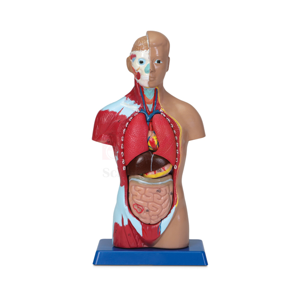
Human Micro Sexless Torso Model
Human Micro Sexless Torso Model (Product Code: SLE/A/11) is a desk-sized torso that packs removable organs, exposed right-side musculature and a partly opened head into a model only 26 cm across its longest side. Because it is small enough to pass around a group, it suits middle and secondary school classes, coaching centres and home-study tutors who want a hands-on human body model for students in India without the storage demands of a full-size torso. It is manufactured by Jain Scientific Equipments Pvt Ltd (ScienceLab) in Ambala and comes on a blue base with a numbered, laminated key.
Specifications
| Parameter | Detail |
|---|---|
| Type | Miniature sexless torso |
| Size (with base) | 10-1/4 x 5 x 3-1/4 in (26 x 12.7 x 8.3 cm) |
| Base | Blue base |
| Exposed structures | Musculature on the right side of the body; right side of the brain; bones of the face |
| Removable organs | Lungs, heart, liver, stomach and intestines |
| Key | Numbered, laminated key identifying 27 anatomical features and 11 major muscles |
Typical Use
The small format changes how the model is used: rather than one teacher demonstrating at the front, several models can circulate so each group lifts out the lungs, heart, liver, stomach and intestines, names them from the key and then reassembles the torso in the right order. That reassembly step is a quick check of whether students understand where each organ sits and which organs overlap. The exposed right-side muscles and facial bones give a second activity once the organ work is done.
For school biology lab setups in India covering life processes, the model supports lessons on digestion, breathing and circulation in a single session. It also travels easily for science exhibitions and outreach visits. Keep the removable organs together in a small tray during class so none are misplaced.
Manufacturer
The model is made by Jain Scientific Equipments Private Limited, trading as ScienceLab from Ambala, Haryana. It is a registered MSME with manufacturing as its major activity (Udyam UDYAM-HR-01-0003714), and overseas orders are shipped under its Importer-Exporter Code AAECJ8618A issued by the DGFT. See the company profile for more.
Related Human Models
Models that pair with a miniature torso for larger or more detailed study:
- Human Sexless Torso Model — the larger dissectible torso for teacher demonstrations.
- Miniature Human Muscular Figure — another compact model, focused on the superficial muscles of the whole body.
- Human Respiratory System Model, 2-parts — expands on the lungs removed from this torso.
- Human Digestive System Model — follows the stomach and intestines in more detail.
Find more options in the Human Models category.
Frequently Asked Questions
How big is the micro torso?
With its base it measures 10-1/4 x 5 x 3-1/4 inches, or 26 x 12.7 x 8.3 cm, small enough for a student desk.
How many structures does the key identify?
The numbered, laminated key identifies 27 anatomical features and 11 major muscles.
Which organs come out?
The lungs, heart, liver, stomach and intestines are removable for closer inspection.
Should we choose this or a full-size torso?
Choose the micro torso for group work, exhibitions and limited storage; choose a full-size torso when the whole class needs to see detail from a distance. Many labs keep one large torso and several small ones.
Can you share packed dimensions for freight?
Packed size and weight are not listed here; ask through our contact page and we will include them in the quotation.
Is export supply available?
Yes. ScienceLab ships to buyers across India and international markets.
Request a Quote
Tell us how many Human Micro Sexless Torso Models you need via the contact page, or add the item to the enquiry cart and we will respond with a quotation.
Last updated: 24 September 2026
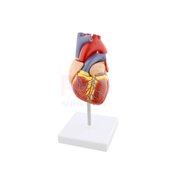
Human Heart Model
Human Heart Model (Product Code: SLE/A/12) is a 3-piece heart enlarged to twice life size, with the front portions of the ventricles and atria lifting away to reveal its internal structures. The outer surface carries the coronary arteries and veins, so one model covers both the external blood supply of the heart muscle and its internal layout. Biology teachers, nursing and paramedical tutors and physiology departments looking for a heart model for anatomy classes in India use it for circulation lessons. It is manufactured by Jain Scientific Equipments Pvt Ltd (ScienceLab), Ambala.
Specifications
| Parameter | Detail |
|---|---|
| Scale | 2x life size |
| Size (with base) | 15.75 x 19 x 17.5 cm |
| Construction | 3-piece model, enlarged for enhanced detail of internal and external structures |
| Removable parts | Anterior portions of the ventricles and the atria |
| Key | Numbered key identifying 30 structures, including the coronary arteries and veins |
Typical Use
A common teaching sequence starts with the closed heart: students follow the coronary arteries and veins across the surface, which leads into a discussion of why a blocked coronary artery damages heart muscle. The teacher then removes the anterior ventricle and atrium pieces and traces blood from the right atrium through the right ventricle to the lungs, and back through the left side to the body. Doubling the size makes this internal detail easier to see from the bench.
The numbered key lets students revise independently after the demonstration, matching the 30 numbered points to their names. The model is a sound fit for double circulation topics in secondary school and for the cardiovascular unit of nursing and allied health courses. After use, refit the removable pieces before storing so the edges are not chipped.
Manufacturer
Jain Scientific Equipments Private Limited makes this heart model in Ambala, Haryana and sells it under the ScienceLab brand, one of many anatomy and biology items it produces for teaching labs. Company information is on the ScienceLab profile.
Related Human Models
Choose from these for a complete circulation unit:
- Heart 3 times Natural Size Model — a larger alternative for big lecture rooms.
- Human Heart on Diaphragm Model — shows how the heart rests in the chest on the diaphragm.
- Human Circulatory System Model — links the heart to arteries and veins throughout the body.
- Human Heart, Pumping Model — demonstrates the pumping action to accompany this anatomical view.
More heart and body models are in the Human Models category.
Frequently Asked Questions
How many pieces does the heart come apart into?
Three. The anterior portions of the ventricles and the atria are removable from the main body.
Are the coronary vessels shown?
Yes, the coronary arteries and veins are among the 30 structures identified on the numbered key.
What is the size of the model?
It is 2x life size and measures 15.75 x 19 x 17.5 cm including the base.
Is this model suitable for a large lecture hall?
It works well for benches and small groups. For viewing from far back in a large hall, consider pairing it with a larger heart model or a document camera.
How should it be cleaned?
Dust with a soft brush and wipe with a slightly damp cloth; do not soak the pieces or use solvent cleaners.
What details do you need for an institutional quotation?
Product code SLE/A/12, quantity, billing and delivery address, and any tender reference. Send them through the contact page.
Request a Quote
Get a quotation for the Human Heart Model through our contact page, or add it to the enquiry cart; we supply institutions in India and international markets.
Last updated: 24 September 2026
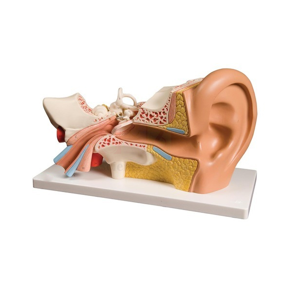
Human Ear Model
Human Ear Model (Product Code: SLE/A/13) is a one-piece ear enlarged to 1-1/2 times life size, showing external and internal structures together with the major blood vessels that supply them. With no loose parts to lose, it is a practical ear model for anatomy lab use in India, where the same model may pass through many classes in a week. Science teachers, audiology and ENT nursing tutors and physiology lecturers use it to explain hearing and balance, and it is made by Jain Scientific Equipments Pvt Ltd (ScienceLab) in Ambala.
Specifications
| Parameter | Detail |
|---|---|
| Scale | 1-1/2x life size |
| Size (with base) | 16.5 x 13 x 13 cm |
| Construction | 1-piece model, enlarged to show greater detail in internal structures |
| Vasculature | Major vasculature included |
| Key | Numbered key identifying 21 external and internal structures |
Typical Use
The model is typically used to trace the path of sound: students start at the outer ear, follow the canal inward and then work through the internal structures while the teacher explains how vibrations are converted into nerve signals. Because the numbered key covers both external and internal structures, a worksheet can ask learners to sort the 21 points into outer, middle and inner ear groups, which reinforces the three-part division that exam questions rely on.
The included vasculature is useful in nursing and allied health classes when discussing ear infections and why inflammation in this region is taken seriously. Its single-piece construction makes it well suited to open display in a biology lab, as there are no removable parts to go missing between classes. A soft brush keeps dust out of the recesses.
Manufacturer
ScienceLab is the trading name of Jain Scientific Equipments Private Limited, an Ambala, Haryana company incorporated under CIN U29309HR2020PTC087597. Its anatomy models are produced at the company's licensed factory in the HSIIDC Industrial Area, Saha, Ambala, which holds Factory Licence AMB-ONLINE-CHD-J-414. The company profile has further details.
Related Human Models
Add one of these for deeper study of hearing and balance:
- Human Inner Ear Model, 18x — a strongly magnified view of the inner ear for senior classes.
- Human Inner Ear Model, 4x — a moderate enlargement of the inner ear alone.
- Human Ear Model, 5 Parts — a take-apart alternative when students should dismantle the ear.
- Human Nasal Cavity Model — completes an ear, nose and throat teaching set.
The full range is in the Human Models category.
Frequently Asked Questions
Does the ear come apart?
No. It is a 1-piece model, enlarged so the internal structures can be seen without dismantling. If you prefer a take-apart version, see the 5-part ear model listed above.
How many structures are identified?
The numbered key identifies 21 external and internal structures.
What are the dimensions?
The model is 1-1/2x life size and measures 16.5 x 13 x 13 cm with its base.
Are blood vessels shown?
Yes, the major vasculature is included on the model.
Can you confirm warranty and replacement terms?
These are not listed here. Ask through the contact page and we will confirm them in the quotation.
Do you accept orders from outside India?
Yes, ScienceLab supplies institutions and distributors in India and international markets.
Request a Quote
Send your requirement for the Human Ear Model (SLE/A/13) through the contact page, or collect it with other items in the enquiry cart before submitting.
Last updated: 24 September 2026
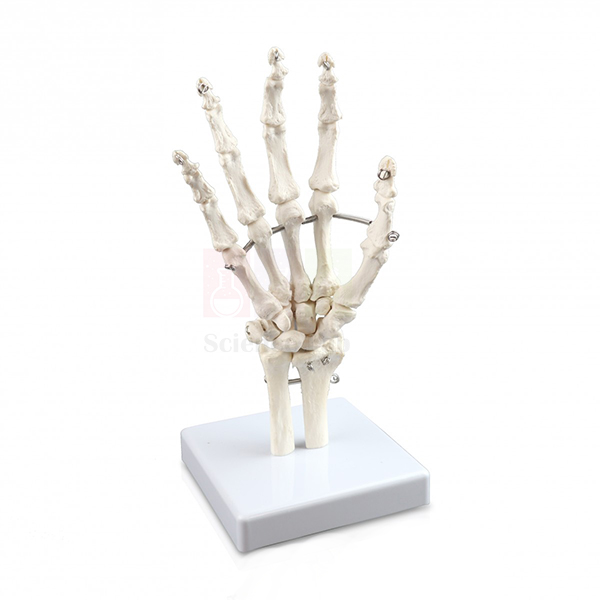
Human Skeletal Hand Model
Human Skeletal Hand Model (Product Code: SLE/A/16) is an approximately life-size plastic skeleton of the right hand, with the bones of the fingers, palm and wrist wired so the joints can move a little, and with the lower ends of the ulna and radius attached. It gives students a close, hands-on look at the small bones that a full skeleton displays only from a distance, which is why it is often bought to complement a human skeleton model in India's school and college labs. It is manufactured by Jain Scientific Equipments Pvt Ltd (ScienceLab), Ambala, and supplied as one piece on a blue base with a laminated key.
Specifications
| Parameter | Detail |
|---|---|
| Material | Plastic |
| Scale | Approximately life size |
| Side | Right hand |
| Construction | One piece, on a blue base |
| Joints | Bones of the fingers, hand and wrist wired to allow some movement |
| Forearm bones | Distal ends of the ulna and radius included |
| Key | Laminated key included |
Typical Use
Anatomy students usually meet the hand as three groups of bones: the carpals of the wrist, the metacarpals of the palm and the phalanges of the fingers. On this model they can count and name each group with the laminated key, then gently flex the wired joints to see how the fingers bend and why the thumb moves differently. Including the ends of the radius and ulna lets the teacher show where the forearm meets the wrist, the region involved in the common wrist fracture discussed in first-aid and physiotherapy classes.
Physiotherapy, occupational therapy, nursing and sports science courses use the hand for palpation practice, holding it alongside their own hand to locate landmarks. Senior school biology classes use it when covering joints and movement. Handle the wired joints gently and avoid forcing them beyond their natural range, since the wires allow only limited movement.
Manufacturer
Jain Scientific Equipments Private Limited produces the model in Ambala, Haryana, under its ScienceLab trade name. For tender files and vendor onboarding, the company's registration identifiers are GSTIN 06AAECJ8618A1ZT and Legal Entity Identifier 3358004HRD1JIVXKYX49. Read about the company on the ScienceLab profile page.
Related Laboratory Equipment
Models that extend a study of the hand and upper limb:
- Human Hand Joint Model — another view of the hand joints for movement studies.
- Human Hand Joint Model with Ligaments — adds the ligaments that hold these bones together.
- Human Arm and Hand model — places the hand in the context of the whole arm.
- Human Skeletal Foot Model — the matching right-foot skeleton for comparing hand and foot bones.
More anatomy models are in the Human Models category.
Frequently Asked Questions
Is this a left or right hand?
It is a model of the right hand.
Can the fingers move?
Yes, to a limited extent. The finger, hand and wrist bones are wired to allow some movement of the joints.
What material is it made from?
Plastic, which is lighter and easier to clean than real bone specimens.
Are forearm bones included?
The distal ends of the ulna and radius are included, enough to show the wrist joint.
Can a left hand be supplied instead?
The listed model is the right hand. Ask through our contact page if you need a different version and we will tell you what is possible.
Do you handle bulk and export orders?
Yes, for schools and colleges in India and international markets; include quantity and destination in your enquiry.
Request a Quote
For pricing on the Human Skeletal Hand Model (SLE/A/16), contact us via the contact page or add it to the enquiry cart.
Last updated: 24 September 2026
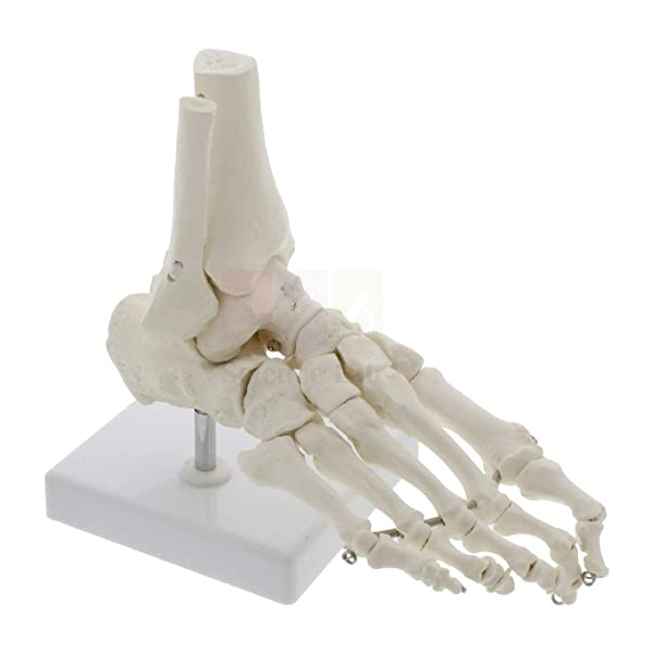
Human Skeletal Foot Model
Human Skeletal Foot Model (Product Code: SLE/A/17) reproduces the bony skeleton of the right foot at approximately life size, from the toes through the midfoot to the ankle, and includes the lower ends of the tibia and fibula where the leg meets the foot. The bones are wired so the joints can move slightly, letting students see how the foot flexes when walking. Physiotherapy, sports science, nursing and biology departments use it to teach the arches and the ankle joint, and as a practical piece of biology lab equipment in India it is compact enough for bench work. Jain Scientific Equipments Pvt Ltd (ScienceLab), Ambala, manufactures it in plastic with a laminated key.
Specifications
| Parameter | Detail |
|---|---|
| Material | Plastic |
| Scale | Approximately life size |
| Side | Right foot |
| Bones represented | Toes, foot and ankle, plus the distal ends of the tibia and fibula |
| Joints | Wired joints allow some movement |
| Key | Laminated key included |
Typical Use
The foot carries the whole body weight, and this model helps explain how it does so. Teachers point out the tarsal bones of the ankle and heel, the metatarsals that form the long arch, and the phalanges of the toes, then use the tibia and fibula ends to show how the ankle joint locks the foot to the leg. Gently moving the wired joints shows how the foot adapts to uneven ground, a useful starting point for discussing flat feet, footwear and common ankle sprains.
In physiotherapy and sports courses, students hold the model beside their own foot to find bony landmarks before practical examination sessions. School biology classes studying the skeletal system can compare it with the hand skeleton to see how similar bone groups are adapted for grasping in one limb and for weight bearing in the other. Support the model on a flat surface when not in use so the wired joints are not strained.
Manufacturer
This foot skeleton is manufactured by Jain Scientific Equipments Private Limited, an Ambala, Haryana company whose teaching-model and lab-equipment range is sold under the ScienceLab name. Visit the company profile to learn about the business.
Related Laboratory Equipment
Useful additions for studying the lower limb and comparing limbs:
- Human Foot Model Set — a companion set of foot models to study alongside this skeleton.
- Human Skeletal Hand Model — the right-hand counterpart for hand–foot comparison lessons.
- Human Knee Joint Model, Life Size — continues up the leg to the next major weight-bearing joint.
- Human Leg and Portion of Pelvis Model — shows the whole lower limb in which this foot sits.
Browse the Human Models category for more.
Frequently Asked Questions
Which foot does the model represent?
The right foot.
Does it include any leg bones?
Yes, the distal ends of the tibia and fibula are included so the ankle joint can be studied.
Is it rigid or articulated?
The joints are wired and allow some movement, but they are not designed for full range of motion.
Does it come on a stand?
A stand or base is not listed in the product details. Ask through our contact page and we will confirm how it is supplied.
How should the model be cleaned?
Use a soft, dry brush between the small bones and a lightly damp cloth on larger surfaces. Do not immerse it in water, which can affect the wiring.
What should a purchase enquiry include?
Product code SLE/A/17, quantity and delivery location, within India or abroad.
Request a Quote
Ask for a quotation on the Human Skeletal Foot Model through the contact page, or add it to the enquiry cart together with the matching hand skeleton.
Last updated: 24 September 2026
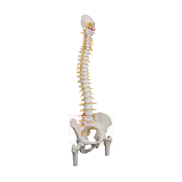
Human Spine with Femur Heads Model
Human Spine with Femur Heads Model (Product Code: SLE/A/18) is a miniature plastic model of the spinal column, pelvis and hip joints, with the tops of the thigh bones seated in articulated hips so students can see how the hip moves. It also represents the spinal cord and its nerve branches, linking the skeletal and nervous systems in one compact piece. Physiotherapy, chiropractic, nursing and school biology classes use it for posture and movement lessons, and as one of the smaller human anatomical models made in India it is easy to keep on a desk or shelf. It is manufactured by Jain Scientific Equipments Pvt Ltd (ScienceLab), Ambala.
Specifications
| Parameter | Detail |
|---|---|
| Material | Plastic |
| Scale | Miniature |
| Regions covered | Spinal column, pelvis and hip joints |
| Nervous structures | Representation of the spinal cord and nerve branches |
| Hip joints | Articulated, to demonstrate hip joint movement |
Typical Use
Physiotherapy and sports science tutors use the articulated hips to demonstrate flexion, extension and rotation of the thigh while students watch how the femur heads turn in their sockets in the pelvis. The same model then turns to the spine: learners follow the curves of the vertebral column and see where nerve branches leave the spinal cord, which helps explain how problems in the lower back can cause pain felt in the leg.
Because it is small, the model is handy for one-to-one explanations, for example when a clinician or trainer shows a patient or athlete what is happening at the hip or lower spine. In school biology lab equipment collections it provides an overview of the axial skeleton and pelvis before students move on to larger single-region models. Move the hip joints through a gentle range only and store the model upright.
Manufacturer
ScienceLab models such as this one are made by Jain Scientific Equipments Private Limited in Ambala, Haryana, a manufacturer of anatomy, biology and general science teaching equipment for institutions. Find out more on the ScienceLab company profile.
Related Laboratory Equipment
Larger and more focused models for the spine, pelvis and hip:
- Human Pelvis with Lower Lumbar and Femur Heads — concentrates on the pelvis and lower back region.
- Human Vertebral Model With Pelvis & Femur Heads — an alternative combined spine, pelvis and hip model.
- Human Femur Bone Model — the whole thigh bone whose head appears in the hip joint here.
- Economy Human Flexible Vertebral Column, Color Coded — for studying spinal regions and flexibility in detail.
Explore the Human Models category for the complete range.
Frequently Asked Questions
Do the hip joints move?
Yes. The hips are articulated to demonstrate hip joint movement.
Is the spinal cord shown?
A representation of the spinal cord and nerve branches is included.
How large is the model?
It is a miniature model; exact dimensions are not listed here. Please ask through our contact page for the measured size.
Is a key or label chart provided?
A key is not mentioned in the product details. Contact us if a key is required for your course and we will confirm what is supplied.
Who typically buys this model?
Physiotherapy and nursing colleges, sports science departments, clinics that explain conditions to patients, and schools teaching the skeletal system.
Can you deliver to institutions abroad?
Yes. ScienceLab supplies India and international markets; share quantity and destination for a delivered quotation.
Request a Quote
Request a price for the Human Spine with Femur Heads Model (SLE/A/18) via the contact page, or add it to the enquiry cart.
Last updated: 24 September 2026
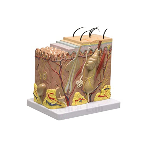
Enlarged Skin Model
Enlarged Skin Model (Product Code: SLE/A/19) magnifies a small block of human skin so that its layers, a hair follicle and a range of sensory receptors can be studied at a size no microscope slide can offer a whole class. The layers appear both as a cross section on the sides and as cutaways on the top surface, and the stratum lucidum is included to represent thick skin such as that of the palms and soles. Teachers of biology, cosmetology, dermatology nursing and first-aid courses use it, and it rounds out a human biology model set for the integumentary system. It is manufactured by Jain Scientific Equipments Pvt Ltd (ScienceLab), Ambala.
Specifications
| Parameter | Detail |
|---|---|
| Size | 20 x 22.5 x 15.5 cm |
| Base | Mounted on a blue base |
| Views | Skin layers shown as a cross section and as cutaways at the top of the model |
| Structures shown | A variety of sensory receptors; structure of a hair follicle |
| Thick skin feature | Stratum lucidum included |
| Key | Laminated key included |
Typical Use
Start with the top cutaways, where students can peel back the layers visually from the surface downwards, then turn the model to the side to see the same layers stacked in cross section. The hair follicle leads into discussion of hair growth and oil secretion, and the sensory receptors let the teacher explain how skin detects touch, pressure, temperature and pain. Pointing out the stratum lucidum answers the classic exam question of why the skin on the palms and soles is thicker than elsewhere.
Beauty and wellness training programmes use the model to explain how products and treatments act on different layers, and first-aid courses use it to explain burn depth in general terms. A useful practical is to show the model first and then have students find the same layers on a prepared skin slide under the microscope. Keep the model away from heat and strong sunlight to preserve its colours.
Manufacturer
Jain Scientific Equipments Private Limited, known to customers as ScienceLab, manufactures this skin model at Ambala in Haryana as part of its range of biology and anatomy teaching aids. The company profile describes the business in more detail.
Related Human Models
Useful additions for a complete lesson on skin and the senses:
- Human Skin Section Model — a second skin section model for comparison or group work.
- Human Skin with Burn Pathologies Model — moves from healthy skin to burn injuries at different depths.
- Prepared Microscope Slide Set – Skin and Excretion — real tissue sections to compare with the enlarged model.
- Human Nerve Ending and Synapse Model — explains how signals from skin receptors travel onward.
See more in the Human Models category.
Frequently Asked Questions
What are the model's dimensions?
It measures 20 x 22.5 x 15.5 cm and is mounted on a blue base.
Does it show thick or thin skin?
The stratum lucidum is included to represent thick skin, the type found on the palms and soles.
Which structures can students identify?
The skin layers, a hair follicle and a variety of sensory receptors, all listed on the laminated key.
What magnification is the model?
The enlargement factor is not listed on this page. Contact us through the contact page if you need it for a specification sheet.
How do we keep it clean?
Dust regularly with a soft brush and wipe with a barely damp cloth; avoid alcohol or solvent wipes on painted areas.
Can you supply training institutes in bulk?
Yes, for institutes in India and international markets. Include the quantity and delivery location in your enquiry.
Request a Quote
To buy the Enlarged Skin Model (SLE/A/19), reach us via the contact page or add it to the enquiry cart and send your full list together.
Last updated: 24 September 2026
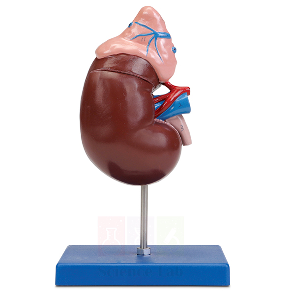
Human Kidney Model
Human Kidney Model (Product Code: SLE/A/20) is a life-size, 2-piece anatomical model of the right human kidney and adrenal gland used to teach renal structure in school biology classrooms, nursing schools and medical colleges. Lifting off its anterior portion exposes the internal structures of the kidney, while the renal artery, renal vein and proximal ureter stay in view, making it dependable biology lab equipment in India. It is made by Jain Scientific Equipments Pvt Ltd (ScienceLab), an OEM manufacturer, at its own factory in Ambala, India.
Specifications
| Parameter | Detail |
|---|---|
| Type | Anatomical model of the right human kidney with adrenal gland |
| Scale | Life size |
| Construction | 2 pieces; anterior portion removable |
| Internal view | Internal structures of the kidney revealed when the anterior portion is removed |
| Vessels and duct | Renal artery, renal vein and proximal end of the ureter |
| Key | Laminated key included |
| Mounting | Mounted on blue base |
| Product code | SLE/A/20 |
| Manufacturer | Jain Scientific Equipments Pvt Ltd (ScienceLab) — OEM |
| Country of origin | India (made in Ambala, Haryana) |
What Is the Human Kidney Model Used For?
With the front piece in place, teachers use the model to show the kidney's outer form, the adrenal gland capping its upper pole, and the vessels and ureter at the hilum. Removing the anterior portion lets the class move inward to the cortex, the medulla and the collecting system that drains into the ureter. Because the adrenal gland is part of the same model, one lesson can bridge excretion and hormonal control.
Schools that keep zoology lab equipment for practicals often place the model beside a dissected mammalian kidney, so students can compare a preserved organ with a clean, labelled reference. The laminated key works well for identification quizzes, and it is worth pointing out that this is the right kidney, which usually sits slightly lower than the left because the liver lies above it. Refit the anterior piece after each class and wipe the model with a soft, dry cloth.
OEM Manufacturer
ScienceLab is the trading name of Jain Scientific Equipments Private Limited, the OEM (original equipment manufacturer) of this kidney model, which is produced at the company's own licensed factory at Saha, Ambala, Haryana, India, rather than bought in from a trader. Schools, colleges, institutions, dealers and importers receive it factory-direct. If a laminated key or base part ever needs replacing, or you want a repeat order, ask us and the manufacturer itself will respond. Read more on our company profile.
Related Human Models
These models complement a life-size kidney in renal and endocrine lessons:
- Human Kidney, Nephron, and Glomerulus Model Set — enlarges the nephron and glomerulus that a life-size organ cannot show.
- Human Kidney with Adrenal Gland Model — a frontal-section alternative that opens into 2 parts.
- Human Kidney, Nephron and Renal Corpuscle Model — a plaque series running from the organ down to the renal corpuscle.
- Kidney Stone Model — shows stones in the opened calices, renal pelvis and ureter.
- Human Endocrine Organs Model — places the adrenal gland among the other hormone-producing glands.
Browse more organ and body models in our Human Models category.
Frequently Asked Questions
Is the Human Kidney Model life size?
Yes. The Human Kidney Model (SLE/A/20) represents the right human kidney and its adrenal gland at life size, so students see the organ at its true proportions rather than enlarged, which makes it a realistic reference beside textbook diagrams.
What does the Human Kidney Model show when it is opened?
Removing the anterior portion of the Human Kidney Model reveals the internal structures of the kidney. The renal artery, renal vein and the proximal end of the ureter are also represented, so the interior can be related to the vessels and duct at the hilum.
Is a key included with the Human Kidney Model?
Yes. A laminated key is included with the Human Kidney Model, so structures can be identified without a separate chart, and the model is mounted on a blue base that keeps it steady on a bench or display shelf.
Who manufactures the Human Kidney Model?
Jain Scientific Equipments Pvt Ltd, trading as ScienceLab, is the OEM manufacturer of the Human Kidney Model. It is made at the company's own licensed factory at Saha, Ambala, Haryana, India, and supplied factory-direct to institutions, dealers and importers.
What are the material and dimensions of the Human Kidney Model?
The material and exact dimensions of the Human Kidney Model are not stated in our current product details. Please ask us through the contact page and we will confirm them for your tender or purchase file before you order.
Can the Human Kidney Model be supplied in bulk or for export?
Yes. The Human Kidney Model is quoted for single units, class sets and distributor quantities for buyers in India and international markets. Mention product code SLE/A/20, the quantity and the delivery destination in your enquiry.
Request a Quote
Quotations for the Human Kidney Model come straight from the manufacturer. Send your quantity through the contact page, or add the model to the enquiry cart together with any other renal models you need.
Last updated: 24 September 2026
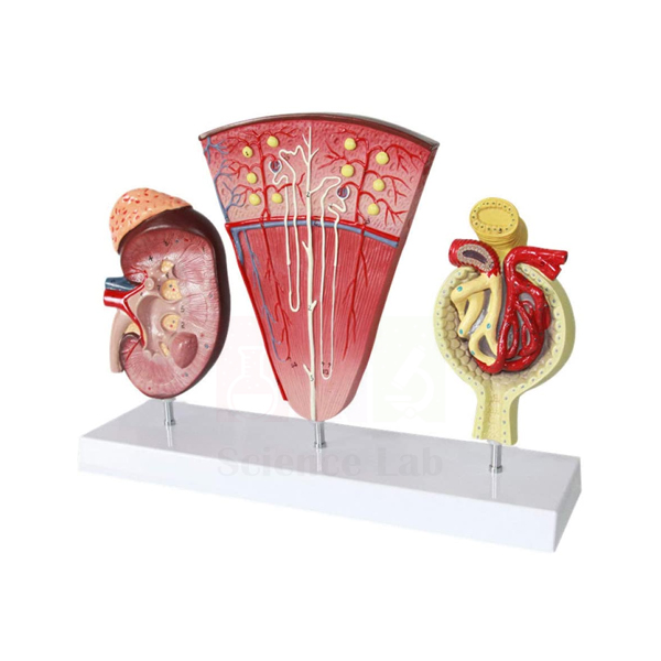
Human Kidney, Nephron, and Glomerulus Model Set
Human Kidney, Nephron, and Glomerulus Model Set (Product Code: SLE/A/21) is a three-part renal model set used to teach kidney structure and urine formation at three magnifications in colleges, medical schools and senior secondary labs. A sectioned kidney at 3× life size, 2 nephrons at 120× and a glomerulus at 700× share one base measuring 65 × 9 × 30 cm. It is made by Jain Scientific Equipments Pvt Ltd (ScienceLab), an OEM manufacturer, at its own factory in Ambala, India.
Specifications
| Parameter | Detail |
|---|---|
| Type | Three-part renal model set |
| Sectioned kidney | 3× life size |
| Nephrons | 2 nephrons, 120× life size |
| Glomerulus | 700× life size |
| Size | 65 × 9 × 30 cm |
| Mounting | Mounted on base |
| Product code | SLE/A/21 |
| Manufacturer | Jain Scientific Equipments Pvt Ltd (ScienceLab) — OEM |
| Country of origin | India (made in Ambala, Haryana) |
What Is the Kidney, Nephron and Glomerulus Model Set Used For?
A typical lesson starts with the enlarged kidney section to locate the cortex, medulla and pelvis, moves to the nephrons to follow filtrate along the tubule and collecting duct, and ends at the glomerulus, where the capillary tuft meets its capsule and filtration begins. Seeing the three scales side by side helps students grasp that the organ in front of them is built from vast numbers of microscopic filtering units.
Pharmacology teachers use the nephron to point out the tubule segments on which different diuretics act, and nursing or physiotherapy courses use the set when covering fluid balance and kidney failure. As the renal centrepiece of a human biology model set, it also suits group work: give each team one scale and ask them to explain their part to the class in sequence. Keep the base on a level surface and dust the models with a soft brush.
OEM Manufacturer
Jain Scientific Equipments Private Limited, which trades as ScienceLab, is the OEM (original equipment manufacturer) of this renal model set. The three magnified models and their base are produced at the company's own licensed factory at Saha, Ambala, Haryana, India, so colleges, dealers and importers buy them factory-direct rather than through a trading intermediary. For repeat orders or a replacement part, ask us and the manufacturer will answer directly. The company profile describes the business and its range.
Related Human Models
Extend a renal physiology unit with these models:
- Human Kidney, Nephron and Renal Corpuscle Model — a plaque series that adds the juxtaglomerular apparatus.
- Human Kidney Model — a life-size right kidney with adrenal gland for true-scale reference.
- Human Kidney with Adrenal Gland Model — a life-size frontal section showing pyramids and papillae.
- Kidney Stone Model — applies the anatomy to a common clinical problem.
- Denoyer-Geppert Free-Standing Human Urinary System Model — shows the kidneys within the complete urinary tract.
See the full range in our Human Models category.
Frequently Asked Questions
What does the Human Kidney, Nephron, and Glomerulus Model Set contain?
The Human Kidney, Nephron, and Glomerulus Model Set contains three models on one base: a sectioned kidney at 3× life size, 2 nephrons at 120× life size and a glomerulus at 700× life size, covering the organ, tubule and filtration levels.
How much space does the model set need?
The Human Kidney, Nephron, and Glomerulus Model Set measures 65 × 9 × 30 cm on its base, so it fits on a standard laboratory bench, lecture table or display shelf without taking over the teaching area.
Why are the parts shown at different magnifications?
Each part of the Human Kidney, Nephron, and Glomerulus Model Set is enlarged to suit the detail being taught: 3× for the whole organ, 120× for the nephrons and 700× for the glomerulus. Viewing the scales together links structure to function.
Is a key card supplied with the model set?
A key is not mentioned in the product details for the Human Kidney, Nephron, and Glomerulus Model Set. If your department needs labelled identification material, confirm it with us through the contact page before ordering.
Who manufactures the Human Kidney, Nephron, and Glomerulus Model Set?
Jain Scientific Equipments Pvt Ltd (ScienceLab) is the OEM manufacturer of the Human Kidney, Nephron, and Glomerulus Model Set, producing it at its own licensed factory at Saha, Ambala, Haryana, India, and supplying it factory-direct.
Can you quote several sets or ship abroad?
Yes. The Human Kidney, Nephron, and Glomerulus Model Set is quoted for institutions and distributors in India and international markets. Include product code SLE/A/21, the number of sets and the destination so the quotation covers packing and freight.
Request a Quote
For a quotation on the Human Kidney, Nephron, and Glomerulus Model Set direct from the manufacturer, contact us through the contact page or add it to the enquiry cart.
Last updated: 24 September 2026
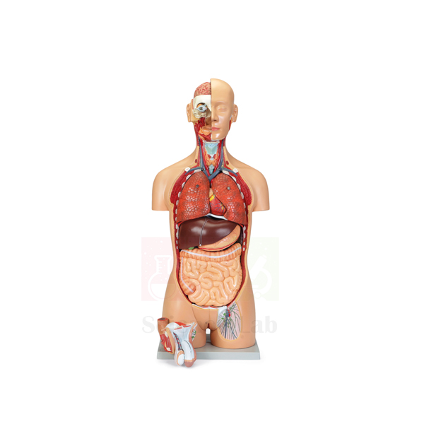
Deluxe Human Male and Female Torso Model
Deluxe Human Male and Female Torso Model (Product Code: SLE/A/22) is a life-size, 27-part torso that teaches from both front and back: the open back exposes musculature, the vertebral column and paravertebral nerve branches, while interchangeable male and female urogenital inserts let one model cover both sexes. A partially dissected head shows the buccal cavity, and half of the brain lifts out to reveal internal structure. Medical, nursing and allied health colleges, and senior schools planning a complete biology lab setup, use it as a central demonstration piece. It is manufactured by Jain Scientific Equipments Pvt Ltd (ScienceLab), Ambala.
Specifications
| Parameter | Detail |
|---|---|
| Scale | Life size |
| Number of parts | 27 |
| Back | Open back with musculature, vertebral column and paravertebral nerve branches |
| Spine | Removable vertebra with a section of spinal cord |
| Head | Partial dissection showing the anatomy of the buccal cavity, including vasculature, muscles, glands and osseous structures |
| Brain | Half of the brain removable to view internal structure |
| Urogenital inserts | Interchangeable male and female inserts |
Typical Use
With 27 parts, the torso supports a full term of lessons rather than a single topic. A typical sequence begins with the removable half brain for the nervous system, then turns the model around to study the back muscles and the vertebral column. Lifting out the vertebra with its segment of spinal cord shows how the cord is protected, and the paravertebral nerve branches show where spinal nerves run. The dissected head gives dental, ENT and speech-therapy students a view of the mouth, its glands and blood supply.
Swapping the male and female urogenital inserts lets teachers compare the two reproductive systems directly without buying a second torso. For viva and practical exams, examiners often remove several parts and ask candidates to replace and name them. Store the torso on a stable table, keep the smaller parts in a labelled box, and cover it when not in use.
Manufacturer
The torso is made by Jain Scientific Equipments Private Limited, which operates under the ScienceLab brand from Ambala, Haryana, and produces full-size and miniature anatomy models for teaching institutions. Company background is available on the ScienceLab profile page.
Related Human Models
Other torsos and system models buyers often compare or add:
- Human Male and Female Torso Model, 20 Parts — a dual-sex torso with fewer parts for simpler courses.
- Human Sexless Torso Model — a 16-part option without urogenital inserts.
- Human Male and Female Urogenital Systems Model — separate detail of the systems represented by the inserts.
- Human Muscular Torso with Head and Open Back Model — another open-back torso for musculature study.
The full range is in the Human Models category.
Frequently Asked Questions
How many parts does the torso have?
It is a 27-part, life-size torso.
Can it show both male and female anatomy?
Yes. Interchangeable male and female urogenital inserts are included.
What can be seen from the back?
The open back shows musculature, the vertebral column and paravertebral nerve branches, with a removable vertebra carrying a section of spinal cord.
Is there a numbered key and a stand?
These are not listed in the product details. Please confirm through our contact page what accompanies the torso before ordering.
What are the packed dimensions and weight?
They are not published here; we will state them in your quotation so you can plan freight and storage.
What should a college include in a tender enquiry?
Product code SLE/A/22, quantity, delivery address, required documents and your tender timeline. We supply institutions across India and international markets.
Request a Quote
For a quotation on the Deluxe Human Male and Female Torso Model, write to us through the contact page, or add the torso to the enquiry cart.
Last updated: 24 September 2026
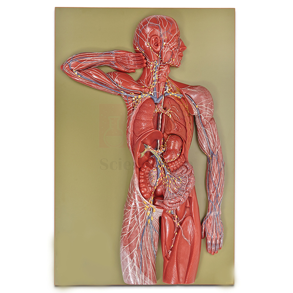
Human Lymphatic System Model
Human Lymphatic System Model (Product Code: SLE/A/23) is a relief model of the human lymphatic system at two-thirds life size, giving learners a whole-body view of a system that is hard to picture from textbook diagrams alone. At 84 x 54 x 12 cm it is large enough for a class to study together, and a numbered key accompanies it for identifying the structures shown. Biology teachers, nursing and paramedical tutors, and physiology departments sourcing biology lab equipment in India for immunity and circulation topics use it. It is manufactured by Jain Scientific Equipments Pvt Ltd (ScienceLab), Ambala.
Specifications
| Parameter | Detail |
|---|---|
| Type | Relief model of the human lymphatic system |
| Scale | 2/3x life size |
| Size | 84 x 54 x 12 cm |
| Key | Numbered key |
Typical Use
The lymphatic system is often taught as an afterthought to blood circulation, yet it explains swelling, the spread of infection and the body's immune defences. A relief model lets the teacher trace how lymph collects from the tissues, passes through lymph nodes where it is filtered, and returns to the bloodstream near the heart. Students can then use the numbered key to locate each labelled point themselves, turning a lecture into a short identification exercise.
Nursing and physiotherapy tutors use the model to explain in general terms why lymph nodes enlarge during infection and why swelling can follow surgery that affects lymph drainage. In schools, it supports lessons on immunity and on how the circulatory and lymphatic systems work together. Because it is a large, flat relief, dust it regularly with a soft brush and keep it where it will not be knocked.
Manufacturer
Jain Scientific Equipments Private Limited manufactures this lymphatic relief under the ScienceLab name at Ambala, Haryana, where the company builds anatomy models, biology apparatus and general laboratory equipment for education. More information is on the company profile.
Related Human Models
Models that connect naturally with a lymphatic system lesson:
- Human Circulatory System Model — shows the blood vessels that lymph eventually drains back into.
- Human Spleen, Duodenum, and Pancreas Model — covers the spleen, an organ central to immunity.
- Human Digestive System Model — supports teaching on fat absorption, which relies on lymph vessels in the gut.
- Human Nervous System Model — another whole-body system model for a matching display set.
Browse all options in the Human Models category.
Frequently Asked Questions
What scale and size is the model?
It is 2/3x life size and measures 84 x 54 x 12 cm.
Is it a free-standing figure or a relief?
It is a relief model of the human lymphatic system.
Does it come with a key?
Yes, a numbered key is supplied for identifying the structures on the model.
Can it be hung on a wall?
The mounting method is not listed in the product details. Ask through our contact page and we will confirm whether it suits wall or table display.
How many structures does the key identify?
The number of keyed structures is not stated here; we can confirm it on request before you place an order.
Do you ship large models like this overseas?
Yes. ScienceLab supplies India and international markets; share the destination so we can quote suitable packing and freight.
Request a Quote
Send your enquiry for the Human Lymphatic System Model (SLE/A/23) through the contact page, or add it to the enquiry cart with any related models.
Last updated: 24 September 2026
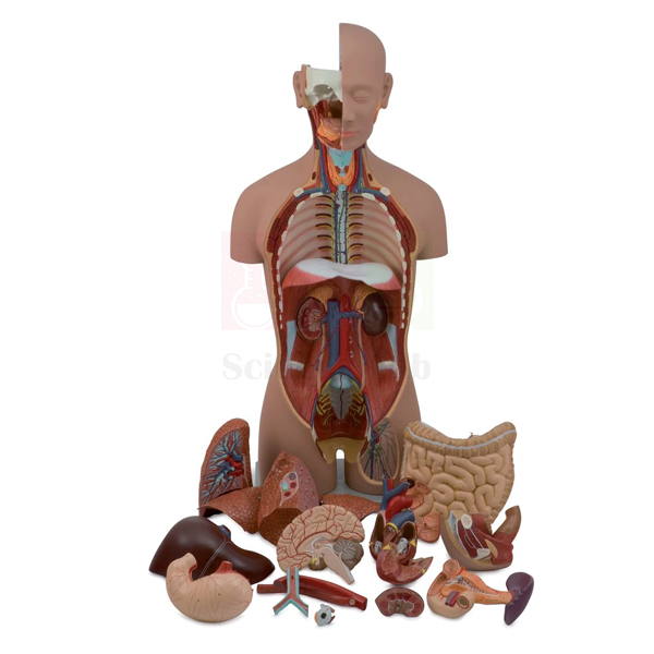
Human Sexless Torso Model
Human Sexless Torso Model (Product Code: SLE/A/24) is a dissectible torso that comes apart into 16 parts, taking students from the brain and eye down through the chest and abdomen to the kidneys and urinary bladder. Open dissections of the neck and thigh expose further anatomical features, and a reinforced polymer base keeps the 90 cm model steady on the demonstration bench. Because it has no reproductive organs, it keeps the focus on the major body systems, which makes it a common choice as the core human biology model for secondary schools, nursing schools and paramedical institutes. It is manufactured by Jain Scientific Equipments Pvt Ltd (ScienceLab), Ambala.
Specifications
| Parameter | Detail |
|---|---|
| Dissectible parts | 16 |
| Open dissections | Neck and thigh |
| Head | Half brain (sectioned longitudinally); eyeball |
| Chest | Both lungs; 2-part heart; descending thoracic aorta and esophagus |
| Abdomen | Stomach; pancreas and duodenum; liver; small and large intestines (one piece) with removable cecum |
| Urinary structures | Half of one kidney (sectioned along the frontal plane); kidney with urethra and urinary bladder |
| Base | Reinforced polymer base |
| Size | 90 x 33 x 23 cm |
Typical Use
The torso is built for step-by-step dissection in front of a class. Teachers usually lift out the lungs first, then open the 2-part heart, follow the aorta and esophagus down through the chest, and remove the stomach, liver, pancreas and duodenum in turn before taking out the intestines and their separate cecum. Each removal reveals the next layer, so students see not only what each organ looks like but also what lies behind it. The longitudinally sectioned half brain and the frontally sectioned kidney show internal structure that a closed organ cannot.
For practical exams, the parts can be laid out on a tray for students to identify and then return to the correct position, a quick test of their understanding of organ placement. The model covers the digestive, respiratory, circulatory, urinary and nervous systems in one piece of equipment, which is why institutions placing a biology model kit bulk order often include at least one torso of this kind. Reassemble it after every session so parts are not misplaced.
Manufacturer
This torso comes from Jain Scientific Equipments Private Limited, incorporated in Haryana under CIN U29309HR2020PTC087597 and trading as ScienceLab. The company's anatomy models are made at its Ambala factory in the HSIIDC Industrial Area, Saha, which operates under Factory Licence AMB-ONLINE-CHD-J-414. See the ScienceLab company profile for more.
Related Human Models
Complementary torsos and organ models for the same course:
- Human Micro Sexless Torso Model — a desk-sized version so student groups can practise while the full torso is demonstrated.
- Deluxe Human Male and Female Torso Model — the step up when reproductive anatomy and an open back are needed.
- Human Kidney Model — enlarges the kidney that appears in section on this torso.
- Human Heart on Diaphragm Model — more detail on the heart and its position above the abdomen.
View the full range in the Human Models category.
Frequently Asked Questions
How many parts does the torso separate into?
It is dissectible into 16 parts, including the half brain, eyeball, both lungs, the 2-part heart and the main abdominal organs.
What is the overall size?
It measures 90 x 33 x 23 cm on its reinforced polymer base.
Does it include reproductive organs?
No. It is a sexless torso; for interchangeable male and female anatomy, see the deluxe torso linked above.
Is a numbered key included?
A key is not listed in the product details. Ask via our contact page and we will confirm what documentation accompanies the model.
How should the torso be cleaned and stored?
Wipe parts with a soft, slightly damp cloth, let them dry, reassemble the torso and keep it covered to protect it from dust and sunlight.
Can you quote for several torsos or for export?
Yes. We supply schools and colleges across India and international markets; tell us the quantity and destination.
Request a Quote
For a price on the Human Sexless Torso Model (SLE/A/24), use our contact page or add it to the enquiry cart.
Last updated: 24 September 2026
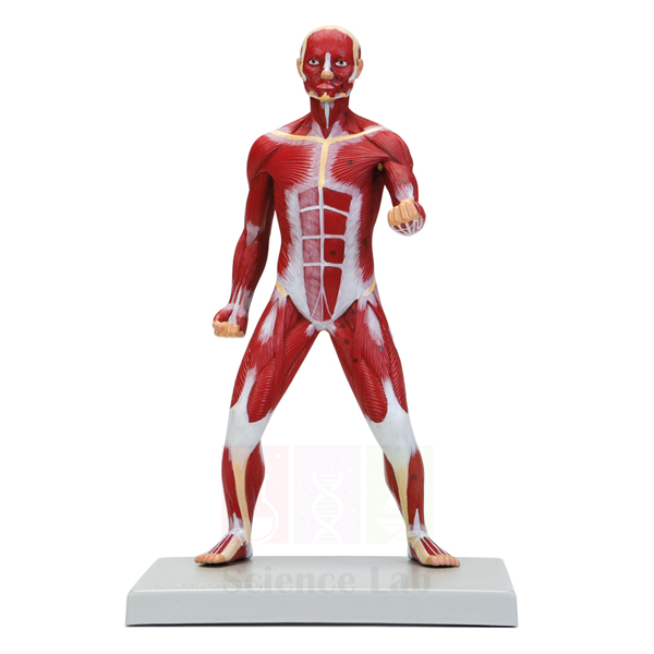
Miniature Human Muscular Figure
Miniature Human Muscular Figure (Product Code: SLE/A/25) is a compact full-body figure at about a quarter of life size that shows the superficial muscles of the whole body, each positioned and sized to correct scale. A numbered key identifies 51 structures, so the figure works as both a demonstration piece and a self-study tool. Sports science, physical education, physiotherapy, fitness training and school biology classes use it to learn muscle names and locations, and it is a space-saving addition to any human biology model set in India. It is manufactured by Jain Scientific Equipments Pvt Ltd (ScienceLab), Ambala, and stands on a durable polymer base.
Specifications
| Parameter | Detail |
|---|---|
| Scale | About 1/4x life size |
| Size | 25 x 18 x 40 cm |
| Base | Durable polymer base |
| Coverage | Overview of the superficial musculature of the whole figure |
| Accuracy of layout | All muscles positioned and sized to correct scale |
| Key | Numbered key with 51 identified structures |
Typical Use
Physical education and fitness instructors use the figure to connect exercises with the muscles they work: pointing to the chest, shoulder, thigh or calf muscles while describing a movement makes the link clear. Because the muscles are to scale, students can compare the relative size of large prime movers with smaller stabilising muscles and understand why some injuries are more common than others. The 51-point key suits quick quizzes, flash-card style revision and label-the-diagram homework.
At this size the figure fits on a desk or a shelf in a sports office, clinic reception or classroom, where a full-size muscular model would be impractical. It also transports easily for workshops and outreach sessions. Dust it with a soft brush and avoid placing it near windows where strong sunlight could fade the colouring.
Manufacturer
Jain Scientific Equipments Private Limited, trading as ScienceLab, produces this miniature figure in Ambala, Haryana, alongside full-size muscular and skeletal teaching models for schools, colleges and training centres. Visit the ScienceLab profile to learn more about the company.
Related Human Models
Pair the miniature figure with these for deeper muscle study:
- Human Muscular Figure Model — a larger muscular figure for whole-class demonstrations.
- Human Sexless Muscular Figure — an alternative whole-body muscle figure to compare.
- Human Muscular Arm Model — closer detail of the upper-limb muscles seen on this figure.
- Skeletal Muscle Fiber Model — explains the fibre structure inside each muscle.
Browse more in the Human Models category.
Frequently Asked Questions
How big is the figure?
It is about 1/4x life size and measures 25 x 18 x 40 cm.
How many structures does the key name?
The numbered key identifies 51 structures.
Does it show deep muscles or internal organs?
It gives an overview of the superficial musculature. For deeper layers or organs, consider a full-size muscular figure or a dissectible torso.
Are the muscles in proportion?
Yes, all muscles are positioned and sized to correct scale.
Is there a warranty on the model?
Warranty terms are not listed here. Please ask through the contact page and we will state them in writing.
Can gyms, clinics and overseas distributors order?
Yes. ScienceLab supplies educational and training buyers in India and international markets.
Request a Quote
For a quotation on the Miniature Human Muscular Figure (SLE/A/25), contact us through the contact page or add it to the enquiry cart.
Last updated: 24 September 2026
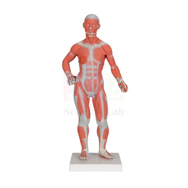
3B Human Muscular Male and Female Model
3B Human Muscular Male and Female Model (Product Code: SLE/A/26) is a half life-size dissectible figure that lets one model serve for both sexes: its male and female genital organs are interchangeable, so a single figure can support lessons on either reproductive anatomy. The figure maps superficial and deep muscle layers together with blood vessels, nerves and internal organs, which makes it a practical human body model kit in India for medical, nursing, physiotherapy and senior biology classrooms. It is supplied by Jain Scientific Equipments Pvt Ltd (ScienceLab), Ambala, India.
Specifications
| Parameter | Detail |
|---|---|
| Type | Dissectible muscular figure with male and female genital organs |
| Scale | 1/2× life size |
| Height | 84 cm tall |
| Number of parts | Dissectible into 33 parts |
| Structures shown | Superficial and deep muscles, blood vessels and nerves, internal organs |
| Internal organs | Can be partially dissected |
| Genital organs | Male and female genital organs are interchangeable |
| Mounting | On stand with base |
Typical Use
In a gross-anatomy or kinesiology session the instructor can begin with the surface musculature, naming muscles region by region and tracing where they attach, then lift away parts to expose the deeper layer and the organs beneath. Because the figure stands 84 cm tall on its own stand and base, it sits comfortably on a demonstration bench where a small group can gather round and point to structures while blood vessels and nerves are followed from one region to the next.
Swapping the genital organs lets the same figure cover male and female reproductive anatomy, which saves bench space in departments that would otherwise keep two separate figures. This makes it a flexible choice among biology teaching aids in India for institutes that teach both school and college levels. A practical habit: lay removed parts on a soft cloth in the order they came off, and count them back against the 33-part total before the model goes back into storage.
Supplier
This figure is supplied by Jain Scientific Equipments Private Limited, the Ambala, Haryana company that trades under the name ScienceLab and equips schools, colleges and training institutes in India and international markets. Because the product name carries the 3B designation, please ask us to confirm brand and origin details when you request a quotation. Background on the company is available on our company profile.
Related Human Models
These models work alongside a dissectible muscular figure when a course moves between muscles, skeleton and organ systems:
- Human Male Muscular Model, Removable Arms — a male muscular figure whose arms can be taken off for focused upper-limb study.
- Human Sexless Muscular Figure — an option for classes that need muscle anatomy without reproductive organs.
- Human Male and Female Urogenital Systems Model — carries the genital-organ lesson on into the urinary and reproductive tracts.
- Human Muscular and Ligamentous Skeleton — links muscle layers to the bones and ligaments they act on.
- Human Male and Female Torso Model, 20 Parts — a torso for closer study of internal organs in both sexes.
See every figure, torso and organ model in our anatomy models category.
Frequently Asked Questions
How many parts does the figure come apart into?
It is dissectible into 33 parts. The internal organs can be partially dissected, so students can see how they sit within the body cavity.
Can one figure be used for both male and female anatomy?
Yes. The male and female genital organs are interchangeable, so the same figure serves both lessons; just fit the set you need before class.
How large is the model?
It is 1/2× life size and stands 84 cm tall on a stand with base, which suits a bench or table rather than floor display.
Is this a genuine 3B-branded product?
Please confirm brand, origin and packing details with us through the contact page before ordering; the quotation will state exactly what is being supplied.
How should the figure be cleaned?
Wipe it with a soft cloth, slightly dampened with mild soapy water if needed, and dry it fully before reassembly. Avoid solvents and abrasive pads, which can dull painted detail.
What should an institutional quotation request include?
Mention the product code SLE/A/26, the quantity, the delivery location and any paperwork your purchase procedure needs, so we can reply with a complete offer.
Can you supply several units or ship overseas?
Yes, we supply institutions in India and international markets. Share quantities and destination and we will advise on packing and dispatch.
Request a Quote
To order the 3B Human Muscular Male and Female Model or ask about availability, send your requirement through our contact page, or add it to the enquiry cart with other models for one combined quotation.
Last updated: 24 September 2026
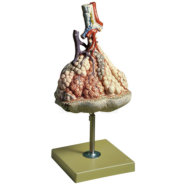
Somso Human Lung Model
Somso Human Lung Model (Product Code: SLE/A/27) is an enlarged anatomical model used to teach the structure of the lung lobule in medical, nursing and senior school biology classrooms. Magnified about 150× life size and mounted on a stand with base, it lets a whole group study lobule structure by eye before moving to the microscope. It is supplied by Jain Scientific Equipments Pvt Ltd (ScienceLab), Ambala, India, to institutions sourcing biology teaching aids in India.
Key Details
| Parameter | Detail |
|---|---|
| Type | Enlarged anatomical teaching model of lung tissue |
| Scale | About 150× life size |
| Structure shown | Structure of the lung lobule |
| Mounting | On stand with base |
| Brand in product name | Somso (brand and origin confirmed at quotation) |
| Product code | SLE/A/27 |
| Supplied by | Jain Scientific Equipments Pvt Ltd (ScienceLab), Ambala, India |
What Is the Somso Lung Model Used For?
The lobule is where the finest air passages end and gas exchange takes place, a level of the lung that is hard to teach from flat diagrams alone. Lecturers set this model on the bench at the start of a respiratory histology session so students see the three-dimensional arrangement first, then look for the same pattern in stained lung sections under the microscope. It also supports discussion of why the lung's internal surface is so large and how that surface aids the diffusion of oxygen and carbon dioxide.
Because it shows tissue rather than the whole organ, it is most useful as the detailed companion to a gross lung model within a human biology model set. Keep it on its stand, point to features with a pointer instead of pressing on the surface, and dust it with a soft, dry cloth after use.
Supplier
Jain Scientific Equipments Private Limited trades as ScienceLab and supplies this lung model from Ambala, Haryana, to colleges, schools and laboratories in India and international markets. Because the product name refers to the Somso brand, please ask us to confirm brand and origin details when you request a quotation. For vendor registration, the supplying company holds Importer-Exporter Code AAECJ8618A from the DGFT and GSTIN 06AAECJ8618A1ZT. More background is available on our company profile.
Related Human Models
Respiratory models that are commonly taught alongside a lung lobule model:
- Human Segmented Lungs Model — a life-size view of both lungs that places the lobule in the context of the whole organ.
- Segmented Lung — a take-apart lung whose lobes separate into individual segments.
- Pathology of the Human Lung Model — for comparing normal lung tissue with infection-related changes.
- Human Lung Model in Bell Jar — demonstrates the breathing mechanics that move air into the lobules.
More respiratory and organ models are listed in our Human Models category.
Frequently Asked Questions
What does "about 150× life size" mean for the Somso Human Lung Model?
It means the Somso Human Lung Model shows lung tissue roughly 150 times larger than it is in the body, so the fine structure of a lobule becomes large enough to point out and discuss in front of a whole class.
Does the Somso Human Lung Model show a complete lung?
No. The Somso Human Lung Model focuses on the structure of the lung lobule rather than the whole organ. For gross anatomy, pair it with a whole-lung model such as the Human Segmented Lungs Model listed above.
Is a stand included with the Somso Human Lung Model?
Yes. The Somso Human Lung Model comes on a stand with base, so it can be displayed upright on a bench, a lecture table or in a display cabinet between respiratory anatomy sessions.
Does the Somso Human Lung Model come with a key?
A numbered key or labelling guide is not listed for the Somso Human Lung Model. Please contact us before ordering if your department needs identification material confirmed as part of the supply.
Who supplies the Somso Human Lung Model?
Jain Scientific Equipments Pvt Ltd (ScienceLab), Ambala, India, supplies the Somso Human Lung Model. The name refers to the Somso brand, so brand, origin and any accompanying documents are confirmed in the quotation; ask us through the contact page.
How should the Somso Human Lung Model be cleaned?
Wipe the Somso Human Lung Model gently with a soft, dry or slightly damp cloth. Avoid solvents and abrasive cleaners, and store it away from strong direct sunlight to protect the painted surface.
Can the Somso Human Lung Model be supplied in quantity or exported?
Yes. Institutional and export enquiries for the Somso Human Lung Model are welcome from India and international markets. State product code SLE/A/27, the quantity and the destination, and the quotation will set out packing and delivery terms.
Request a Quote
To price this lung lobule model for your department, send your quantity and delivery location through the contact page, or add it to your enquiry cart together with other anatomy models you need.
Last updated: 24 September 2026

Larynx Human Model
Larynx Human Model (Product Code: SLE/A/32) is an enlarged anatomical model of the human voice box used to teach voice production and airway protection in biology, nursing and speech-science classrooms. Built at about 2 1/2× life size, it has a removable arytenoid cartilage, vocal cords and epiglottis that students can lift out and refit. It is manufactured by Jain Scientific Equipments Pvt Ltd (ScienceLab), an OEM manufacturer, at its own factory in Ambala, India, for colleges assembling a human biology model set.
Specifications
| Parameter | Detail |
|---|---|
| Type | Enlarged human larynx model |
| Scale | About 2 1/2× life size |
| Size | 12 × 14 × 28 cm (height 28 cm) |
| Removable parts | Arytenoid cartilage, vocal cords and epiglottis |
| Mounting | On base |
| Product code | SLE/A/32 |
| Manufacturer | Jain Scientific Equipments Pvt Ltd (ScienceLab) — OEM |
| Country of origin | India (made in Ambala, Haryana) |
What Is the Larynx Model Used For?
The model is most useful when explaining how voice is produced and how swallowing is kept separate from breathing. With the epiglottis in place, a teacher can show how it folds over the entrance to the larynx; lifting it off then reveals the vocal cords, and removing the arytenoid cartilage shows the point at which the cords are tensioned and moved. Speech-therapy and audiology students often use this sequence to connect anatomy with the sounds they assess, while nursing learners relate it to airway care.
In school biology lab equipment in India the larynx is usually taught within the respiratory system, and a model 28 cm tall stands well on a front bench for group viewing. Ask students to replace each removable part themselves after the demonstration; putting the epiglottis back in the correct position is a quick check that they have understood its orientation.
OEM Manufacturer
ScienceLab is the trading name of Jain Scientific Equipments Private Limited, the OEM (original equipment manufacturer) of this larynx model, which is made at the company's own licensed factory at Saha, Ambala, Haryana, India. Medical and nursing colleges, schools, dealers and importers buy it factory-direct rather than through a trader, and if a small removable part goes missing, a replacement can be requested from the manufacturer itself — just ask us. Find out more on our company profile.
Related Human Models
Pair the larynx with these models to follow the airway above and below the voice box:
- Human Larynx, Trachea, and Bronchi Model — continues the airway down into the trachea and bronchial tree.
- Human Larynx Model — a second larynx design for comparison or for equipping more benches.
- Human Lungs with Heart and Larynx Model — places the larynx in context with the lungs and heart.
- Anatomical Human Nasal Cavity Model — covers the upper airway that leads to the larynx.
More respiratory and organ models are listed in our Human Models category.
Frequently Asked Questions
Which parts of the Larynx Human Model can be removed?
The Larynx Human Model has three removable parts: the arytenoid cartilage, the vocal cords and the epiglottis. Each can be lifted out, examined on its own and then refitted, which helps students see how the parts relate to one another.
How big is the model?
The Larynx Human Model is about 2 1/2× life size and measures 12 × 14 × 28 cm, with a height of 28 cm. It is mounted on a base, so it stands securely on a front bench for group viewing.
Does it show the trachea and lungs as well?
No. The Larynx Human Model concentrates on the voice box itself. For the lower airway, pair it with the Human Larynx, Trachea, and Bronchi Model or the Human Lungs with Heart and Larynx Model listed above.
Is it suitable for speech therapy and ENT teaching?
Yes. The removable vocal cords and epiglottis make the Larynx Human Model a practical aid for explaining voice production and airway protection in speech-therapy, audiology, ENT and nursing teaching, as well as in school biology.
Who manufactures the Larynx Human Model?
The Larynx Human Model is manufactured by Jain Scientific Equipments Pvt Ltd (ScienceLab), an OEM, at its own licensed factory at Saha, Ambala, Haryana, India. Quotations and requests for replacement removable parts go directly to the manufacturer through our contact page.
How should the small removable parts be looked after?
Refit the removable parts of the Larynx Human Model after each session, keep the model on its base away from direct sun, and clean it only with a soft, slightly damp cloth so the surface stays in good condition.
Request a Quote
Prices for the Larynx Human Model come direct from the manufacturer. Write to us via the contact page with your quantity and location, or place it in the enquiry cart while you select other anatomy models.
Last updated: 24 September 2026
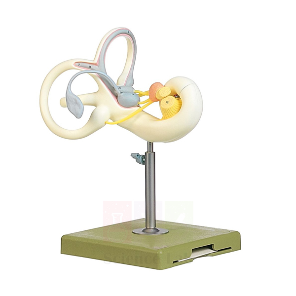
Human Inner Ear Model, 18x
Human Inner Ear Model, 18x (Product Code: SLE/A/33) magnifies the tiny organs of hearing and balance about 18 times, turning structures only millimetres across in the skull into a model that a whole class can study. Opened sections reveal the saccule and utricle, and a median section of the cochlea shows its internal spiral. Audiology, ENT, physiology and advanced biology departments use it as an ear model for the anatomy lab in India, and it is manufactured by Jain Scientific Equipments Pvt Ltd (ScienceLab), Ambala.
Specifications
| Parameter | Detail |
|---|---|
| Type | Enlarged human inner ear model |
| Magnification | About 18× life size |
| Size | 24 × 18 × 33 cm |
| Opened structures | Superior semicircular canal and auricle opened to show the saccule and utricle |
| Cochlea | Includes a median section of the cochlea |
| Mounting | On stand with base |
Typical Use
The inner ear is hard to teach from diagrams because its canals curve in three planes. At this magnification a lecturer can trace the semicircular canals with a pointer, then move to the opened region to show where the saccule and utricle sit and how they relate to balance. The median cochlear section lets students follow the spiral turns and discuss how sound waves are converted into nerve signals.
Audiology and speech-and-hearing students often use the model before clinical postings to connect test results with anatomy, and physiology classes pair it with a whole-ear model to show where the inner ear lies. For a high school biology lab that needs one clear sense-organ model, the stand keeps it upright at the front of the room; turn it slowly so each group sees the opened side and the cochlear section in turn.
Manufacturer
This inner ear model is produced by Jain Scientific Equipments Private Limited, which operates as ScienceLab from Ambala in Haryana and supplies anatomy and science teaching equipment to buyers across India and international markets. Visit our company profile to learn how the company is organised.
Related Human Models
Combine the enlarged inner ear with these models for a complete sense-organ lesson:
- Human Inner Ear Model, 4x — a smaller-scale inner ear for bench work or where space is limited.
- Human Ear Model, 5 Parts — shows outer, middle and inner ear together so students can locate the inner ear.
- Human Ear Model — an alternative whole-ear model for general classes.
- Human Eye Model with Eyelids — completes a special-senses teaching set.
All ear, eye and other organ models are in our anatomy models category.
Frequently Asked Questions
How much is the inner ear enlarged?
About 18× life size, which is large enough for lecture-hall viewing.
Does the model show the inside of the cochlea?
Yes, it includes a median section of the cochlea so its internal turns can be seen.
Which balance organs are visible?
The superior semicircular canal and auricle are opened to show the saccule and utricle.
What space does it need?
It measures 24 × 18 × 33 cm and stands on its own stand with base, so a shelf or bench corner is enough for storage.
Does it include the outer and middle ear?
It is an inner ear model. For the full ear, add one of the whole-ear models listed above.
Can a test report or calibration certificate be supplied?
As a teaching model it has no measuring function; if your purchase process needs specific documents, please discuss them with us through the contact page.
Request a Quote
Ask for a quotation on the Human Inner Ear Model, 18x by messaging us on the contact page, or collect it in the enquiry cart with any other sense-organ models you need.
Last updated: 24 September 2026
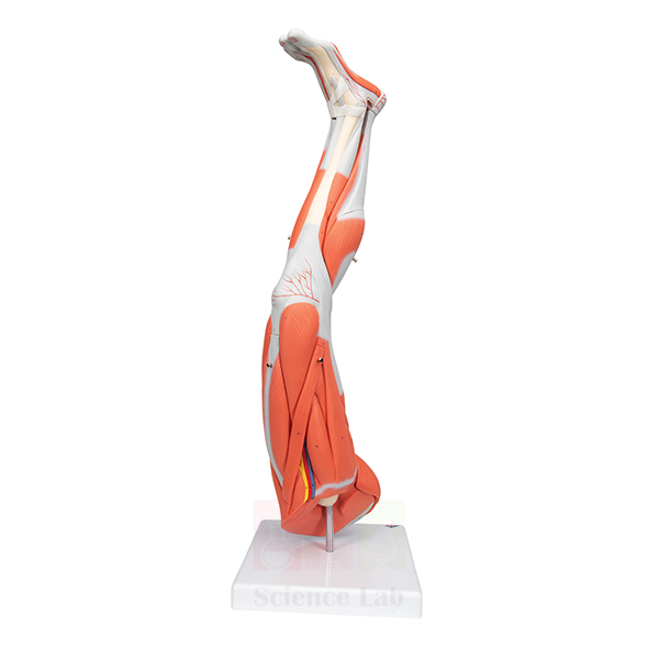
3B Human Left Leg Model
3B Human Left Leg Model (Product Code: SLE/A/35) is a stand-mounted model of the left lower limb at about 2/3× life size, roughly 75 cm long, that can be taken apart into 9 parts. It gives anatomy, physiotherapy, sports-science and nursing classes a single leg they can study region by region, from thigh to foot, without needing a full figure. Institutions looking for lab equipment for college teaching in India can order it from Jain Scientific Equipments Pvt Ltd (ScienceLab), Ambala, which supplies this model.
Specifications
| Parameter | Detail |
|---|---|
| Type | Dissectible model of the human left leg |
| Scale | About 2/3× life size |
| Length of leg | About 75 cm |
| Number of parts | Dissectible into 9 parts |
| Mounting | On stand |
Typical Use
A dedicated limb model lets a lecturer spend a full session on the lower extremity. Students can work through the thigh, knee, lower leg and foot in order, removing parts as each region is covered and naming what they see before moving on. Physiotherapy and sports-science learners find this especially useful when linking anatomy to movement and common injuries of the leg.
Because the model stands upright on its stand at about 75 cm, it can be placed beside a full skeleton or muscular figure so that the class can compare scale and position. For practical examinations, remove one or two of the 9 parts and ask candidates to identify them and replace them correctly; this is quick to set up and easy to mark.
Supplier
Jain Scientific Equipments Private Limited, trading as ScienceLab and based in Ambala, Haryana, supplies this leg model to teaching institutions in India and international markets. As the product name includes the 3B designation, ask us to confirm brand and origin details at the time of quotation. More about the company is on our company profile.
Related Human Models
These items fit the same lower-limb and locomotor lessons:
- Human Leg and Portion of Pelvis Model — shows how the leg connects to the pelvis above.
- Human Knee Joint Model, Life Size — a close-up of the knee for joint and ligament teaching.
- Human Femur Bone Model — the main thigh bone on its own for osteology practicals.
- Human Skeletal Foot Model — completes the limb with the bones of the foot.
- 3B Human Muscular Male and Female Model — a whole-body figure for placing the leg in context.
Explore more limb, joint and torso models in our anatomy models category.
Frequently Asked Questions
Is this a left or right leg?
It is a left leg model. Please contact us if you need to discuss other limb models.
How many parts does it separate into?
It is dissectible into 9 parts, which keeps assembly manageable for students.
What is the scale and length?
About 2/3× life size, with a leg length of about 75 cm, mounted on a stand.
Is this a genuine 3B-branded model?
Brand and origin should be confirmed with us before ordering; ask through the contact page and the quotation will state what is supplied.
Which courses is it suited to?
It fits anatomy, physiotherapy, sports science, nursing and senior biology classes that study the lower limb in detail.
How do I keep the parts in good condition?
Reassemble the leg on its stand after each class, avoid forcing parts into place, and clean with a soft cloth only.
Request a Quote
Tell us the quantity and delivery address for the 3B Human Left Leg Model through our contact page, or add it to the enquiry cart and request one quotation for your whole list.
Last updated: 24 September 2026
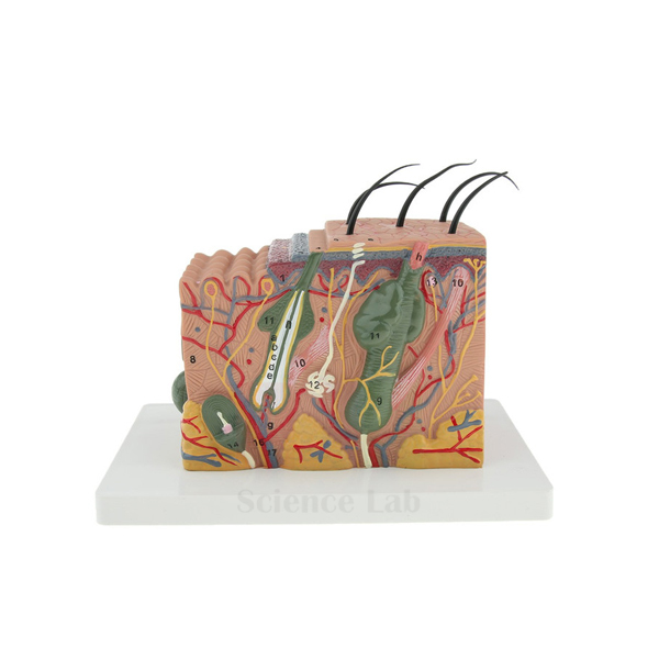
Human Skin Section Model
Human Skin Section Model (Product Code: SLE/A/37) enlarges a block of scalp skin about 70× so that hairs, glands and nerve endings, normally seen only under a microscope, can be pointed out on a solid model. The section is cast in one piece and mounted on a sturdy base, which makes it robust enough for repeated handling in class. Biology, dermatology, cosmetology and nursing teachers use it to explain skin layers and appendages, and schools sourcing from a biology lab equipment supplier in India can order it directly from its manufacturer, Jain Scientific Equipments Pvt Ltd (ScienceLab), Ambala.
Specifications
| Parameter | Detail |
|---|---|
| Type | Enlarged section through human scalp skin |
| Magnification | About 70× life size |
| Size | 21 × 20 × 11 cm |
| Construction | 1-piece section |
| Structures shown | Tissue components of the scalp, with detailed hairs, sebaceous glands, sweat glands and sensory receptors |
| Mounting | Mounted on a sturdy base |
Typical Use
The model is typically used when introducing the integumentary system. Starting at the surface, students can follow a hair shaft down to its root, locate the sebaceous gland that oils it, find the sweat glands and then identify the sensory receptors that respond to touch, pressure and temperature. Because it is a single piece, there is nothing to lose or reassemble, so it can be passed between groups during a practical.
Cosmetology and trichology courses find the scalp section particularly relevant because it shows hair in its natural setting. In a school or college biology lab it works well next to prepared microscope slides of skin: students view the slide first, then use the model to make sense of the three-dimensional structure behind what they saw under the lens.
Manufacturer
The model is made by Jain Scientific Equipments Private Limited, a company in Ambala, Haryana whose products carry the ScienceLab name and reach schools, colleges and training centres in India and international markets. Our company profile gives an overview of the business.
Related Human Models
These items extend a lesson on skin structure and the senses:
- Enlarged Skin Model — another magnified skin model for comparison or additional benches.
- Human Skin with Burn Pathologies Model — moves from healthy skin to the effects of burns.
- Prepared Microscope Slide Set – Skin and Excretion — real tissue sections to view alongside the model.
- Human Head Section Model — places the scalp in the context of the whole head.
Browse further organ and tissue models in our anatomy models category.
Frequently Asked Questions
Which part of the body does the section come from?
It is a section through the human scalp, so hair and associated glands are prominent.
How much is it enlarged?
About 70× life size, giving an overall size of 21 × 20 × 11 cm on its base.
Does the model come apart?
No. It is a 1-piece section, which makes it quick to use and easy to store.
Are sensory receptors shown?
Yes, sensory receptors are shown along with hairs, sebaceous glands and sweat glands.
How should it be cleaned?
Dust it with a soft brush or dry cloth; if needed, wipe gently with a slightly damp cloth and let it dry before storing.
Can you supply it in quantity for several labs?
Yes. Send your quantity and delivery locations through the contact page and we will quote for the full order.
Request a Quote
For a price on the Human Skin Section Model, reach us on the contact page or put it in the enquiry cart together with the slides and models you plan to buy.
Last updated: 24 September 2026
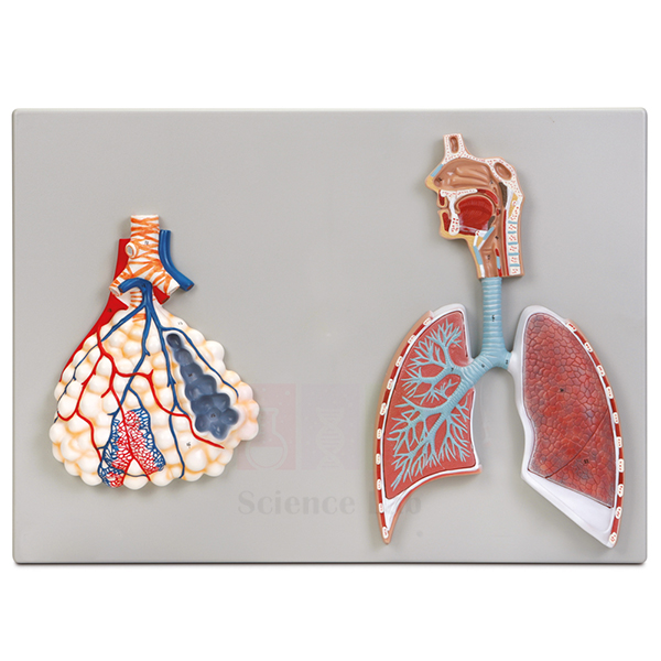
Human Respiratory System Model, 2-parts
Human Respiratory System Model, 2-parts (Product Code: SLE/A/38) follows the path of inhaled air from the nasopharynx down to the terminal bronchioles of the right lung, and adds a highly magnified view of the pulmonary alveoli where gas exchange happens. The left lung is shown in surface view with its superior and inferior lobes, and every structure is numbered against an accompanying key. It is a clear, labelled option for biology lab equipment in schools in India, nursing colleges and physiology departments, and it is manufactured by Jain Scientific Equipments Pvt Ltd (ScienceLab), Ambala.
Specifications
| Parameter | Detail |
|---|---|
| Type | Two-part model of the human respiratory system |
| Size | 53 × 38 cm |
| Airway shown | From the nasopharynx to the terminal bronchioles of the right lung |
| Left lung | Surface view showing superior and inferior lobes |
| Alveoli detail | Highly magnified pulmonary alveoli showing their association with terminal bronchioles and the capillary bed |
| Labelling | Structures numbered and referenced on the accompanying key |
Typical Use
Teachers generally walk the class along the airway in sequence: nasopharynx, larynx and trachea, then the branching bronchi of the right lung down to the terminal bronchioles. The magnified alveoli detail then answers the natural next question of how oxygen reaches the blood, because it shows the air sacs wrapped in their capillary bed. Contrasting the dissected right side with the surface view of the left lung helps students see both the outer lobes and the inner branching on one model.
The numbered key makes the model convenient for self-study and assessment. Give students the key with the names covered and ask them to identify each number, or use the model as a revision station where learners check their labelled diagrams against it. It also supports health education lessons on breathing, smoking and lung care at school level.
Manufacturer
Jain Scientific Equipments Private Limited manufactures this respiratory model under its ScienceLab brand in Ambala, Haryana, and ships teaching equipment to customers in India and international markets. For a short history of the company and its product range, visit our company profile.
Related Human Models
Build a complete breathing and gas-exchange unit with these related models:
- Human Lung Model in Bell Jar — demonstrates the mechanics of breathing to complement the anatomy.
- Human Larynx, Trachea, and Bronchi Model — a three-dimensional view of the upper part of the same airway.
- Pathology of the Human Lung Model — shows how infections change lung tissue.
- Lung Demonstration Model — another hands-on option for lung function lessons.
- Anatomical Human Nasal Cavity Model — details the entry point of the airway.
Our complete list of organ-system models is in the anatomy models category.
Frequently Asked Questions
What does the two-part model include?
It shows the airway from the nasopharynx to the terminal bronchioles of the right lung, a surface view of the left lung and a magnified view of the alveoli, with a numbered key.
How large is it?
The model measures 53 × 38 cm.
Are the structures labelled?
They are numbered and referenced on the accompanying key rather than printed with names, which suits testing as well as teaching.
Does it show gas exchange?
It shows the anatomy of gas exchange: magnified alveoli with the terminal bronchioles and capillary bed around them.
Which classes is it suitable for?
It suits secondary biology, nursing, paramedical and first-year physiology teaching.
What details should we send for a tender or institutional order?
Send the product code SLE/A/38, quantity, delivery address and any required documents through the contact page.
Request a Quote
Request pricing for the Human Respiratory System Model, 2-parts through our contact page, or add it to the enquiry cart if you are assembling a full respiratory teaching set.
Last updated: 24 September 2026
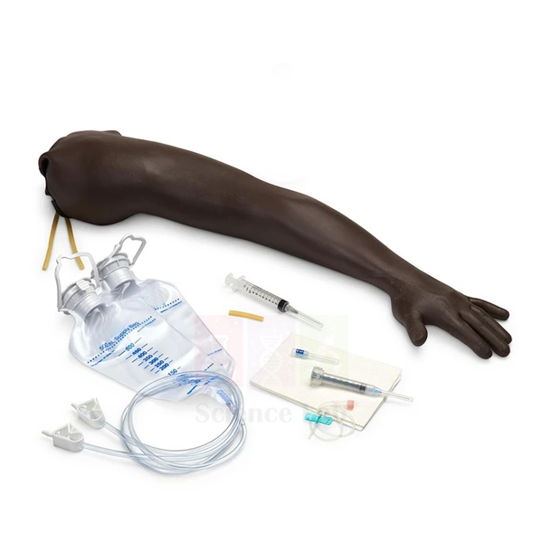
Adult Venipuncture and Injection Training Arm, Dark
Adult Venipuncture and Injection Training Arm, Dark (Product Code: SLE/A/39) is a skills-lab arm for practising intravenous access and intramuscular injection before learners work with patients. It simulates starting simple IVs and placing over-the-needle IV catheters, with veins along the forearm and on the back of the hand, and a foam pad that stands in for muscle tissue for injection practice. The dark simulated skin helps trainees practise vein location on darker skin tones. Nursing schools, paramedical institutes and hospital training units buy it from Jain Scientific Equipments Pvt Ltd (ScienceLab), Ambala, a vocational training lab equipment manufacturer in India.
Specifications
| Parameter | Detail |
|---|---|
| Type | Adult venipuncture and injection training arm |
| Dimensions | 35 × 12 × 6 in |
| Skin | Simulated dark skin; the vein rolls as it is palpated |
| Needle feedback | Characteristic “pop” can be felt as the needle penetrates the vein |
| Vein access sites | Along the forearm and at the back of the hand |
| IV procedures simulated | Starting simple IVs and over-the-needle IV catheters |
| Injection practice | Intramuscular injection using a foam pad that simulates muscle tissue |
Typical Use
In a nursing skills lab the arm is used for step-by-step practice of peripheral venous access: applying a tourniquet, palpating for a suitable vein, anchoring it against the rolling that happens in real patients, inserting the needle and recognising the “pop” that signals entry into the vein. Trainees then progress to over-the-needle catheters on the forearm and hand sites. The separate foam muscle pad allows intramuscular injection technique to be taught in the same session without switching trainers.
Instructors in paramedical and allied-health colleges often run timed stations with the arm for assessment, and it is a practical addition to lab equipment for college skills centres in India. Always follow your institution’s sharps-disposal procedure during practice, and rotate puncture sites between groups to spread wear across the simulated veins.
Manufacturer
The training arm is manufactured by Jain Scientific Equipments Private Limited, whose trading name is ScienceLab. From its base in Ambala, Haryana, the company provides training and laboratory equipment to nursing, medical and technical institutions in India and international markets. Read more on our company profile.
Related Human Models
Clinical-skills and anatomy items that are often ordered with an injection training arm:
- Advanced Adult CPR Training Manikin — adds resuscitation practice to the same nursing skills lab.
- Human Muscular Arm Model — helps trainees learn the arm muscles before choosing injection sites.
- Human Circulatory System Model — explains the venous system that venipuncture relies on.
- Human Arm and Hand model — the skeletal landmarks of the arm and hand for surface-anatomy teaching.
See all trainers and models in our anatomy models category.
Frequently Asked Questions
Which procedures can be practised on this arm?
Starting simple IVs, placing over-the-needle IV catheters and giving intramuscular injections into the foam muscle pad.
Where are the veins located?
Veins are accessible along the forearm and at the back of the hand.
Does it give realistic feedback?
Yes. The vein rolls as it is palpated, and a characteristic “pop” can be felt when the needle enters the vein.
What are the dimensions?
The arm measures 35 × 12 × 6 inches.
Are replacement skins, veins or simulated blood included?
The supplied contents and the availability of spare parts should be confirmed before ordering; please ask through our contact page.
Is there a lighter skin-tone version?
This listing is the dark-skin arm. Contact us if you need to discuss other skin tones for a mixed training set.
What warranty is offered?
Warranty terms are stated in each quotation; ask us for the current terms when you enquire.
Request a Quote
To equip a skills lab with the Adult Venipuncture and Injection Training Arm, Dark, send your requirement via the contact page or add it to the enquiry cart with any manikins and models you also need.
Last updated: 24 September 2026
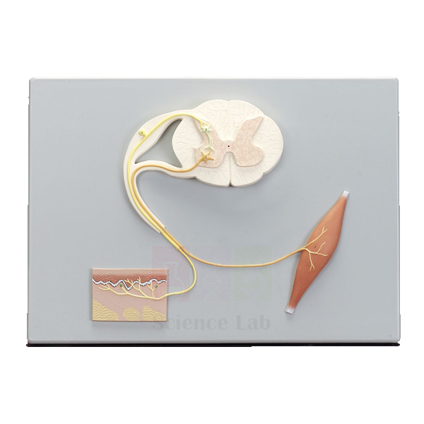
Human Reflex Pathway Model
Human Reflex Pathway Model (Product Code: SLE/A/40) is a plaque-mounted model that lays out a typical reflex arc from start to finish: the sensory side that detects a stimulus, the neural connections in the spinal cord and the motor side that produces the response. Each structure is numbered and matched to an accompanying key, so the pathway can be read like a labelled diagram in relief. Physiology, psychology, nursing and senior biology teachers use it among their biology teaching aids in India, and it is manufactured by Jain Scientific Equipments Pvt Ltd (ScienceLab), Ambala.
Specifications
| Parameter | Detail |
|---|---|
| Type | Plaque-mounted model of a human reflex pathway |
| Size | 53 × 38 cm |
| Components shown | Sensory, neural and motor components of a typical reflex arc |
| Labelling | Pertinent structures numbered and referenced on the accompanying key |
| Mounting | Plaque mount |
Typical Use
Reflexes are one of the first topics where students must connect structure with function in the nervous system. With this model a teacher can trace the route of a signal in order: the receptor picks up the stimulus, the sensory neuron carries it to the spinal cord, connections there pass it on, and the motor neuron carries the command to the effector muscle. Demonstrating a knee-jerk or withdrawal reflex on a volunteer immediately afterwards makes the pathway memorable.
Because the plaque is flat, it can be hung on a wall or propped on an easel as a reference throughout a neurology unit. The numbered key also makes it convenient for practical tests: cover the key and ask students to name each numbered structure and state whether it belongs to the sensory, neural or motor part of the arc.
Manufacturer
Made in Ambala, Haryana by Jain Scientific Equipments Private Limited (trading as ScienceLab), this model is one of many teaching items the company provides to schools, universities and training institutes in India and international markets. Our company profile has further details.
Related Human Models
These models help students see where each part of the reflex arc sits in the body:
- Human Spinal Cord Section Model — enlarges the grey and white matter where reflex connections are made.
- Human Vertebral And Partial Spinal Cord Model — shows the cord inside the vertebral column with its nerve roots.
- Human Nervous System Model — places the reflex arc within the whole nervous system.
- Human Skin Section Model — shows the sensory receptors where many reflexes begin.
Find more nervous-system and organ models in our anatomy models category.
Frequently Asked Questions
What does the model show?
It shows the sensory, neural and motor components of a typical reflex arc on a single plaque.
How are structures identified?
Pertinent structures are numbered and referenced on the accompanying key.
What size is the plaque?
It measures 53 × 38 cm, large enough for front-of-class viewing.
Can it be wall-mounted?
It is a plaque mount, so it is designed to be displayed flat. Please confirm the fixing arrangement with us if you plan to hang it permanently.
Which courses use it?
It suits secondary and senior secondary biology, nursing, physiotherapy, psychology and first-year medical physiology.
Can you quote for several departments at once?
Yes. List the items and quantities for each department through our contact page and we will send one consolidated quotation.
Request a Quote
Price and delivery details for the Human Reflex Pathway Model are available on request through our contact page; you can also save it to the enquiry cart while you choose other nervous-system models.
Last updated: 24 September 2026
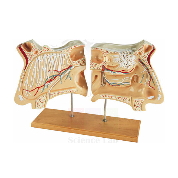
Human Nose Model
Human Nose Model (Product Code: SLE/A/41) enlarges the external nose and nasal cavity about 3× life size and separates into 5 parts, so that the intricate bones and the mucous-membrane lining of the cavity can be opened up and examined. Individual cartilages and bones are coloured, making it easy to tell the flexible framework of the nose apart from its bony support. ENT, anatomy, nursing and senior biology classes use it for the upper respiratory tract and the sense of smell. It is made by Jain Scientific Equipments Pvt Ltd (ScienceLab), Ambala, among the human anatomical models manufacturers in India.
Specifications
| Parameter | Detail |
|---|---|
| Type | Dissectible model of the human nose and nasal cavity |
| Scale | About 3× life size |
| Size | 38 × 72 × 8 cm |
| Number of parts | Dissectible into 5 parts |
| Colour coding | Individual cartilage and bone are coloured |
| Structures shown | Bones of the nose and the nasal cavity covered by mucous membrane |
| Mounting | Mounted on base |
Typical Use
The nasal cavity is a complicated space of thin bones and folded lining that is almost impossible to appreciate from a flat drawing. Opening this model part by part lets students see how the external cartilages give the nose its shape, where bone takes over, and how the mucous membrane covers the cavity walls to warm and moisten incoming air. The colour coding helps learners separate cartilage from bone at a glance.
ENT and nursing tutors use the model when explaining nasal packing, nosebleeds and common obstructions, while school teachers bring it into lessons on breathing and smell. Keep the 5 parts together on the base between classes, and check that each piece is seated correctly when reassembling so the model presents the cavity in its true orientation.
Manufacturer
ScienceLab is the trading name of Jain Scientific Equipments Private Limited (CIN U29309HR2020PTC087597), an Ambala, Haryana company that produces anatomical and science teaching equipment at its licensed factory at 449, HSIIDC Industrial Area, Saha, Ambala (Factory Licence AMB-ONLINE-CHD-J-414). It serves institutions in India and international markets; see our company profile for more.
Related Human Models
Other nose and airway models that suit the same ENT and respiratory lessons:
- Human Nose Model on Base — an alternative nose model for additional benches.
- Human Nose Model on Stand — a stand-mounted version for display and comparison.
- Anatomical Human Nasal Cavity Model — concentrates on the interior of the nasal passages.
- Human Tongue Model — pairs smell with taste in a special-senses lesson.
More head, sense-organ and respiratory models are in the anatomy models category.
Frequently Asked Questions
How many parts does the nose model have?
It is dissectible into 5 parts that reveal the bones and the lined nasal cavity.
What is the scale and overall size?
About 3× life size, measuring 38 × 72 × 8 cm on its base.
Are cartilage and bone distinguished?
Yes. Individual cartilage and bone are coloured so they can be told apart easily.
Is it suitable for ENT teaching?
Yes. It is used in ENT, nursing and anatomy classes, as well as in school biology for breathing and smell.
How should it be cleaned?
Wipe the parts gently with a soft, dry or lightly damp cloth, and avoid alcohol or solvent cleaners on the painted surfaces.
Can you supply a biology model kit bulk order that includes this model?
Yes. Send us your full list and quantities through the contact page and we will quote the items together.
Request a Quote
For a quotation on the Human Nose Model, contact us through the contact page or add it to the enquiry cart and submit your list when ready.
Last updated: 24 September 2026
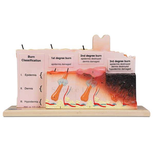
Human Skin with Burn Pathologies Model
Human Skin with Burn Pathologies Model (Product Code: SLE/A/42) is an anatomical teaching model of human skin designed to illustrate how burns damage the skin. It gives nursing, paramedical, first-aid and dermatology instructors a physical reference for discussing burn injuries, which are difficult to teach from photographs alone. It is manufactured by Jain Scientific Equipments Pvt Ltd (ScienceLab), Ambala, for colleges and schools looking for school biology lab equipment in India and for clinical-skills teaching. Because the detailed specification for this listing is being reviewed, please confirm scale, size, mounting and the pathologies represented with us before ordering.
Key Details
| Parameter | Detail |
|---|---|
| Type | Anatomical teaching model of human skin |
| Purpose | Illustrating burn pathologies of the skin |
| Supplied as | Single teaching model; confirm current specification on enquiry |
| Suitable for | Nursing, paramedical, first-aid, dermatology and senior biology teaching |
Typical Use
Burn care is usually taught by first reviewing healthy skin and then showing how heat, chemicals or electricity destroy tissue to different depths. A burn pathology model supports that second step: instructors can point to the damaged areas while explaining how depth of injury relates to pain, blistering, healing time and the need for specialist care. Placing it next to a normal skin section model makes the contrast between healthy and injured tissue clear.
In first-aid and emergency-care courses the model helps learners understand why prompt cooling, covering and referral matter, while nursing students use it to link wound assessment with the underlying skin layers. Ask us which burn stages are shown so you can align the model with your syllabus before purchase.
Manufacturer
Jain Scientific Equipments Private Limited, known to customers as ScienceLab, is an Ambala, Haryana company making models and laboratory equipment for education and training in India and international markets. Our team can answer detailed questions about this model; company background is on our company profile.
Related Human Models
Items that help students compare healthy and injured skin:
- Human Skin Section Model — a healthy scalp skin section to study before discussing burns.
- Enlarged Skin Model — another reference for normal skin layers.
- Prepared Microscope Slide Set – Skin and Excretion — microscope slides of skin tissue for practical work.
Explore all skin, organ and clinical teaching models in our anatomy models category.
Frequently Asked Questions
What does this model teach?
It is a skin model focused on burn pathologies, used to explain how burns affect the skin. Please ask us for the exact pathologies represented.
What are its size and scale?
The specification for this listing is being confirmed; contact us through the contact page and we will send current dimensions before you order.
Who is it intended for?
Nursing and paramedical colleges, first-aid and emergency-care trainers, dermatology teaching and senior biology classes.
Can it be used without a normal skin model?
Yes, but many teachers find the lesson clearer when a healthy skin model is used alongside it for comparison.
How should the model be cared for?
Keep it out of direct sunlight, dust it regularly and clean only with a soft, slightly damp cloth.
What should an institutional enquiry include?
Quote the product code SLE/A/42, the quantity and the delivery location, plus any documents your purchase process requires.
Request a Quote
To check the current specification and price of the Human Skin with Burn Pathologies Model, message us through the contact page or add it to the enquiry cart with related skin models.
Last updated: 24 September 2026
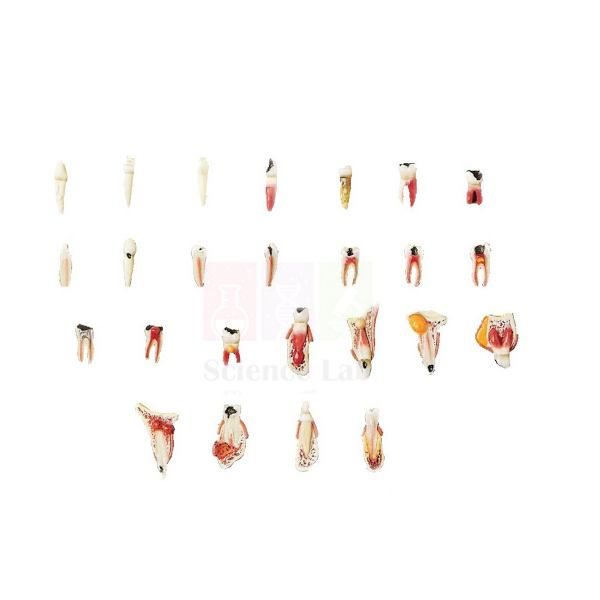
Odontopathies Model
Odontopathies Model (Product Code: SLE/A/44) is a life-size collection of 25 diseased teeth mounted on a green base under a removable transparent cover. Together they show periodontitis, necrosis and varying degrees of dental caries and dental pulp inflammation, so students can compare early and advanced disease side by side on real-size teeth. Dental hygiene schools, dental and nursing colleges and school health-education programmes use it, and it is manufactured by Jain Scientific Equipments Pvt Ltd (ScienceLab), Ambala, an anatomical models manufacturer.
Specifications
| Parameter | Detail |
|---|---|
| Type | Dental pathology display model |
| Scale | Life size |
| Size | 26 × 32 × 4 cm |
| Contents | 25 diseased teeth |
| Diseases depicted | Periodontitis, necrosis, and varying degrees of dental caries and dental pulp inflammation |
| Mounting | Green base with removable transparent cover |
Typical Use
Because the teeth are life size, learners see disease exactly as it appears in a patient’s mouth rather than on an enlarged model. A tutor can arrange a lesson around the progression of decay: from shallow caries through deeper lesions that reach the pulp and cause inflammation, on to necrosis, and then contrast these with periodontitis, where the supporting tissues rather than the tooth itself are affected. The transparent cover keeps the set dust-free on display and lifts off when students need a closer look.
Dental hygiene and assistant courses use the set for identification practice, asking trainees to classify each tooth by condition. In schools it fits oral health education, where showing real-size decayed teeth is often more persuasive than a chart. Replace the cover after every session so the small teeth stay clean and in position.
Manufacturer
This dental model comes from Jain Scientific Equipments Private Limited, an Ambala, Haryana manufacturer that sells under the ScienceLab name to dental, medical and general education buyers in India and international markets. To learn about the company, visit our company profile.
Related Human Models
Use these items with the Odontopathies Model for a complete oral-health unit:
- Dental Caries Model — focuses on the decay process in more detail.
- Human Teeth (Denture) — a healthy denture model for brushing demonstrations and comparison.
- Human Teeth — a wall chart to reinforce tooth names and structure.
- Human Skull — shows how teeth sit in the upper and lower jaws.
- Human Tongue Model — covers the other main structure of the oral cavity.
Browse all anatomy and pathology models in our anatomy models category.
Frequently Asked Questions
How many teeth are included?
The model has 25 diseased teeth mounted on one base.
Which dental diseases are shown?
Periodontitis, necrosis, and varying degrees of dental caries and dental pulp inflammation.
Are the teeth enlarged?
No, they are life size, which helps students recognise conditions as they appear clinically.
How is the model protected?
The teeth sit on a green base under a removable transparent cover. The whole unit measures 26 × 32 × 4 cm.
Can individual teeth be removed?
The teeth are described as mounted on the base; please confirm with us through the contact page if you need removable teeth for your course.
Do you supply several sets for a dental college?
Yes. Tell us how many sets you need and where they should be delivered, and we will send a combined quotation.
Request a Quote
Enquire about the Odontopathies Model through our contact page, or add it to the enquiry cart alongside other dental and oral-health teaching aids.
Last updated: 24 September 2026
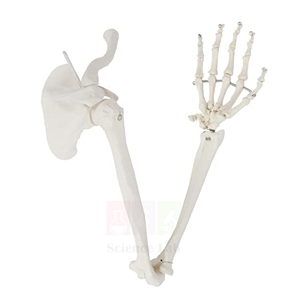
Human Arm and Hand model
Human Arm and Hand model (Product Code: SLE/A/51) is a life-size skeletal model of the complete upper limb, running from the shoulder girdle, with its scapula and clavicle, down through the arm to the bones of the hand. The bones are wire mounted with flexible elbow and shoulder joints, so students can move the limb through its range and see how the bones glide at each joint. At 0.6 kg it is light enough to pass around a class. It serves osteology practicals in medical, physiotherapy, nursing and biology courses as a focused alternative to a full human skeleton model in India, and it is manufactured by Jain Scientific Equipments Pvt Ltd (ScienceLab), Ambala.
Specifications
| Parameter | Detail |
|---|---|
| Type | Skeletal model of the human arm and hand |
| Scale | Life size |
| Bones included | Arm and hand, including the scapula and clavicle |
| Assembly | Wire mounted |
| Joints | Flexible elbow and shoulder joints |
| Weight | 0.6 kg |
Typical Use
Osteology sessions on the upper limb usually start at the shoulder girdle. With this model the tutor can show how the clavicle braces the scapula, then move the flexible shoulder joint to demonstrate flexion, extension and rotation before bending the elbow to illustrate hinge movement. Students can then trace the long bones of the arm and forearm into the wrist and fingers, naming bony landmarks where muscles attach.
Physiotherapy and sports-medicine learners value the movable joints for relating anatomy to exercise and injury, and nursing tutors use the limb to point out landmarks for injections and blood-pressure measurement. Handle the wire joints gently and avoid bending them beyond a natural range, which keeps the articulation working smoothly over years of classes.
Manufacturer
Jain Scientific Equipments Private Limited manufactures this skeletal limb in Ambala, Haryana and markets it under its trading name, ScienceLab, to teaching institutions in India and international markets. You can read about the company on our company profile.
Related Human Models
Models that extend upper-limb teaching from bones to joints and muscles:
- Human Skeletal Hand Model — a separate hand for detailed study of carpals and finger bones.
- Human Shoulder Joint Model — shows the shoulder joint in close-up.
- Human Elbow Joint Model — a closer look at the elbow articulation.
- Human Muscular Arm Model — adds the muscles that move these bones.
- Human Skeleton, Rod-Supported — a full skeleton for placing the upper limb in context.
See all skeletal and anatomy models in our anatomy models category.
Frequently Asked Questions
Does the model include the shoulder girdle?
Yes. The scapula and clavicle are included along with the bones of the arm and hand.
Can the joints be moved?
The model is wire mounted with flexible elbow and shoulder joints, so both can be moved to demonstrate motion.
Is it life size?
Yes, it is a life-size skeletal model.
How heavy is it?
It weighs 0.6 kg, which makes it easy to hand around during practicals.
Does it come with a stand?
A stand is not listed in the specification; please confirm display options with us through the contact page.
What is needed for a college purchase order?
Quote the product code SLE/A/51, the number of limbs required and your delivery location, together with any vendor documents your institution asks for.
Request a Quote
Ask us for a price on the Human Arm and Hand model via the contact page, or add it to the enquiry cart if you are building an upper-limb set.
Last updated: 24 September 2026
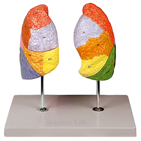
Human Segmented Lungs Model
Human Segmented Lungs Model (Product Code: SLE/A/53) is a life-size model of the left and right lungs used to teach segmental lung anatomy in anatomy departments, nursing and physiotherapy colleges and school biology labs. Its lobes are painted in different colours to highlight their locations, and a numbered key comes with the 33 x 23 x 31 cm model on its stand. It is made by Jain Scientific Equipments Pvt Ltd (ScienceLab), an OEM manufacturer, at its own factory in Ambala, India.
Specifications
| Parameter | Detail |
|---|---|
| Scale | Life size |
| Size | 33 x 23 x 31 cm |
| Anatomy shown | Segmental anatomy of the left and the right lungs |
| Colour coding | Lobes painted in different colours to highlight their locations |
| Key | Numbered key included |
| Mounting | On stand with base |
| Product code | SLE/A/53 |
| Manufacturer | Jain Scientific Equipments Pvt Ltd (ScienceLab) — OEM |
| Country of origin | India (made in Ambala, Haryana) |
What Is the Segmented Lungs Model Used For?
Segmental anatomy matters because each bronchopulmonary segment has its own airway and blood supply, which is why chest findings are described by segment and why physiotherapists position patients according to the segment they want to drain. Tutors use this model to show where each region of the lungs sits while students work through the numbered key, matching every number to the area it marks. The colour-painted lobes stay readable from the back of a lecture room as well as at close range.
The model suits respiratory anatomy practicals, revision before viva examinations and school lessons on lung structure, and it is a natural centrepiece for a human biology model set covering the respiratory system. Lift it by the stand rather than by the lungs, and clean it with a soft cloth.
OEM Manufacturer
ScienceLab is the trading name of Jain Scientific Equipments Private Limited, the OEM (original equipment manufacturer) of this lung model. The company (CIN U29309HR2020PTC087597) produces it at its own licensed factory at 449, HSIIDC Industrial Area, Saha, Ambala, Haryana, India (Factory Licence AMB-ONLINE-CHD-J-414). Colleges, schools, dealers and importers buy it factory-direct, and repeat orders or questions about the numbered key go to the manufacturer itself; just ask us. Read more on our company profile.
Related Human Models
Models that extend a segmental lung lesson from the whole organ down to tissue level:
- Segmented Lung — a take-apart alternative whose lobes separate into individual segments.
- Somso Human Lung Model — a greatly enlarged view of the lung lobule for histology teaching.
- Pathology of the Human Lung Model — shows how infections alter lung tissue, for comparison with healthy anatomy.
- Human Thoracic Organs Model — places the lungs among the heart, trachea, diaphragm and other organs of the chest.
The model is listed with our other anatomical models in the Human Models category.
Frequently Asked Questions
Does the Human Segmented Lungs Model show both lungs?
Yes. The Human Segmented Lungs Model shows the segmental anatomy of both the left and the right lungs at life size, with the lobes painted in different colours so each one's location is easy to pick out.
How large is the Human Segmented Lungs Model?
The Human Segmented Lungs Model measures 33 x 23 x 31 cm and stands on its own base, so plan for that footprint on a bench, a lecture table or in a display cabinet.
Can the Human Segmented Lungs Model be taken apart?
The Human Segmented Lungs Model is described as a life-size display on a stand with colour-painted lobes; detachable pieces are not stated. If students must separate lobes and segments by hand, the Segmented Lung listed above is designed for that.
What is the numbered key used for?
The numbered key supplied with the Human Segmented Lungs Model matches numbers on the model, so students can name each marked structure without a separate textbook diagram, which is useful for self-testing before practical examinations.
Who manufactures the Human Segmented Lungs Model?
Jain Scientific Equipments Pvt Ltd, trading as ScienceLab, is the OEM manufacturer of the Human Segmented Lungs Model and makes it at its own licensed factory at Saha, Ambala, Haryana, India, supplying institutions and dealers directly.
What should an institutional enquiry include?
For the Human Segmented Lungs Model, give product code SLE/A/53, the number of units, the delivery address and any tender format you follow. We supply buyers in India and international markets and quote directly as the manufacturer.
Request a Quote
For pricing and delivery details on the Human Segmented Lungs Model, straight from the manufacturer, reach our team via the contact page or place the model in your enquiry cart and submit one combined request.
Last updated: 24 September 2026
Human Hand
Human Hand (Product Code: SLE/A/57) is a life-size replica of the bones of the human hand, articulated with wire so the fingers keep a slight flexibility. Weighing only 0.1 kg, it can sit on a desk, be held in one palm and compared directly with the student’s own hand, which makes bone names and joint positions far easier to remember. It is a compact choice for osteology practicals, physiotherapy and occupational-therapy courses and school biology, made by Jain Scientific Equipments Pvt Ltd (ScienceLab), Ambala, one of the high school biology lab equipment manufacturers serving Indian institutions.
Specifications
| Parameter | Detail |
|---|---|
| Type | Replica of the bones of the human hand |
| Scale | Life size |
| Articulation | Articulated with wire |
| Movement | Slight flexibility through the fingers |
| Weight | 0.1 kg |
Typical Use
The hand contains many small bones in a compact space, and students often confuse the carpals of the wrist. A life-size replica solves this by letting each learner lay the model beside their own hand and match bone to finger and knuckle. Tutors typically work from the wrist outwards: carpals first, then the metacarpals of the palm, then the phalanges of each finger and the thumb.
The slight flexibility in the fingers is enough to show how the joints bend without risking the wire articulation. Occupational-therapy and physiotherapy students use the model when discussing grip, fractures and hand rehabilitation, and several can be ordered so that small groups each have one on the bench. Store it flat or in a box so the fingers are not bent under other equipment.
Manufacturer
The model is produced by Jain Scientific Equipments Private Limited, the Ambala, Haryana company behind the ScienceLab brand. It is a registered MSME enterprise (Udyam UDYAM-HR-01-0003714), and its export orders are shipped under Importer-Exporter Code AAECJ8618A issued by the DGFT, supporting customers in India and international markets. More is available on our company profile.
Related Human Models
Pair this hand with models that add ligaments, soft tissue or the rest of the limb:
- Human Skeletal Hand Model — an alternative skeletal hand for extra benches or comparison.
- Human Hand Joint Model with Ligaments — shows how ligaments hold the hand bones together.
- 3-D Printed Human Superficial Hand Dissection Model — adds the soft-tissue layer over the bones.
- Human Arm and Hand model — the full upper limb from shoulder girdle to fingers.
- Human Skeletal Foot Model — lets students compare the bones of the hand and foot.
Our full range of skeletal and anatomy models is in the anatomy models category.
Frequently Asked Questions
Is this a full hand skeleton?
Yes, it is a life-size replica of the bones of the human hand, articulated with wire.
Do the fingers move?
There is slight flexibility through the fingers, enough to show how they bend.
How much does it weigh?
0.1 kg, so it is easy to handle and pass around.
How does it differ from the Human Arm and Hand model?
This item is the hand only; the arm and hand model also includes the arm bones, scapula and clavicle with movable elbow and shoulder joints.
Is the model left or right?
The listing does not specify the side; please ask through the contact page if you need a particular one.
Can you supply class sets?
Yes. Let us know how many you need for small-group work and we will quote accordingly.
Request a Quote
Send your quantity for the Human Hand through our contact page, or place it in the enquiry cart to request a single quotation with other skeletal models.
Last updated: 24 September 2026
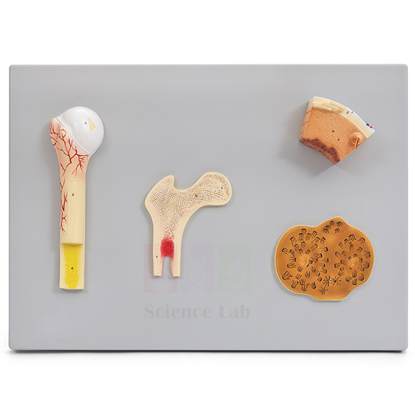
Human Bone Structures Model
Human Bone Structures Model (Product Code: SLE/A/58) is a plastic model that uses the femur to show what a living bone looks like both outside and in. It displays the periosteum covering the surface, the blood vessels that supply the bone, the spongy bone inside, the dense compact bone of the shaft and the Haversian system that organises compact bone at the microscopic level. Students of histology, physiology, nursing and school biology use it to connect bone anatomy with growth, repair and blood supply. It is manufactured by Jain Scientific Equipments Pvt Ltd (ScienceLab), Ambala, for institutions that need school biology lab equipment in India and college-level teaching aids.
Specifications
| Parameter | Detail |
|---|---|
| Type | Bone structure model based on the femur |
| Material | Plastic |
| External structures | Periosteum and blood vessels |
| Internal structures | Spongy bone, compact bone and the Haversian system |
Typical Use
Most students first meet bone as a dry skeleton, which hides the fact that bone is living tissue. This model corrects that picture. A teacher can begin with the periosteum and blood vessels on the outside, then move inward to show why the ends of a long bone are filled with spongy bone while the shaft is built of compact bone, and finally explain how the Haversian system carries vessels through the compact layer.
The model is a natural bridge between gross anatomy and histology: after studying it, learners find bone slides under the microscope much easier to interpret. It also supports lessons on fractures and healing, osteoporosis and why bones need a blood supply. Because it is plastic, it can be handled by students in practical sessions and wiped clean afterwards.
Manufacturer
Based in Ambala, Haryana, Jain Scientific Equipments Private Limited makes this bone model and sells it under its trading name ScienceLab to schools, colleges and universities in India and international markets. Visit our company profile to find out more about the company.
Related Human Models
Combine this model with the following for a full lesson on bone from whole skeleton to tissue:
- Human Femur Bone Model — the whole femur, showing where the illustrated structures sit.
- 3B Bone Microanatomy Model — an alternative focus on bone at the microscopic level.
- Microslide Animal Tissue Set 2 Muscle, Bone, Connective — prepared slides to compare real bone tissue with the model.
- Disarticulated Human Skeleton — individual bones for wider osteology practicals.
Find more bone and anatomy models in our anatomy models category.
Frequently Asked Questions
Which bone does the model represent?
It shows the external and internal structures of the femur.
What structures can students identify?
The periosteum, blood vessels, spongy bone, compact bone and the Haversian system.
What is it made of?
The model is made of plastic, so it is durable and easy to clean with a damp cloth.
What are its dimensions and scale?
These are not given in the current listing; please ask us through the contact page and we will confirm before you order.
Is it suitable for school students?
Yes. It works for senior school biology as well as nursing, physiotherapy and first-year medical histology.
Can it be included in an export order?
Yes, we supply institutions in India and international markets. Share the destination and quantity and we will advise on packing and shipping.
Request a Quote
Get a quotation for the Human Bone Structures Model by writing to us on the contact page, or add it to the enquiry cart with the slides and skeletal models on your list.
Last updated: 24 September 2026
Atlas, Axis, and Occipital Bones Set
Atlas, Axis, and Occipital Bones Set (Product Code: SLE/A/59) is a life-size representation of the base of the skull, the occipital plate, with the first two cervical vertebrae, the atlas and axis, held in their natural positions. It isolates the craniovertebral junction, the region that lets the head nod and turn, so it can be studied without the distraction of a whole skull or spine. The set is rod-mounted on a durable polymer base and is used in anatomy, physiotherapy, chiropractic, radiography and nursing teaching. It is manufactured by Jain Scientific Equipments Pvt Ltd (ScienceLab), Ambala, as a companion to a full human skeleton model in India.
Specifications
| Parameter | Detail |
|---|---|
| Type | Occipital plate with atlas and axis vertebrae in situ |
| Scale | Life size |
| Size (without stand) | 12 × 9 × 7 cm |
| Mounting | Rod-mounted on a durable polymer base |
Typical Use
The joints between the skull, atlas and axis are unlike any others in the spine, and they are hard to visualise from textbook images. With the bones shown in situ, a lecturer can explain how the occipital condyles rest on the atlas to allow nodding, and how the atlas pivots around the axis to turn the head from side to side. Students can then identify features such as the ring shape of the atlas and the upward projection of the axis.
Physiotherapy and chiropractic courses use the set when discussing neck movement and upper cervical injuries, while radiography students compare it with images of the region. Because it is small and firmly rod-mounted, it can stand on a bench throughout a practical; lift it by the base rather than by the bones to protect the mounting.
Manufacturer
This set is made by Jain Scientific Equipments Private Limited, a manufacturer in Ambala, Haryana that trades as ScienceLab and supplies medical, paramedical and school institutions in India and international markets. For company information, see our company profile.
Related Human Models
These skull and spine models extend a lesson on the head and neck:
- Human Skull with Cervical Spine Model — shows the full skull with the whole neck region.
- Human Cervical Spinal Column Model — continues below the axis through the rest of the cervical vertebrae.
- Human Skull Bones Model — places the occipital bone among the other bones of the skull.
- Human Vertebral Column Model With Skull — the complete spine and skull for context.
- Human Vertebral And Partial Spinal Cord Model — relates vertebrae to the spinal cord they protect.
More skeletal and anatomy models are listed in our anatomy models category.
Frequently Asked Questions
Which bones are included?
The occipital plate of the skull with the atlas and axis vertebrae in situ.
Is the set life size?
Yes, it is a life-size representation.
How big is it?
It measures 12 × 9 × 7 cm without the stand.
How is it mounted?
The bones are rod-mounted on a durable polymer base.
Can the atlas and axis be removed?
The listing describes them as mounted in situ; please check with us through the contact page if your course needs separate vertebrae.
What information helps us prepare an institutional quotation?
The product code SLE/A/59, required quantity, delivery address and any registration or tender documents you need from the supplier.
Request a Quote
To buy the Atlas, Axis, and Occipital Bones Set, contact our team via the contact page or add it to the enquiry cart along with other skull and spine models.
Last updated: 24 September 2026
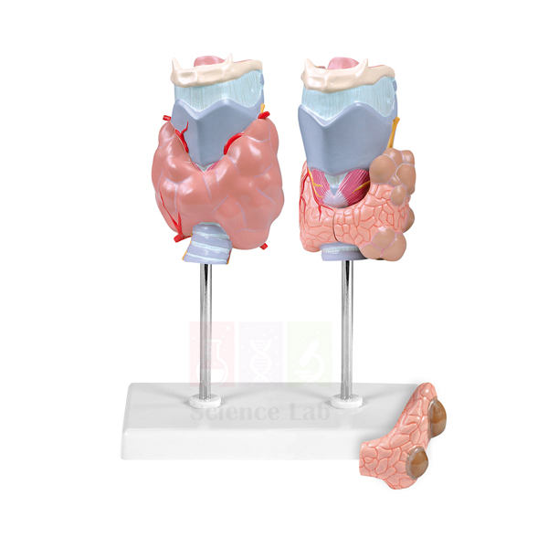
Pathology of the Human Thyroid Model
Pathology of the Human Thyroid Model (Product Code: SLE/A/60) sets two thyroid glands side by side so that learners can compare a gland affected by autoimmune disease with one carrying tumour changes next to healthy tissue. It is designed for pathology, physiology and nursing lessons in which students must recognise how common thyroid disorders alter the organ. The model is made by Jain Scientific Equipments Pvt Ltd (ScienceLab), Ambala, within its range of human anatomical models for schools, colleges and medical training institutes.
Specifications
| Parameter | Detail |
|---|---|
| Model type | Thyroid pathology teaching model |
| Contents | 2 thyroid models, divided 4 ways |
| First thyroid | Illustrates Graves' autoimmune disease |
| Second thyroid | Shows thyroid adenoma and carcinoma, compared with the normal right side of the thyroid |
| Mounting | On stand with base |
| Size | 33 x 23 x 25 cm |
Typical Use
A pathology tutor can open the lesson with the normal right side of the second gland, move across to the adenoma and carcinoma on the same piece, and finish with the Graves' specimen. Working from healthy to diseased tissue in this order lets students put the differences they observe into their own words, which is useful for viva preparation, practical records and case-based discussion.
The model also serves nursing and allied-health classes that cover thyroid swelling and hormone disorders, and it can stay on the demonstration bench as a reference throughout an endocrine unit. As a biology teaching aid it works well after students have located the thyroid on a general endocrine or neck model, because they then know where the gland sits before studying what goes wrong with it. Keep the model on its base while it is being examined and wipe it with a soft, dry cloth after the session.
Manufacturer
This thyroid pathology model comes from Jain Scientific Equipments Private Limited, the Ambala, Haryana company that trades under the ScienceLab name and produces anatomical and laboratory teaching equipment for institutions in India and international markets. Background on the business is available on the company profile page.
Related Human Models
Teachers covering the thyroid and its surroundings often order these models alongside it:
- Human Endocrine Organs Model — shows where the thyroid fits among the other hormone-producing glands.
- Anatomy of the Human Neck Model — helps students locate the gland within the neck before they study its diseases.
- Larynx Human Model — the thyroid lies just below the larynx, so this model shows its immediate neighbour.
- Pathology of the Human Trachea and Bronchi Model — a second pathology model for comparing disease changes in a nearby organ system.
See the complete range in the Human Models category.
Frequently Asked Questions
What does the Pathology of the Human Thyroid Model include?
Two thyroid models, divided 4 ways, on a stand with base. One illustrates Graves' autoimmune disease; the other shows adenoma and carcinoma beside the normal right side of the gland.
How much bench space does it need?
The model measures 33 x 23 x 25 cm, so it fits on a standard demonstration table or display shelf.
Is healthy thyroid tissue shown for comparison?
Yes. The second model keeps the normal right side of the thyroid as a reference against the adenoma and carcinoma. A separate complete healthy gland is not listed in the product details.
Does it come with a key or teaching guide?
A key is not mentioned in the product details. Please confirm the current supply contents with us through the contact page before you order.
Which courses is it most suited to?
It is aimed at pathology, physiology, nursing and paramedical teaching, where thyroid disorders are part of the syllabus. Senior biology classes can also use it with teacher guidance.
What should an institutional quotation request include?
Quote product code SLE/A/60, the quantity, the delivery address and any other human models you need on the same order, so that everything can be quoted and packed together.
Can you deliver outside India?
Yes. ScienceLab supplies institutions in India and international markets; tell us the destination and we will advise on packing and shipment.
Request a Quote
For pricing and availability of SLE/A/60, write to us through the contact page or add the model to your enquiry cart with any other items for your laboratory.
Last updated: 24 September 2026
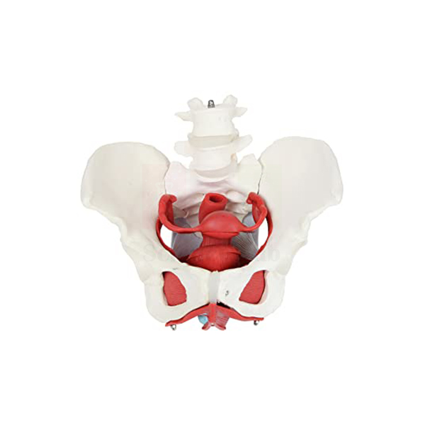
Female Pelvis with Organs
Female Pelvis with Organs (Product Code: SLE/A/61) combines a life-size skeleton of the complete female pelvis with the ovaries, uterus, urinary bladder and rectum in their natural positions, so the bony ring and the organs it protects can be studied together. Midwifery, nursing, gynaecology and anatomy teachers use it to explain the pelvic floor and the perineal region, which are hard to visualise from diagrams alone. It is made by Jain Scientific Equipments Pvt Ltd (ScienceLab), Ambala, an anatomical models manufacturer serving schools, colleges and medical institutes.
Specifications
| Parameter | Detail |
|---|---|
| Scale | Life-size skeleton of a complete female human pelvis |
| Organs included | Ovaries, uterus, urinary bladder and rectum |
| Pelvic floor | Muscular and urogenital features shown in great detail |
| Perineal region | Structures especially well rendered and numbered |
| Key | Included, with 26 identified structures |
| Size | 26 x 17 x 17 cm |
Typical Use
Because the organs sit inside a life-size bony pelvis, students can see how the bladder lies in front of the uterus and the rectum behind it, and how the pelvic floor muscles close the outlet from below. The numbered perineal structures and the 26-point key make the model well suited to identification tests: the tutor calls out a number and students name the structure, or the reverse.
In midwifery and obstetric nursing courses it supports discussion of pelvic floor support, continence and the effects of childbirth on the perineum, while general anatomy classes use it to link the reproductive, urinary and digestive systems in one region. As a human body model for small-group work it can be passed between benches; hold it by the bony pelvis rather than the organs, and wipe it with a slightly damp cloth when needed.
Manufacturer
ScienceLab is the trading name under which Jain Scientific Equipments Private Limited makes and ships teaching models from its base in Ambala, Haryana. Institutions that want to know more about the company before placing an order can read the ScienceLab company profile.
Related Human Models
For a complete pelvic and reproductive anatomy set, buyers often add:
- Female Half Pelvis Model — a sectioned view that complements the whole-pelvis presentation.
- Human Female Urogenital Model — follows the urinary and genital tracts beyond the pelvic region.
- Female Skeletal Pelvis — the bony pelvis on its own for osteology and pelvic measurement lessons.
- Male Pelvis Section Model — lets students compare male and female pelvic organ arrangement.
More options are listed in the Human Models category.
Frequently Asked Questions
Which organs are shown in the Female Pelvis with Organs?
The ovaries, uterus, urinary bladder and rectum, set within a life-size skeleton of the complete female pelvis.
Is a key supplied?
Yes. A key identifying 26 structures is included, and the perineal structures on the model are numbered to match.
What are the dimensions?
The model measures 26 x 17 x 17 cm.
Is it suitable for midwifery and nursing training?
Yes. The detailed pelvic floor and perineal region make it a practical aid for obstetric nursing and midwifery lessons, as well as for general anatomy classes.
Can the organs be removed?
The product details do not state that the organs are removable. If separate parts matter for your teaching, please check with us through the contact page.
What warranty applies?
Warranty and after-sales terms are given with each quotation; ask our team for the terms that apply to your order.
Can you supply several units for a nursing college?
Yes. State the quantity and delivery location in your enquiry, and we will quote for the full lot, whether the college is in India or in an international market.
Request a Quote
Send your requirement for SLE/A/61 through our contact form, or place it in the enquiry cart and submit the whole list at once.
Last updated: 24 September 2026
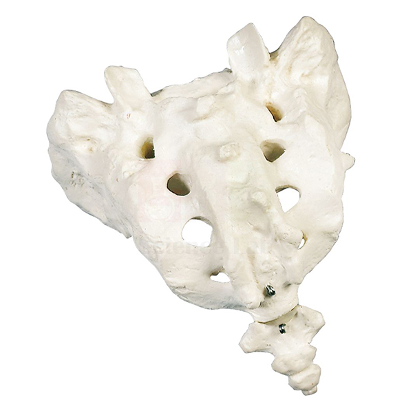
Human Sacrum and Coccyx
Human Sacrum and Coccyx (Product Code: SLE/A/62) is a life-size, one-part model of the lowest section of the vertebral column: the wedge-shaped sacrum and the small tailbone below it. Anatomy, physiotherapy and nursing students use it to learn the landmarks of this region without handling a full skeleton, and it is a convenient desk piece when a complete human skeleton model is shared by a large class. It is made by Jain Scientific Equipments Pvt Ltd (ScienceLab), Ambala.
Specifications
| Parameter | Detail |
|---|---|
| Bones represented | Human sacrum and coccyx |
| Scale | Life-size |
| Construction | 1-part model |
| Size | 20 x 15 x 15 cm |
| Weight | 0.2 kg |
Typical Use
Osteology practicals usually ask students to identify the sacral promontory, the sacral foramina through which nerves pass, the auricular surfaces that meet the hip bones and the joint between sacrum and coccyx. A single-piece, life-size model lets each student turn the bone in the hand, compare the anterior and posterior surfaces and sketch them, then return it for the next group. At 0.2 kg it is light enough for repeated handling during a long practical.
Physiotherapy and sports-science tutors use the same model to explain low back and tailbone pain, the sacroiliac joint and the attachment points of pelvic muscles. It also works as an item of school biology lab equipment when senior students study the regions of the spine, because it shows clearly how several vertebrae fuse into one bone. Store it in a closed cupboard to keep it free of dust.
Manufacturer
The sacrum and coccyx model is part of the anatomy range made by Jain Scientific Equipments Private Limited, a company in Ambala, Haryana that sells its products under the ScienceLab brand. You can read how the company works on the profile page.
Related Human Models
These items extend a lesson on the lower spine and pelvis:
- Human Sacrum and Coccyx-2 — an alternative unmounted plastic version of the same bones.
- Human Pelvis with Lower Lumbar and Femur Heads — shows the sacrum in place between the hip bones.
- Human Lumbar Set Model — the vertebrae that sit directly above the sacrum.
- Female Skeletal Pelvis — for studying the sacrum as part of the pelvic ring.
Explore more in the Human Models category.
Frequently Asked Questions
Is the Human Sacrum and Coccyx life-size?
Yes. It is a life-size model made as a single part.
What are its size and weight?
It measures 20 x 15 x 15 cm and weighs 0.2 kg.
Does the coccyx come apart from the sacrum?
No. This is a 1-part model, so the sacrum and coccyx form one piece.
How does it differ from Human Sacrum and Coccyx-2?
Both are life-size. This version is a one-part model with a listed weight of 0.2 kg and size of 20 x 15 x 15 cm, while the -2 version is an unmounted plastic replica with different dimensions. Compare both pages or ask us which suits your course.
What material is it made of?
The material is not stated in the product details. Please ask through the contact page if you need this for a tender specification.
Can it be ordered in class sets?
Yes. Tell us how many units you need and the delivery location, and we will quote for the set.
Request a Quote
To receive a quotation for SLE/A/62, use the contact page or collect items in the enquiry cart and send them to us together.
Last updated: 24 September 2026
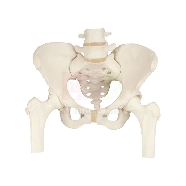
Human Pelvis with Lower Lumbar and Femur Heads
Human Pelvis with Lower Lumbar and Femur Heads (Product Code: SLE/A/63) is a life-size plastic replica that joins the pelvic bones, sacrum and coccyx to the heads of both femurs and to the last two lumbar vertebrae, L4 and L5, with their intervertebral discs. Showing the lumbosacral junction and both hip joints on one piece makes it a practical reference for orthopaedics, physiotherapy, chiropractic and anatomy teaching. The replica is made by Jain Scientific Equipments Pvt Ltd (ScienceLab), Ambala, and is widely used as lab equipment for college anatomy departments.
Specifications
| Parameter | Detail |
|---|---|
| Scale | Life-size replica |
| Material | Plastic |
| Pelvic structures | Pelvic bones, sacrum and coccyx |
| Femur | Femur heads |
| Spine | Lower lumbar vertebrae L4 and L5 with intervertebral discs |
Typical Use
Low back pain, disc problems and hip disorders all involve the zone this model covers. A physiotherapy lecturer can point to the L4 and L5 discs to explain why this level is so often involved in disc prolapse, then move down to the sacrum and across to the femur heads to discuss how load passes from the spine through the pelvis into the legs. Because the femur heads sit in their sockets, students can also see the orientation of the hip joint relative to the pelvis.
In general anatomy and sports-science classes the model supports lessons on the pelvic girdle, posture and the differences between the lumbar spine and the fused sacral vertebrae. It is also useful at patient-education desks in clinics, where a life-size replica helps explain a diagnosis. Keep it on a flat surface when not in use and clean the plastic with a mild soap solution and a soft cloth.
Manufacturer
Jain Scientific Equipments Private Limited, which operates as ScienceLab from Ambala, Haryana, makes this pelvis replica as part of its skeletal and anatomical model line. The company is a registered MSME enterprise (Udyam UDYAM-HR-01-0003714), and its export shipments are handled under Importer-Exporter Code AAECJ8618A issued by the DGFT. Further details are on the company profile.
Related Human Models
Useful companions for spine and hip teaching include:
- Human Spine with Femur Heads Model — extends the view upward to the full vertebral column.
- Human Hip Joint Model — a closer look at the joint formed by the femur head and pelvis.
- Human Lumbar Set Model — covers the lumbar vertebrae above L4 and L5.
- Human Sacrum and Coccyx — the sacral bones as a separate piece for handling.
- Male Skeletal Pelvis — for comparing pelvic shape between the sexes.
The full selection is in the Human Models category.
Frequently Asked Questions
Which vertebrae are included?
The lower lumbar vertebrae L4 and L5, each with its intervertebral disc, attached above the sacrum.
What material is the model made from?
Plastic, which makes it suitable for regular handling in practical classes.
Are both femur heads included?
Yes. The replica includes the femur heads with the pelvic bones, so the hip joints can be shown.
Is it life-size?
Yes, it is a life-size replica. Overall dimensions are not listed; ask us if you need them for storage planning.
Is there a stand or mounting?
A stand is not mentioned in the product details. Please confirm the supplied configuration through the contact page.
What vendor details can you provide for a college purchase?
We can share company registration details such as our Udyam and IEC numbers along with the quotation. Send your purchase requirements and we will include what your institution needs.
Request a Quote
Ask for a quotation for SLE/A/63 on the contact page, or add it to the enquiry cart along with other skeletal models you plan to buy.
Last updated: 24 September 2026
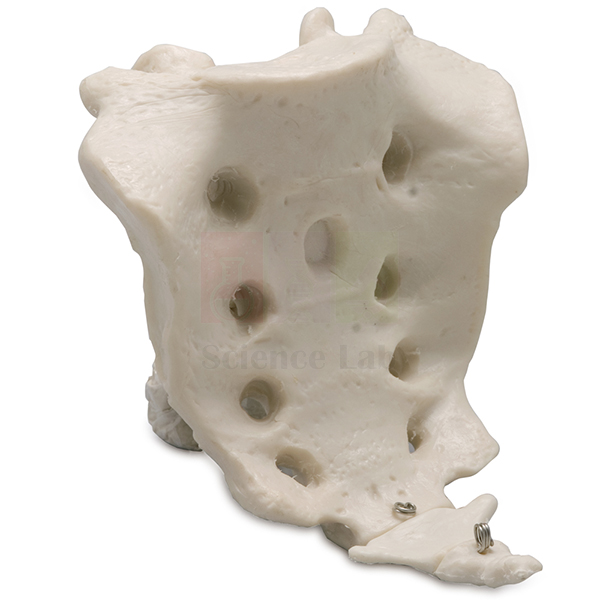
Human Sacrum and Coccyx-2
Human Sacrum and Coccyx-2 (Product Code: SLE/A/64) is an unmounted, life-size plastic replica of the sacral vertebrae together with the coccyx. With no stand attached, it can be picked up, turned over and laid flat on a tray, which suits hands-on bone identification and station-based practical exams. It is made by Jain Scientific Equipments Pvt Ltd (ScienceLab), Ambala, whose school and college lab equipment range includes a wide choice of skeletal models.
Specifications
| Parameter | Detail |
|---|---|
| Structures | Sacral vertebrae with the coccyx |
| Scale | Life-size replica |
| Material | Plastic |
| Mounting | Unmounted |
| Size | 20 x 11 x 6 cm |
Typical Use
The sacrum forms when five sacral vertebrae fuse during development, and a loose replica is the easiest way to show students the traces of that fusion: the transverse ridges on the front surface, the rows of foramina and the median crest at the back. Because the piece is unmounted, examiners can place it at a practical station in any orientation and ask candidates to decide which side is anterior, which end is the base and where the coccyx articulates.
In zoology lab equipment collections it can be set beside animal skeletons to compare how the tail region is reduced in humans. Nursing and physiotherapy tutors also use it when discussing sitting posture, coccyx injuries and the location of sacral injection sites. The plastic surface can be wiped clean after each class, and the compact size allows several pieces to be stored in one drawer.
Manufacturer
This unmounted replica is produced by Jain Scientific Equipments Private Limited, known to customers as ScienceLab, which designs and supplies anatomy teaching aids from Ambala in Haryana. The company profile gives an overview of its work.
Related Human Models
To build a lower-spine practical station, consider adding:
- Human Sacrum and Coccyx — the one-part version of the same region, useful as a second specimen.
- Human Pelvis Structural Model With 5 Pcs Lumbar Vertebraes — places the sacrum within the pelvis and below the lumbar spine.
- Human Lumbar Spinal Column Model — the section of spine that rests on the sacrum.
- Disarticulated Human Skeleton — for full-body bone identification sessions.
Browse other skeletal and organ models in the Human Models category.
Frequently Asked Questions
Does Human Sacrum and Coccyx-2 come on a stand?
No. It is supplied unmounted so it can be handled freely.
How big is the replica?
20 x 11 x 6 cm, reproducing the bones at life size.
What is it made of?
Plastic, which is easy to clean between practical groups.
Which one should I choose, this or the one-part Human Sacrum and Coccyx?
Choose this version if you want an unmounted plastic piece of 20 x 11 x 6 cm for handling and practical stations. If you are unsure, send us your course needs and we will suggest the better fit.
How should it be cleaned?
Wipe it with a soft cloth dampened with mild soapy water, then dry it. Avoid solvents that may mark the surface.
Is a bulk order possible for a medical or nursing college?
Yes. Give us the quantity and delivery details through the contact page and we will prepare a quotation for the batch.
Request a Quote
Price and delivery details for SLE/A/64 are available on request: reach us via the contact page or save the item in your enquiry cart.
Last updated: 24 September 2026
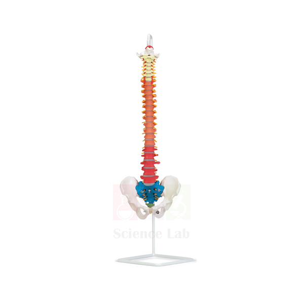
Economy Human Flexible Vertebral Column, Color Coded
Economy Human Flexible Vertebral Column, Color Coded (Product Code: SLE/A/65) is a fully flexible, life-sized plastic spine that runs from the occipital plate at the base of the skull down through the cervical, thoracic and lumbar vertebrae to the sacrum, coccyx and complete pelvis. Each region is color coded, and the model adds vertebral arteries, spinal nerves and a prolapsed L3–L4 disc, so one piece covers normal anatomy, movement and a common clinical problem. It is made by Jain Scientific Equipments Pvt Ltd (ScienceLab), Ambala, for schools, colleges, physiotherapy departments and clinics that need a spine without buying a full human skeleton model.
Specifications
| Parameter | Detail |
|---|---|
| Scale | Life-sized |
| Material | Plastic |
| Flexibility | Fully flexible vertebral column |
| Skeletal parts | Occipital plate; cervical, thoracic and lumbar vertebrae; sacrum; coccyx; complete pelvis |
| Colour coding | Regions of the vertebral column color coded for easy identification |
| Soft structures | Vertebral arteries and spinal nerves |
| Pathology | Prolapsed L3–L4 intervertebral disc |
Typical Use
Because the column bends, a teacher can flex, extend and side-bend it in front of the class to show how the curves of the neck, chest and lower back change with movement. The colour coding lets beginners count and name the regions quickly, and the spinal nerves leaving the column make it easy to explain why a disc prolapse can cause pain or numbness further down the body. Pointing out the prolapsed L3–L4 disc next to the healthy levels turns an abstract diagnosis into something students can see.
Physiotherapists and chiropractors use the model at the treatment desk to explain posture and back problems to patients, and sports-science, nursing and school biology lab setups use it for units on the skeletal and nervous systems. When storing it, hang or lay it straight so the column does not stay bent for long periods, and dust it with a soft cloth.
Manufacturer
This vertebral column is part of the budget-friendly anatomy line from Jain Scientific Equipments Private Limited, which trades as ScienceLab and is based in Ambala, Haryana. For tender and vendor-registration paperwork, the company's details are GSTIN 06AAECJ8618A1ZT and LEI 3358004HRD1JIVXKYX49. Read more on the company profile page.
Related Human Models
Spine and nervous-system lessons often use these models alongside the flexible column:
- Human Spinal Cord Section Model — shows the cord that the vertebral column protects.
- Human Lumbar Spinal Column Model — a focused look at the lower back region.
- Human Cervical Spinal Column Model — detail of the neck vertebrae for comparison.
- Human Advanced Osteoporosis Model — adds bone-loss pathology to a lesson on spinal disorders.
- Human Vertebral Column Model With Skull — an alternative that includes the skull.
Other anatomy models are listed in the Human Models category.
Frequently Asked Questions
Is the whole column really flexible?
Yes. The product details describe it as a fully flexible vertebral column, so it can be bent to demonstrate spinal movement.
Which parts of the spine and pelvis are included?
The occipital plate, cervical, thoracic and lumbar vertebrae, sacrum, coccyx and a complete pelvis.
What does the colour coding show?
Each region of the vertebral column is coloured differently, so students can identify cervical, thoracic, lumbar and sacral sections at a glance.
Does it show any pathology?
Yes. It includes a prolapsed intervertebral disc at L3–L4, together with vertebral arteries and spinal nerves.
Is a stand included?
A stand is not listed in the product details. Please ask through the contact page about stand options before ordering.
What does “Economy” mean for this model?
It indicates the budget version in our vertebral column range. For a side-by-side comparison with other spine models, tell us your requirements and we will advise.
Can you supply it for a government or institutional tender?
Yes. Share the tender quantity and delivery location; we can provide our GSTIN and LEI details with the quotation.
Request a Quote
Request current terms for SLE/A/65 through the contact page, or add the column to your enquiry cart with other items for the same order.
Last updated: 24 September 2026
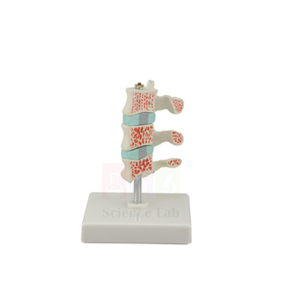
Human Advanced Osteoporosis Model
Human Advanced Osteoporosis Model (Product Code: SLE/A/66) sets three life-size vertebrae in a row: one healthy, one with first-stage pathological changes and one with advanced-stage changes. Seeing the stages together helps learners understand how osteoporosis weakens the spine step by step, which is why the model is used in medical, nursing, pharmacy and physiotherapy teaching as well as in patient counselling. It is made by Jain Scientific Equipments Pvt Ltd (ScienceLab), Ambala, a biology lab equipment supplier in India for education and healthcare training.
Specifications
| Parameter | Detail |
|---|---|
| Scale | Life size |
| Number of vertebrae | 3 |
| Vertebra 1 | Healthy vertebra |
| Vertebra 2 | Pathological changes at first stage |
| Vertebra 3 | Pathological changes at advanced stage |
| Size | 12 x 12 x 15 cm |
Typical Use
Osteoporosis is easier to explain when learners can compare a normal vertebra directly with diseased ones. A lecturer can ask students to describe the healthy vertebra first, then note what has changed at the first stage and how much further the advanced vertebra has deteriorated. This progression links well to discussions of risk factors, bone density testing, fracture risk and prevention through diet and exercise.
In pharmacy and pharmacology courses the model gives context to lectures on drugs used to treat bone loss, while in physiotherapy it supports advice on safe movement for patients with a fragile spine. Clinics and community health programmes also use it at counselling desks, and it fits neatly into a human biology model set for senior school classes studying the skeleton. Its compact size means it can sit on a desk permanently; wipe it occasionally with a dry cloth.
Manufacturer
Jain Scientific Equipments Private Limited, trading as ScienceLab, produces this osteoporosis model in Ambala, Haryana, alongside skeletal, organ and pathology models for teaching. Visit the company profile to learn more about the business.
Related Human Models
Pair this model with the following for a fuller lesson on bone health:
- Human Bone Structures Model — explains the internal structure that osteoporosis thins out.
- Economy Human Flexible Vertebral Column, Color Coded — shows where the affected vertebrae sit in the whole spine.
- Human Hip Joint Model — the hip is another common site of osteoporotic fracture.
- Microslide Animal Tissue Set 2 Muscle, Bone, Connective — prepared slides for viewing bone tissue under the microscope.
See more in the Human Models category.
Frequently Asked Questions
How many vertebrae does the Human Advanced Osteoporosis Model show?
Three: one healthy vertebra, one at the first stage of pathological change and one at an advanced stage.
Are the vertebrae life size?
Yes. The model is life size, and the whole set measures 12 x 12 x 15 cm.
Is it suitable for patient education?
Yes. The side-by-side stages are easy for non-specialists to understand, so clinics and physiotherapy practices use it when explaining bone loss.
Can the vertebrae be separated from each other?
The product details do not say whether the vertebrae are removable. Please check with us through the contact page if this matters for your use.
What material is it made from?
The material is not specified in the product details; we can confirm it with your quotation.
Do you ship to hospitals and colleges outside India?
Yes. We supply India and international markets; include the destination country in your enquiry so we can quote shipping.
Request a Quote
For a quotation on SLE/A/66, contact us through the contact page or list it in the enquiry cart together with any other bone or joint models you need.
Last updated: 24 September 2026
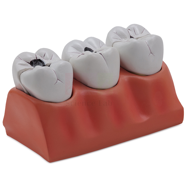
Dental Caries Model
Dental Caries Model (Product Code: SLE/A/67) enlarges three teeth to 4x life size, each seated in its socket, to show how dental caries progressively destroys tooth structure. The model can be taken apart into 7 parts, so students can look inside the teeth as well as at their surfaces. Dental, dental-hygiene and nursing tutors use it, and it is equally useful as a biology teaching aid for schools in India running oral-health lessons. It is made by Jain Scientific Equipments Pvt Ltd (ScienceLab), Ambala.
Specifications
| Parameter | Detail |
|---|---|
| Scale | 4x life size |
| Dissectible | Into 7 parts |
| Teeth | 3 teeth, seated in their sockets |
| Shows | Progressive deterioration of tooth structure by dental caries |
Typical Use
Tooth decay usually begins in the outer enamel and, if untreated, moves inward toward the dentine and the pulp. With three enlarged teeth showing successive degrees of damage, the model lets a tutor walk through that sequence and relate each step to symptoms such as sensitivity and pain, and to treatments ranging from a simple filling to more extensive work. Opening the dissectible parts shows the inside of the teeth, which is where the later stages become visible.
School health-education programmes use the model to show children why brushing, limiting sugary snacks and regular dental check-ups matter, while dental-hygiene students use it to practise explaining caries to patients. Store the 7 parts together after each lesson so none are lost, and clean them with a damp cloth rather than immersing them in water.
Manufacturer
The Dental Caries Model is produced by Jain Scientific Equipments Private Limited. The company operates under the ScienceLab name from Ambala, Haryana, making models for dental colleges, schools and health-training centres. Its background is described on the company profile.
Related Laboratory Equipment
For a complete oral-health or dentition lesson, these items work well with the caries model:
- Human Teeth (Denture) — shows the full set of teeth so students can name each type before studying decay.
- Human Teeth — an additional dentition model for group work.
- Human Skull — places the teeth within the upper and lower jaws.
- Human Tongue Model — completes a lesson on the mouth and the start of digestion.
- Mini Science Kit – Teeth and Eating — hands-on activities for younger students.
Find more anatomy teaching models in the Human Models category.
Frequently Asked Questions
How large is the Dental Caries Model compared with real teeth?
It is 4x life size, so details of the tooth structure are visible from a distance in a classroom.
How many parts does it come apart into?
7 parts, allowing the interior of the teeth to be examined.
How many teeth are shown?
Three teeth, each seated in its socket, illustrating the progressive stages of caries.
Is it suitable for primary and secondary schools?
Yes. Teachers use it for oral-hygiene and health-education lessons; for younger children, it is best handled by the teacher because of the small removable parts.
Is a stand, key or storage box included?
These are not listed in the product details. Please confirm the supplied accessories with us through the contact page.
Can a dental college order several units?
Yes. Send the required quantity and delivery location and we will quote for the full requirement.
Request a Quote
To get a quotation for SLE/A/67, reach our team on the contact page or put the model in your enquiry cart.
Last updated: 24 September 2026
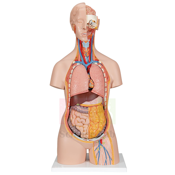
Human Muscular Torso with Head and Open Back Model
Human Muscular Torso with Head and Open Back Model (Product Code: SLE/A/68) is a life-size torso that combines the head, the musculature of the trunk and an open back in one large teaching piece. It separates into 32 parts and comes with interchangeable female and male genital organs (4 parts), so a single model can serve lessons on both sexes. Medical colleges, nursing schools, physiotherapy departments and senior science classes use it as a central demonstration model. It is made by Jain Scientific Equipments Pvt Ltd (ScienceLab), Ambala, and is among the most detailed of its human anatomical models.
Specifications
| Parameter | Detail |
|---|---|
| Model type | Muscular torso with head and open back |
| Scale | Life size |
| Number of parts | Separates into 32 parts |
| Genital organs | Interchangeable female and male genital organs (4 parts) |
| Size | 90 x 39 x 26 cm |
Typical Use
A torso of this size works best as the anchor of a systems-based course. The lecturer can start with the surface muscles of the trunk and head, then remove parts one at a time to move deeper, asking students to name each structure before it comes off and to replace the parts in the correct order afterwards. The open back gives access to the posterior side of the trunk, so back and spinal anatomy can be taught on the same model rather than on a separate piece.
Swapping the female and male genital organs lets the same torso support reproductive-system lessons for both sexes, which is economical for institutions buying school and college lab equipment on a fixed budget. With 32 parts, it is a good idea to keep a checklist beside the model and count the parts back in at the end of each practical; store it under a dust cover away from direct sunlight.
Manufacturer
Jain Scientific Equipments Private Limited makes this muscular torso under its ScienceLab brand in Ambala, Haryana, and supplies it to teaching hospitals, colleges and schools in India and international markets. You can find out more about the company on the profile page.
Related Human Models
These models complement a muscular torso in anatomy teaching:
- Human Muscular Figure Model — adds the limbs, so muscles of the whole body can be taught.
- Human Male and Female Torso Model, 20 Parts — an organ-focused torso for internal anatomy lessons.
- Human Head and Neck Model — detailed study of the head and neck region.
- Human Muscular Arm Model — covers the upper limb muscles that a torso leaves out.
Browse all options in the Human Models category.
Frequently Asked Questions
How many parts does the torso separate into?
32 parts, including interchangeable female and male genital organs made up of 4 parts.
What are the overall dimensions?
90 x 39 x 26 cm, at life size. Plan for a sturdy table or trolley when positioning it.
Can one model be used for both male and female anatomy?
Yes. The genital organs are interchangeable, so the torso can show either sex.
What is the model made from, and is a stand included?
Material and stand are not specified in the product details. Please ask through the contact page and we will confirm them with your quotation.
Is a key to the numbered structures supplied?
A key is not listed in the product details; let us know if you need one and we will advise on what is available.
How is a large model like this packed for shipping?
Packing depends on destination and quantity. Tell us the delivery address, in India or abroad, and we will explain packing and freight options.
Request a Quote
For price and lead time on SLE/A/68, write to us through the contact page or add it to your enquiry cart with the rest of your anatomy order.
Last updated: 24 September 2026
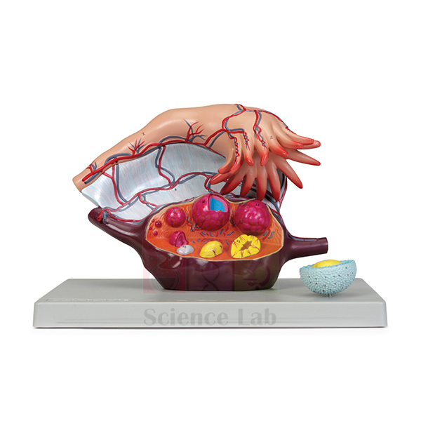
Human Ovary Model
Human Ovary Model (Product Code: SLE/A/70) enlarges the ovary to about 5x life size and sections it to reveal follicles at successive stages of maturation, from the primary ovarian follicle through to the corpus luteum and corpus albicans. A removable fallopian tubule and the vascular and suspensory structures complete the picture, and a numbered key is included. It serves reproductive biology, histology, gynaecology and nursing teaching, and suits high school biology lab equipment lists for classes covering the menstrual cycle. It is made by Jain Scientific Equipments Pvt Ltd (ScienceLab), Ambala.
Specifications
| Parameter | Detail |
|---|---|
| Scale | About 5x life size |
| Size | 33 x 23 x 21 cm |
| Base | Mounted on a durable polymer base |
| Removable part | Fallopian tubule |
| External features | Vascular and suspensory structures |
| Sectioned ovary | Follicles at various stages of maturation, from the primary ovarian follicle to the corpus luteum and corpus albicans |
| Follicle detail | Primary follicle cross-sectioned to reveal the oocyte and zona pellucida |
| Key | Numbered key included |
Typical Use
The model turns the ovarian cycle into a sequence students can follow with a finger: begin at the primary follicle, trace the growing follicles across the cut surface, reach the corpus luteum that forms after ovulation, and end at the corpus albicans that remains when it regresses. The enlarged cross-section of the primary follicle then shows the oocyte and its surrounding zona pellucida, linking the gross model to what students later see under the microscope.
Removing the fallopian tubule makes it easier to explain where the released egg travels and where fertilisation normally takes place. In college histology and embryology classes the model is often shown before slide work, and in nursing and midwifery courses it supports teaching on fertility and hormonal changes. Keep the model on its base and clean it with a soft, slightly damp cloth.
Manufacturer
ScienceLab is the brand of Jain Scientific Equipments Private Limited, an Ambala, Haryana company that makes reproductive, skeletal and organ models for teaching. For vendor registration, the company's GSTIN is 06AAECJ8618A1ZT and its Legal Entity Identifier (LEI) is 3358004HRD1JIVXKYX49. More information is on the company profile.
Related Human Models
For a reproductive-biology teaching set, consider adding:
- Ts Of Ovary (Mammalian) — a transverse section for comparing the model with real tissue under the microscope.
- Human Female Genital Organs Model — shows the ovary in relation to the uterus and other organs.
- Human Female Ovary Model — an alternative ovary model for a second teaching station.
- Human Embryo Development Set — continues the story from fertilisation into early development.
Further models are in the Human Models category.
Frequently Asked Questions
How much is the ovary enlarged?
About 5x life size, with an overall size of 33 x 23 x 21 cm.
Which follicle stages can students see?
Follicles from the primary ovarian follicle through maturing stages to the corpus luteum and corpus albicans, plus a cross-sectioned primary follicle showing the oocyte and zona pellucida.
Which part is removable?
The fallopian tubule can be removed from the ovary.
Is there a key to the numbered features?
Yes, a numbered key is included.
What is the base made of?
The model is mounted on a durable polymer base.
What documents can you provide for a college purchase order?
We can supply the quotation along with company details such as GSTIN and LEI. Tell us what your purchase department requires via the contact page.
Request a Quote
Ask for pricing on SLE/A/70 through the contact page, or add it to the enquiry cart to combine it with other reproductive-system models.
Last updated: 24 September 2026
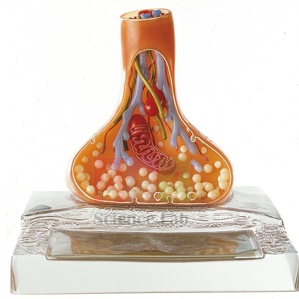
Human Nerve Ending and Synapse Model
Human Nerve Ending and Synapse Model (Product Code: SLE/A/72) magnifies the junction where one nerve cell passes its signal to the next. It shows the axon terminal with its synapse, the postsynaptic membrane with receptive sites, the neurotubules and neurotransmitter vesicles, and the way neurotransmitter is delivered across the synaptic gap. Physiology, neuroscience, psychology and pharmacology teachers use it to make an invisible process concrete. It is made by Jain Scientific Equipments Pvt Ltd (ScienceLab), Ambala.
Specifications
| Parameter | Detail |
|---|---|
| Construction | One-piece model, removable from its base for closer examination |
| Presynaptic side | Axon terminal with synapse, neurotubules and neurotransmitter vesicles |
| Postsynaptic side | Postsynaptic membrane structure with receptive sites |
| Process shown | Mechanism of delivery of neurotransmitter across the synapse |
| Size | 21 x 22 x 22 cm |
Typical Use
Synaptic transmission is usually taught as a chain of events: an impulse reaches the axon terminal, vesicles move toward the membrane and release neurotransmitter, the chemical crosses the gap, and it binds to receptive sites on the next cell. With every stage represented on one model, the teacher can point to each step in order and then ask students to explain it back in sequence. Lifting the model off its base lets small groups inspect the vesicles and receptor sites up close.
In pharmacology lectures the same structures help explain how many drugs act by blocking receptors or altering neurotransmitter release, which makes the model a practical piece of pharmacy college lab equipment. Psychology and senior biology classes also use it when studying reflexes, learning and the effects of substances on the nervous system. Clean it with a dry, soft cloth and return it to its base after handling.
Manufacturer
This synapse model is made by Jain Scientific Equipments Private Limited, which runs the ScienceLab brand from Ambala, Haryana and supplies neuroscience and anatomy teaching models to institutions in India and international markets. The company profile explains more about the business.
Related Human Models
For a nervous-system teaching set, these items are good companions:
- Neuron Model — shows the whole nerve cell before zooming in on its ending.
- Dissectible Human Neuron — a take-apart view of neuron structure for hands-on work.
- Microslide Nerve & Muscle Action — prepared slides that link the model to real tissue.
- Skeletal Muscle Fiber Model — the target tissue for nerve signals at the neuromuscular junction.
- Human Spinal Cord Section Model — places nerve pathways in their anatomical setting.
See the wider range in the Human Models category.
Frequently Asked Questions
What structures does the Human Nerve Ending and Synapse Model show?
The axon terminal with synapse, the postsynaptic membrane with receptive sites, neurotubules, neurotransmitter vesicles and the mechanism by which neurotransmitter crosses the synapse.
Can the model be taken off its stand?
Yes. It is a one-piece model that lifts off its base for closer examination.
How big is it?
21 x 22 x 22 cm, large enough to be seen clearly in a classroom demonstration.
Does it come apart into several pieces?
No. The model itself is a single piece; only the base is separate.
Is a key or description card included?
A key is not mentioned in the product details. Please confirm through the contact page if you need labelling material.
What information do you need to prepare a quotation?
The product code SLE/A/72, quantity, delivery address and any related nervous-system models you want quoted on the same order.
Request a Quote
Send an enquiry for SLE/A/72 through the contact page or add the model to your enquiry cart and submit it with the rest of your list.
Last updated: 24 September 2026
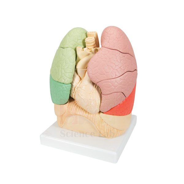
Segmented Lung
Segmented Lung (Product Code: SLE/A/73) is an about-life-size, take-apart lung model used to demonstrate the segmental bronchus of each lung segment in medical, nursing and school anatomy teaching. The lung detaches into 2 lobes that separate into individual segments, and the model depicts the bronchial tree close to the heart, trachea, esophagus and aorta. It is made by Jain Scientific Equipments Pvt Ltd (ScienceLab), an OEM manufacturer, at its own factory in Ambala, India.
Specifications
| Parameter | Detail |
|---|---|
| Scale | About life size |
| Size | 25 x 25 x 35 cm |
| Dissection | Lung detaches into 2 lobes that separate into individual segments |
| Teaching focus | Segmental bronchus of each lung segment in individual lobes |
| Airways depicted | Branching system of the bronchi and bronchioles |
| Neighbouring structures | Heart, trachea, esophagus and aorta; chest cavity organs and structures |
| Product code | SLE/A/73 |
| Manufacturer | Jain Scientific Equipments Pvt Ltd (ScienceLab) — OEM |
| Country of origin | India (made in Ambala, Haryana) |
What Is the Segmented Lung Model Used For?
The Segmented Lung is built for a hands-on disassembly exercise. Students first identify the airway branching beside the heart, trachea, esophagus and aorta, then detach the lobes, separate them into segments and match each segment with its segmental bronchus before rebuilding the lung. Working piece by piece shows why a single diseased segment can be treated on its own and why the airway pattern is described segment by segment in clinical teaching.
For group practicals, lay the separated pieces on a tray in the order they were removed so the lung reassembles quickly, and count them back before storage. Within a department's biology lab equipment, it serves as the interactive partner to a fixed display model used for demonstration.
OEM Manufacturer
Jain Scientific Equipments Private Limited, trading as ScienceLab, is the OEM (original equipment manufacturer) of the Segmented Lung and makes it at the company's own licensed factory at Saha, Ambala, Haryana, India. The company is a registered MSME enterprise (Udyam UDYAM-HR-01-0003714) and ships export orders under Importer-Exporter Code AAECJ8618A issued by the DGFT. Because the model comes from the factory rather than a trader, questions about a missing or damaged segment and repeat orders go straight to the manufacturer; ask us. Our company profile gives more detail.
Related Human Models
Items that support the same respiratory anatomy and lung-function practicals:
- Human Segmented Lungs Model — a life-size display alternative with colour-painted lobes and a numbered key.
- Human Thoracic Organs Model — widens the lesson to the other organs of the chest.
- Human Lungs with Heart and Larynx Model — adds the larynx and trachea above the lungs.
- Pathology of the Human Lung Model — lung abscess, pneumonia and tuberculosis for comparison with healthy segments.
- Lung Volume Bags — simple breathing-volume activities after the anatomy lesson.
Find more organ models in our Human Models category.
Frequently Asked Questions
How does the Segmented Lung come apart?
The Segmented Lung detaches into 2 lobes, and each lobe separates into its individual segments. The exact number of pieces is not stated in our product details, so ask us if you need the count for an inventory record.
What size is the Segmented Lung?
The Segmented Lung is about life size and measures 25 x 25 x 35 cm, so it fits on a practical bench where a small group can take it apart and rebuild it together.
Does the Segmented Lung show only the lung?
No. Besides the lung segments, the Segmented Lung depicts the branching bronchi and bronchioles close to the heart, trachea, esophagus and aorta, illustrating the chest cavity organs and structures around the airways.
How is the Segmented Lung different from the Human Segmented Lungs Model?
The Segmented Lung is designed to be taken apart into lobes and segments for hands-on work, whereas the Human Segmented Lungs Model is a fixed life-size display on a stand with colour-painted lobes and a numbered key.
Who manufactures the Segmented Lung?
Jain Scientific Equipments Pvt Ltd (ScienceLab) is the OEM manufacturer of the Segmented Lung, producing it at its own licensed factory at Saha, Ambala, Haryana, India. Institutions, dealers and importers buy it factory-direct.
Is a stand or key included with the Segmented Lung?
A stand or key is not mentioned in the product details for the Segmented Lung. Please contact us to confirm exactly what is packed with the model before you place a class or departmental order.
Can you supply the Segmented Lung for a whole class or for export?
Yes. The Segmented Lung is quoted for single units and class sets and shipped to buyers in India and international markets. Include product code SLE/A/73, the quantity and the destination in your enquiry.
Request a Quote
Ready to order the Segmented Lung or compare it with other lung models? Send your requirements through our contact page or collect items in the enquiry cart for a single quotation direct from the manufacturer.
Last updated: 24 September 2026
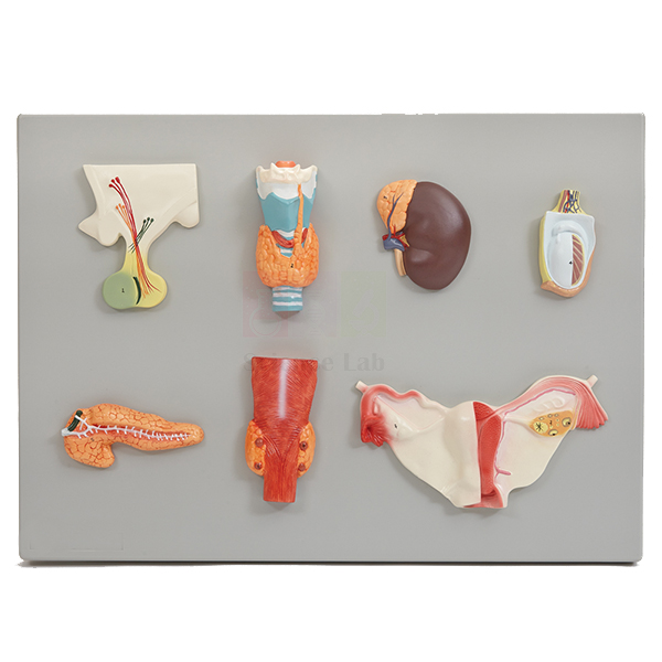
Human Endocrine Organs Model
Human Endocrine Organs Model (Product Code: SLE/A/74) brings the main hormone-producing glands together on a single plaque at about life size. Individual representations of the pituitary, adrenal, thyroid and parathyroid glands appear alongside a testis and a dissected ovary with its fallopian tubes and associated structures, and a key is supplied. Because it is plaque-mounted, it can hang on a wall or stand on a bench, giving biology, physiology and nursing classrooms a permanent endocrine reference. It is made by Jain Scientific Equipments Pvt Ltd (ScienceLab), Ambala.
Specifications
| Parameter | Detail |
|---|---|
| Format | Plaque mount |
| Scale | About life size |
| Glands shown | Pituitary, adrenal, thyroid and parathyroid glands |
| Gonads shown | A testis, and a dissected ovary with fallopian tubes and associated structures |
| Key | Included |
| Size | 53 x 38 cm |
Typical Use
Endocrinology is often taught gland by gland, which makes it hard for students to keep the whole system in view. With the glands displayed side by side, a teacher can introduce each one, name the hormones it releases and explain the feedback links between them, for example how the pituitary controls the thyroid, adrenal glands and gonads. Because the organs are shown at about life size, students also get a realistic sense of how small some of these glands are.
Mounted on a classroom wall, the plaque works like a three-dimensional alternative to a biology chart set, so it can stay on display for revision throughout the term. It is useful in nursing and pharmacy courses covering diabetes, thyroid disorders and hormone therapy, and in senior school biology units on control and coordination. Dust the surface with a soft brush or cloth; avoid hanging it in direct sunlight.
Manufacturer
Jain Scientific Equipments Private Limited produces the endocrine plaque under its ScienceLab trading name in Ambala, Haryana, as one of many models for schools, nursing colleges and medical institutes. Company details are available on the company profile page.
Related Human Models
To teach individual glands in more depth, many buyers add:
- Pathology of the Human Thyroid Model — shows common thyroid disorders once the healthy gland has been introduced.
- Kidney with Adrenal Gland Model — places the adrenal gland on top of the kidney where it belongs.
- Human Ovary Model — an enlarged, sectioned ovary for studying the ovarian cycle.
- Endocrine System Model Activity Set — hands-on activities that reinforce the same topic.
- Human Male Genital Organs Model — context for the testis shown on the plaque.
More models are available in the Human Models category.
Frequently Asked Questions
Which glands appear on the Human Endocrine Organs Model?
The pituitary, adrenal, thyroid and parathyroid glands, plus a testis and a dissected ovary with fallopian tubes and associated structures.
How big is the plaque?
53 x 38 cm, with the organs shown at about life size.
Can it be wall-mounted?
It is a plaque mount, designed for display. Please confirm the fixing arrangement you need for your wall or bench through the contact page.
Is a key included?
Yes, the model is supplied with a key.
Is this suitable for school students?
Yes. It works well for senior secondary biology units on hormones, as well as for nursing, pharmacy and physiology courses.
What warranty is offered?
Warranty terms are stated in our quotation. Contact us for the terms that apply to your order.
Request a Quote
For a quotation on SLE/A/74, get in touch via the contact page or add the plaque to your enquiry cart.
Last updated: 24 September 2026
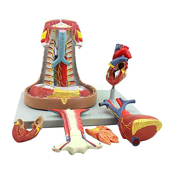
Human Mediastinum Model
Human Mediastinum Model (Product Code: SLE/A/75) is a life-size model of the central compartment of the chest, the space between the lungs that holds the heart, great vessels, trachea and esophagus. It is dissectible into 5 parts: the sternum and thymus lift away to reveal the pericardial sac and the major pulmonary and systemic vessels, and a separate 2-part heart shows the chambers and valves inside. Anatomy, cardiology, respiratory-therapy and nursing courses use it for thoracic anatomy teaching. It is made by Jain Scientific Equipments Pvt Ltd (ScienceLab), Ambala.
Specifications
| Parameter | Detail |
|---|---|
| Scale | Life size |
| Dissectible | Into 5 parts |
| Removable structures | Sternum and thymus, revealing the pericardial sac and major pulmonary and systemic vessels |
| Heart | Separate 2-part heart with internal detailing of chambers and valves |
| Other structures | Esophagus, trachea and diaphragm |
| Base | Mounted on a durable polymer base |
| Key | Included |
| Size | 40 x 26 x 30 cm |
Typical Use
The mediastinum is one of the hardest regions for students to picture because so many structures overlap. This model lets them uncover it in layers, much as in a dissection: first the sternum and thymus at the front, then the pericardial sac wrapping the heart, then the great vessels leaving it, with the trachea and esophagus running behind and the diaphragm forming the floor. Opening the separate heart shows the chambers and valves, so the model also doubles as a heart model for anatomy lessons on blood flow.
Clinical tutors use it to explain chest procedures, the position of the heart behind the sternum and why problems in one structure can affect its neighbours. Surgical and emergency-care trainees find the life-size scale helpful when relating surface landmarks to deeper organs. After each session, check that all 5 parts are back in place on the polymer base and wipe the surfaces with a soft cloth.
Manufacturer
This mediastinum model is manufactured by Jain Scientific Equipments Private Limited, the company behind the ScienceLab brand in Ambala, Haryana, for medical colleges and training institutes in India and international markets. Learn more on the company profile.
Related Human Models
For a full thoracic anatomy session, buyers often combine this model with:
- Human Heart Model — a stand-alone heart for more detailed cardiac lessons.
- Human Larynx, Trachea, and Bronchi Model — follows the airway above and below the mediastinum.
- Human Heart on Diaphragm Model — shows the relationship between the heart and the diaphragm from another angle.
- Human Respiratory System Model, 2-parts — adds the lungs on either side of the mediastinum.
- Economy Human Spine (vertebrae) and Thorax — the bony cage that encloses the chest organs.
Browse the Human Models category for more.
Frequently Asked Questions
How many parts does the Human Mediastinum Model come apart into?
5 parts, including the removable sternum and thymus and the separate 2-part heart.
What can be seen once the sternum and thymus are removed?
The pericardial sac and the major pulmonary and systemic vessels.
Does the heart open to show its interior?
Yes. The separate heart is in 2 parts, with internal detailing of the chambers and valves.
What are the dimensions?
40 x 26 x 30 cm at life size, on a durable polymer base.
Is a key supplied?
Yes, the model comes with a key.
Can you deliver to medical colleges abroad?
Yes. We supply institutions in India and international markets; include your country and quantity in the enquiry and we will quote accordingly.
Request a Quote
Request a quotation for SLE/A/75 on the contact page, or add it to your enquiry cart together with other thoracic models.
Last updated: 24 September 2026
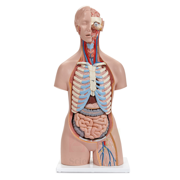
3B Human Sexless Torso Model
3B Human Sexless Torso Model (Product Code: SLE/A/77) is a life-size torso on a base that can be dissected into 13 parts to expose the organs of the chest and abdomen. The lungs and heart each come out in 2 parts, and the liver, stomach, frontal portion of the kidney, one half of the bladder and the intestines are also removable. Because it has no sex-specific organs, it keeps the focus on the respiratory, circulatory, digestive and urinary systems, which suits schools, nursing colleges and first-year medical anatomy. It is supplied by Jain Scientific Equipments Pvt Ltd (ScienceLab), Ambala, a laboratory equipment supplier in India.
Specifications
| Parameter | Detail |
|---|---|
| Scale | Life size |
| Dissectible | Into 13 parts |
| Removable organs | Lungs (2 parts), heart (2 parts), liver, stomach, frontal portion of the kidney, one half of the bladder and the intestines |
| Mounting | On base |
| Size | 38 x 25 x 87 cm |
Typical Use
A sexless torso is the standard choice when the lesson is about the organ systems common to everyone. Students can take out the lungs and heart to see how they fill the chest, lift out the liver and stomach to follow the upper digestive tract, and remove the intestines to reach the kidney and bladder behind them. Putting the 13 parts back in the right order is itself a useful test of how well the class has understood where each organ lies.
Schools use this kind of human body model for units on digestion, breathing and circulation, while nursing and paramedical tutors use it to relate organ positions to physical examination. Opening the 2-part heart and the frontal portion of the kidney lets teachers introduce internal structure without needing separate organ models. Lift the torso by its base, not by the organs, and store it covered to keep dust off the removable parts.
Supplier
This torso is supplied by Jain Scientific Equipments Private Limited, which trades as ScienceLab from Ambala, Haryana, and sources and ships anatomy models. For vendor registration, the supplying company holds GSTIN 06AAECJ8618A1ZT and handles export consignments under Importer-Exporter Code AAECJ8618A from the DGFT. Read more on the company profile.
Related Human Models
Alternatives and companions for torso-based teaching include:
- Human Sexless Torso Model — another sexless torso option for comparison when planning a purchase.
- Human Micro Sexless Torso Model — a small-format torso for individual desks or younger classes.
- Human Digestive System Model — follows the digestive tract in more detail than a torso allows.
- Human Kidney Model — an enlarged look at the organ partly shown in the torso.
See all torsos and organ models in the Human Models category.
Frequently Asked Questions
How many parts does the 3B Human Sexless Torso Model have?
It is dissectible into 13 parts, including 2-part lungs and a 2-part heart.
Which organs can be removed?
The lungs, heart, liver, stomach, the frontal portion of the kidney, one half of the bladder and the intestines.
How tall is it?
The model measures 38 x 25 x 87 cm on its base, at life size.
Is it manufactured by ScienceLab?
This torso is listed as a supplied item. For details of its make and origin, please ask us through the contact page.
Why choose a sexless torso?
It concentrates on the organ systems shared by all learners and avoids reproductive organs, which many schools prefer for general biology classes.
What registration details can you share for an institutional order?
Our IEC and GSTIN details can be provided with the quotation, along with any other vendor information your purchase team requests.
Request a Quote
To check availability and pricing for SLE/A/77, contact us via the contact page or add it to the enquiry cart.
Last updated: 24 September 2026
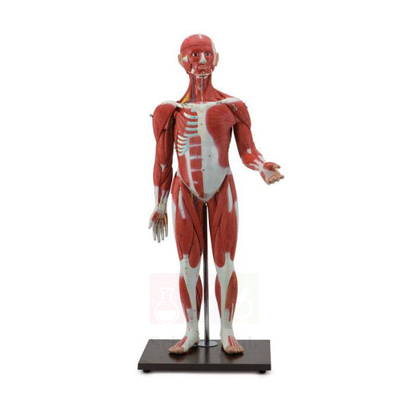
Human Sexless Muscular Figure
Human Sexless Muscular Figure (Product Code: SLE/A/78) is a 30-part, half life-size figure that displays the whole body's muscles together with the internal organs, standing on a rod-mounted, durable base. Superficial and deeper muscles, tendons, ligaments, vessels and organs are all represented, and many pieces come off, from the chest plate and arms to the brain, heart and abdominal organs. It gives anatomy, physiotherapy, sports-science and physical-education classes a full-body reference in a size that fits on a desk. It is made by Jain Scientific Equipments Pvt Ltd (ScienceLab), Ambala, as part of its biology lab equipment in India for schools and colleges.
Specifications
| Parameter | Detail |
|---|---|
| Scale | 1/2x life size |
| Number of parts | 30 |
| Mounting | Rod-mounted on a durable base |
| Size | 33 x 30 x 85 cm |
| Structures shown | Superficial and deeper muscles, tendons, ligaments, vessels and organs |
| Removable structures | Chest plate; arms; heart (2 parts); top of skull; brain (2 parts); front part of both lungs; liver; stomach; large and small intestines; duodenum, pancreas and spleen; many individual muscles |
Typical Use
Because it shows muscles and organs on one figure, this model lets a lesson move naturally from the outside of the body to the inside. Students can first identify the superficial muscles of the limbs and trunk, then remove individual muscles to see the deeper layer and the tendons and ligaments beneath. Taking off the chest plate, the front of the lungs and the abdominal organs then shows how the muscular wall protects the heart, liver, stomach, intestines, pancreas and spleen, while lifting the top of the skull reveals the 2-part brain.
Physical-education and sports-science teachers use the figure to link muscle groups with movements and common strains, and physiotherapy students use it to practise naming origins and actions. At half life size it is easy to carry between rooms and to share among small groups. Keep a list of the 30 parts with the model so everything is returned after each class, and store it where the rod mount will not be knocked.
Manufacturer
The Human Sexless Muscular Figure is one of the full-body models produced by Jain Scientific Equipments Private Limited, a manufacturer in Ambala, Haryana that sells under the ScienceLab name. Its history and activities are summarised on the company profile.
Related Human Models
Muscle and whole-body anatomy lessons often use these items alongside the figure:
- Miniature Human Muscular Figure — a smaller option for individual student desks.
- Human Muscular Torso with Head and Open Back Model — a life-size torso for close study of the trunk muscles.
- Human Muscular and Ligamentous Skeleton — shows how muscles and ligaments attach to the bones.
- Skeletal Muscle Fiber Model — explains muscle structure at the cellular level.
- Human Skeleton, Rod-Supported — the skeleton beneath the muscles, for combined lessons.
Find more in the Human Models category.
Frequently Asked Questions
What scale is the Human Sexless Muscular Figure?
It is 1/2x life size and measures 33 x 30 x 85 cm on its base.
How many parts does it have?
30 parts in total.
Which structures can be removed?
The chest plate, arms, 2-part heart, top of the skull, 2-part brain, front part of both lungs, liver, stomach, large and small intestines, duodenum, pancreas and spleen, and many individual muscles.
How is it mounted?
The figure is rod-mounted on a durable base, so it stands upright for display and demonstration.
Is a numbered key included?
A key is not mentioned in the product details. Please ask through the contact page if you need one.
Can we order it together with other anatomy models in one consignment?
Yes. List all the models you need with quantities and the delivery address, and we will send one combined quotation.
Request a Quote
For a price on SLE/A/78, message us through the contact page or add the figure to your enquiry cart with the rest of your anatomy list.
Last updated: 24 September 2026
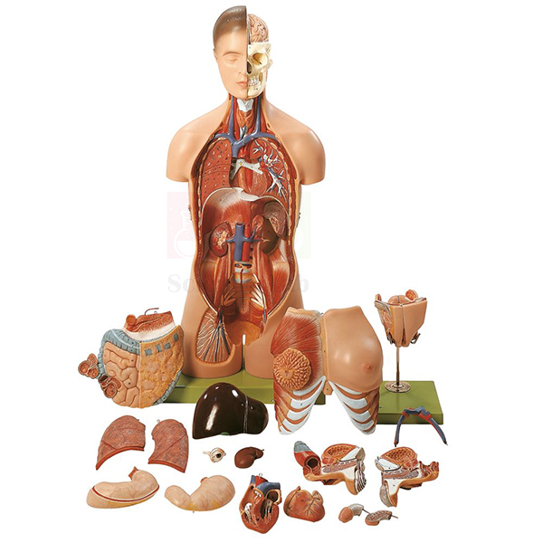
Human Male and Female Torso Model, 20 Parts
Human Male and Female Torso Model, 20 Parts (Product Code: SLE/A/79) is a life-size anatomical torso that includes the head and lets a teacher switch between male and female reproductive anatomy on the same figure. Its 20 separable parts allow students to take the model apart, study each region and rebuild it, which makes it a practical centrepiece for human anatomical models in school biology rooms, nursing schools and medical colleges. The model is made by Jain Scientific Equipments Pvt Ltd (ScienceLab), Ambala, India.
Specifications
| Parameter | Detail |
|---|---|
| Scale | Life size |
| Size | 40 x 26 x 92 cm |
| Number of parts | 20 |
| Region shown | Torso and head, depicted in detail |
| Interchangeable genitalia | Female (2-part) and male (4-part) |
| Identification key | Identifies more than 180 significant structures |
| Presentation | Museum-quality anatomical model |
Typical Use
Teachers use this torso to show where the major organs of the head, chest and abdomen sit and how they relate to one another, then remove parts one at a time so the class can see what lies beneath. Because the genital inserts are interchangeable, a single model covers both male and female reproductive anatomy — useful for senior secondary biology, nursing foundation courses and first-year anatomy practicals where space for two separate torsos is limited.
The key naming more than 180 structures turns the model into a self-study station: students can be given a list of numbers to identify and check against the key. When dismantling, lay the parts out in order on a padded tray so they go back in the right sequence. As part of school biology lab equipment in India, a full-size torso like this is usually shared across sections, so store it covered to keep dust off the detail.
Manufacturer
This torso is produced by Jain Scientific Equipments Private Limited, a company in Ambala, Haryana that trades under the brand name ScienceLab and makes anatomy teaching models alongside a wider range of laboratory equipment. You can learn more about the business on our company profile page.
Related Human Models
Models that pair well with this torso in anatomy and reproduction lessons:
- Deluxe Human Male and Female Torso Model — an alternative dual-sex torso to compare before ordering
- Human Sexless Torso Model — a torso without genitalia for general organ lessons in junior classes
- Human Male and Female Urogenital Systems Model — a closer look at the urinary and reproductive organs introduced on the torso
- Human Brain Model, 15 Parts — extends the head region into detailed brain anatomy
- Human Respiratory System Model, 2-parts — follows the airway and lungs in more depth
See every torso, organ and system model in our human anatomy models range.
Frequently Asked Questions
Can one model show both male and female anatomy?
Yes. It comes with interchangeable female (2-part) and male (4-part) genitalia, so you swap the inserts rather than buying two torsos.
How many structures does the key identify?
The key identifies more than 180 significant structures on the model, which is enough for detailed labelling exercises at school and college level.
How much space does it need?
The model measures 40 x 26 x 92 cm and is life size, so plan for a sturdy bench or a cupboard shelf of at least that height.
Is a stand or base included?
The product listing does not state the mounting arrangement. Please confirm stand or base details with us through the contact page before ordering.
How should the model be cleaned?
Wipe it with a soft cloth dampened with mild soapy water and dry it straight away. Avoid solvents and abrasive pads, which can dull the painted detail.
What warranty applies?
Warranty terms are confirmed with each quotation. Ask our team for the current terms when you request pricing.
Can you supply several units to schools or distributors?
Yes. Bulk orders for institutions and dealers in India and international markets are handled on request; share the quantity and delivery location with us.
Request a Quote
For pricing, lead time or an institutional quotation, write to us on the contact page or add this torso to your enquiry cart with any other models you need.
Last updated: 24 September 2026
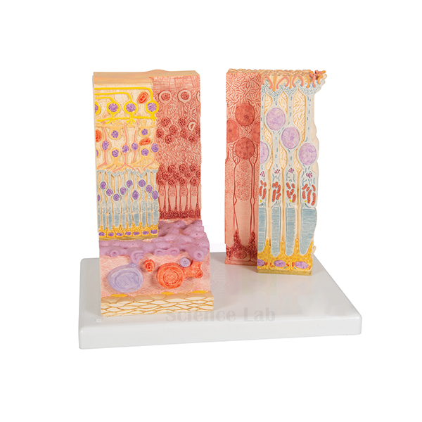
Eye Microanatomy Model
Eye Microanatomy Model (Product Code: SLE/A/80) brings the microscopic layers of the human retina up to a size a whole class can examine, presenting two highly magnified views side by side on one convenient base. It helps students connect what they read about rods, cones and neural layers with a three-dimensional form, and suits higher secondary biology, optometry, nursing and physiology teaching. The model is made by Jain Scientific Equipments Pvt Ltd (ScienceLab), Ambala, India.
Specifications
| Parameter | Detail |
|---|---|
| Subject | Microanatomy of the human retina |
| Views | Two highly magnified views on 1 base |
| Left view | Complete anatomical structure of the retina |
| Right view | Sectional enlargement of the retina |
| Size | 25 x 23 x 18.5 cm |
Typical Use
The retina is hard to picture from a textbook diagram, and this model fills that gap. A lecturer can start with the left-hand view to explain the overall organisation of the retina, then move to the sectional enlargement on the right to trace how light passes through the layers before reaching the photoreceptors. It works well at the start of a vision physiology unit, before students look at stained sections under the microscope, and as a revision aid when discussing conditions that affect the back of the eye.
Because both views share a single base, the model is easy to carry between classrooms and to place on a demonstration desk. Institutions building a set of eye model anatomy teaching aids in India often combine it with a whole-eyeball model, so learners can move from gross structure to the cellular level in one lesson.
Manufacturer
ScienceLab is the trading name of Jain Scientific Equipments Private Limited, which designs and supplies biology models and laboratory apparatus from Ambala in Haryana, India. Details of the company, its facilities and product lines are on our company profile.
Related Human Models
Items that support the same vision and eye-structure lessons:
- Human Eye Model — the whole eyeball, to set the retina in context
- Human Eye in Orbit Model — shows the eye, muscles and optic nerve within the bony orbit
- Cataract Eye Model with 4 Forms of Cataract — links normal eye anatomy to a common clinical condition
- Microslide Basic Human Histology — prepared human tissue slides for the microscope part of a histology practical
More eye, ear and organ models are listed in our anatomy models category.
Frequently Asked Questions
What part of the eye does this model show?
Only the retina. The left view shows its complete anatomical structure and the right view is a sectional enlargement, both highly magnified.
Is this a whole-eyeball model?
No. It is a microanatomy model of the retina. For the full eyeball, pair it with a separate eye model such as the Human Eye Model listed above.
What are the dimensions?
25 x 23 x 18.5 cm, with both views mounted on one base, so it fits easily on a teacher’s desk or display shelf.
What is the exact magnification?
The listing describes the views as highly magnified but does not state a factor. If you need the figure for a tender, ask us through the contact page.
Does it come with a labelled key?
A key is not mentioned in the product details. Please check with our team whether identification material can be provided with your order.
How do I keep the model in good condition?
Dust it with a soft brush, wipe with a slightly damp cloth when needed, and keep it out of direct sunlight to protect the painted layers.
What should an institutional quotation request include?
Mention the product code SLE/A/80, the quantity, the delivery address and any paperwork your purchase department needs, and we will prepare the quote.
Request a Quote
Send your requirement through our contact page, or place the model in the enquiry cart together with the other eye models you are considering.
Last updated: 24 September 2026
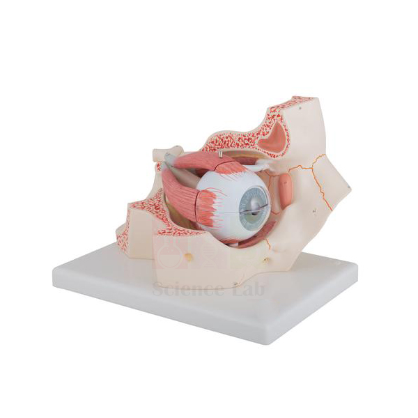
Human Eye in Orbit Model
Human Eye in Orbit Model (Product Code: SLE/A/81) is a large, 3x life size eye that sits in a section of the bony orbit, with the optic nerve shown in its natural position against the orbital floor and medial wall. The eyeball dissects into 7 pieces, so students can open it layer by layer from the sclera inwards to the lens and vitreous humor. It is a dependable biology teaching aid for schools, optometry courses and nursing colleges, made by Jain Scientific Equipments Pvt Ltd (ScienceLab), Ambala.
Specifications
| Parameter | Detail |
|---|---|
| Scale | 3x life size |
| Dissectible into | 7 pieces |
| Sclera | 2-part, with cornea and eye muscle attachments |
| Choroid | 2-part, with iris and retina |
| Other removable parts | Lens; vitreous humor |
| Orbit | Bony orbit (floor and medial wall) with the optic nerve in its natural position |
Typical Use
This model lets a teacher explain the eye both as an organ and as a structure housed in the skull. Lifting away the two halves of the sclera reveals the choroid, iris and retina; removing those exposes the lens and the vitreous humor, which is the natural order for a lesson on the three coats of the eyeball and the path of light to the retina. The muscle attachments on the sclera help when discussing how the eye is moved, and the orbit shows how the optic nerve leaves the eye towards the brain.
In practical exams it works well as a spotting station: students name each removed part and state its function. For school biology lab equipment in India where one eye model must serve many batches, keep the small parts in a labelled box beside the model so the lens and vitreous body are not misplaced between classes.
Manufacturer
The Human Eye in Orbit Model comes from Jain Scientific Equipments Private Limited, better known by its trading name ScienceLab, whose works are located in Ambala, Haryana. An overview of what the company makes and how it operates is available on the company profile page.
Related Human Models
Useful companions for a complete unit on the eye and vision:
- Eye Microanatomy Model — magnified retina views to follow up the retina shown inside this eye
- Human Eye Model with Eyelids — adds the eyelids and external features of the eye
- Human Eye Model — a separate eyeball model for group work at student benches
- Cataract Eye Model with 4 Forms of Cataract — relates the lens to cataract formation
- Human Brain Model in Head — continues the optic nerve story into the brain
Our anatomy models range lists further sense organ and system models.
Frequently Asked Questions
Which parts can be removed?
The 2-part sclera with cornea and muscle attachments, the 2-part choroid with iris and retina, the lens and the vitreous humor — 7 pieces in all.
How large is the model?
The eye is 3x life size, which keeps the lens and inner layers easy to see from the back of a classroom. Ask us for packed dimensions if you need them for storage planning.
Is the optic nerve shown?
Yes. It is shown in its natural position within the bony orbit, formed here by the orbital floor and medial wall.
Is it suitable for junior classes?
Yes, with supervision. The small removable parts need careful handling, so younger students are best shown the dissection by the teacher.
How should it be cleaned?
Wipe the outer surfaces with a soft, barely damp cloth and let each part dry fully before reassembly. Do not soak the pieces.
What warranty is offered?
Warranty terms are given with each quotation; please ask via the contact page for the current conditions.
Can you export it?
Yes. We supply institutions and distributors in India and international markets; send your destination and quantity for a shipping-inclusive quote.
Request a Quote
To order or get a price, message us on the contact page or add the model to the enquiry cart and submit your list in one go.
Last updated: 24 September 2026
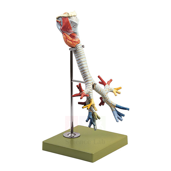
Human Larynx, Trachea, and Bronchi Model
Human Larynx, Trachea, and Bronchi Model (Product Code: SLE/A/82) is a slightly enlarged model of the upper and middle airway, mounted on a stand with base, that follows the air passage from the larynx down the trachea into the bronchial tree and its individual segment bronchi. The thyroid gland and associated muscles are included, and the model dissects into 2 parts. It is used in respiratory system teaching at senior school, nursing, paramedical and medical level, and is made by Jain Scientific Equipments Pvt Ltd (ScienceLab), Ambala.
Specifications
| Parameter | Detail |
|---|---|
| Scale | Slightly enlarged |
| Mounting | On stand with base |
| Dissectible into | 2 parts |
| Size | 20 x 18 x 39 cm H |
| Structures shown | Thyroid gland and muscles; trachea with bronchial tree; individual segment bronchi |
Typical Use
The model is built for lessons on the conducting zone of the respiratory system. Starting at the larynx, a teacher can point out the relationship of the thyroid gland to the airway, then follow the trachea to its division and on into the branching bronchial tree. Showing the individual segment bronchi helps students understand why the lungs are described in segments, a point that matters later in physiology, radiology and clinical subjects.
Standing upright on its base at 39 cm tall, it can be placed on a demonstration table and turned so every group sees the branching pattern. Colleges that are setting up biology lab equipment in India for nursing or paramedical courses often place it next to a full lung model so the airway and the lung tissue can be discussed together.
Manufacturer
Jain Scientific Equipments Private Limited, incorporated under CIN U29309HR2020PTC087597, makes this airway model and trades as ScienceLab. The company works from Ambala, Haryana, where production is carried out at its licensed factory at 449, HSIIDC Industrial Area, Saha (Factory Licence AMB-ONLINE-CHD-J-414). Read more on our company profile.
Related Human Models
Other models used in the same respiratory system teaching sequence:
- Larynx Human Model — a dedicated larynx model for detailed study of the voice box
- Pathology of the Human Trachea and Bronchi Model — contrasts the healthy airway with diseased tissue
- Human Respiratory System Model, 2-parts — places the airway within the complete respiratory system
- Segmented Lung — shows the lung segments supplied by the segment bronchi
- Anatomical Human Nasal Cavity Model — covers the upper airway above the larynx
Browse more organ and system models in the anatomy models section.
Frequently Asked Questions
Does the model include the lungs?
No. It covers the larynx, thyroid gland and muscles, trachea, bronchial tree and segment bronchi. For lung tissue, add a lung model such as the Segmented Lung listed above.
How does it come apart?
The model is dissectible into 2 parts, which lets you open it to show internal features of the airway during a demonstration.
What is the overall height?
It measures 20 x 18 x 39 cm H on its stand and base, so it fits on a standard classroom table or display shelf.
Is it life size?
It is slightly enlarged, which makes the smaller segment bronchi easier to see without distorting their overall arrangement.
How do I clean it?
Use a soft brush for dust in the branching bronchi and a lightly damp cloth on flat surfaces. Keep solvents and alcohol wipes away from the paintwork.
What details do you need for a college purchase order?
Send the product code SLE/A/82, quantity, billing and delivery addresses, and any vendor registration details your office requires, through the contact page.
Request a Quote
Ask for pricing and availability on our contact page, or save the model to your enquiry cart while you select the rest of your respiratory models.
Last updated: 24 September 2026
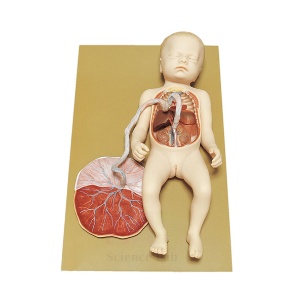
Human Fetal Circulation Model
Human Fetal Circulation Model (Product Code: SLE/A/83) is a life-size model of a female fetus before birth, complete with umbilical cord and placenta, in which the thoracic and abdominal cavities are opened to reveal how blood travels in the unborn child. The venous and arterial ducts that bypass the fetal liver and lungs are shown clearly, and the model separates into 2 parts on its base. It supports embryology, obstetrics, midwifery and senior biology teaching, and is made by Jain Scientific Equipments Pvt Ltd (ScienceLab), Ambala.
Specifications
| Parameter | Detail |
|---|---|
| Scale | Life size |
| Subject | Female fetus before birth, with umbilical cord and placenta |
| Exposed regions | Thoracic and abdominal cavities |
| Circulatory features | Venous and arterial ducts shown |
| Separates into | 2 parts |
| Mounting | On base |
| Size | 48 x 30 x 14 cm |
Typical Use
Fetal circulation is one of the harder topics for students because the pathways change at birth. With this model an instructor can begin at the placenta, follow oxygenated blood along the umbilical cord into the fetus, and show how the venous duct carries much of it past the liver while the arterial duct diverts blood away from the lungs. Comparing these shunts with adult circulation makes it easier to explain what closes after the first breaths and why some congenital heart conditions occur.
The life-size scale also gives learners a realistic sense of the fetus, cord and placenta together, which is useful in midwifery and nursing skills rooms. Among biology teaching aids for reproduction and development, it fits naturally after an embryo development series, closing the unit with the circulatory picture just before birth.
Manufacturer
This model is made by Jain Scientific Equipments Private Limited, an anatomical models manufacturer in Ambala, Haryana that sells under the ScienceLab name to schools, colleges and hospitals. Find out more about the company on our company profile.
Related Human Models
Models that complete a unit on pregnancy, development and circulation:
- Human Placenta Model — enlarges the maternal–fetal exchange surface shown on this model
- Human Embryo Development Set — covers the earlier stages that lead up to the fetus
- Human Heart Model — for explaining the heart chambers the fetal shunts bypass
- Human Circulatory System Model — adult pulmonary and systemic circulation for side-by-side comparison
- Human Birth Demonstration Model — continues the story through delivery
For more developmental and organ models, open our anatomy models category.
Frequently Asked Questions
Which fetal shunts does the model show?
It shows the venous and arterial ducts. For any other feature you need for a syllabus, ask us to confirm before ordering.
Are the placenta and umbilical cord included?
Yes. The fetus is modelled with its umbilical cord and placenta, so the full route from placenta to fetal heart can be traced.
How big is it?
It is life size and measures 48 x 30 x 14 cm on its base, compact enough for a skills-lab shelf.
Does it come apart?
Yes, it separates into 2 parts, allowing the internal circulation to be shown and then closed up for storage.
How should it be stored?
Keep it on its base in a closed cabinet, away from heat and direct sun, and wipe with a soft dry cloth after handling.
Is a warranty provided?
Warranty conditions are stated in our quotation. Contact us via the contact page for details.
Can hospitals and training institutes order more than one?
Yes. Multiple units for institutions in India and international markets can be quoted together; tell us the quantity and delivery city or country.
Request a Quote
Request a price through our contact page or add this model to your enquiry cart along with the other embryology items on your list.
Last updated: 24 September 2026
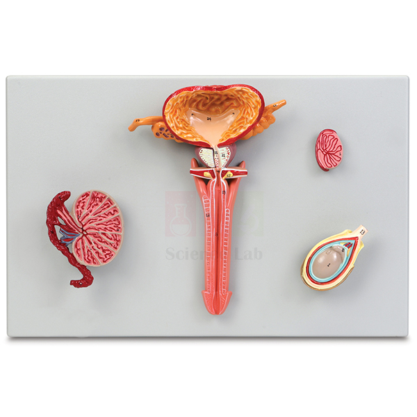
Human Male Reproductive System Model Set
Human Male Reproductive System Model Set (Product Code: SLE/A/84) is a set of 4 plaque-mounted models that together illustrate the interior anatomy of the male reproductive tract. The urinary bladder, prostate gland and penis are sectioned longitudinally, and the testis is shown from 3 perspectives, from its position in the scrotum down to an enlarged dissection of its internal tubules. A numbered key identifies 25 structures. The set serves reproductive biology, urology, nursing and medical teaching, and is made by Jain Scientific Equipments Pvt Ltd (ScienceLab), Ambala.
Specifications
| Parameter | Detail |
|---|---|
| Set contents | 4 plaque-mounted models |
| Mounting | Durable polymer bases |
| Size (each) | 50 x 26 x 8 cm |
| Sectioned organs | Urinary bladder, prostate gland and penis, sectioned longitudinally |
| Testis views | In situ within a life-sized scrotum; life-sized longitudinal dissection; 2x life-sized dissection |
| Enlarged testis detail | Epididymis, seminiferous tubules, tunica albuginea and other significant structures |
| Key | Numbered key with 25 identified structures |
Typical Use
Because the set is split across four plaques, a teacher can move from the big picture to fine detail in a logical sequence: first the sectioned bladder, prostate and penis to show the course of the urethra and the glands that open into it, then the testis in the scrotum, then its life-sized dissection, and finally the 2x enlargement where the seminiferous tubules and epididymis explain where sperm are produced and stored. The plaques can be laid flat on a bench or propped up for display.
Flat, wall-style mounting also makes the set practical for rotating practical stations, since each plaque can go to a different group with the numbered key. For colleges assembling a human biology model set, this pairs well with a pelvis model so students see the reproductive organs in relation to the bony pelvis.
Manufacturer
The set is manufactured by Jain Scientific Equipments Private Limited, the Ambala, Haryana company behind the ScienceLab brand. It is a registered MSME enterprise with manufacturing as its major activity (Udyam UDYAM-HR-01-0003714), and its overseas shipments go out under Importer-Exporter Code AAECJ8618A issued by the DGFT. More background is on our company profile.
Related Human Models
Models often ordered alongside this set for reproduction and urology lessons:
- Human Male Genital Organs Model — a single three-dimensional model to complement the flat plaques
- Human Male and Female Urogenital Systems Model — compares male and female urogenital anatomy
- Male Pelvis Section Model — shows the same organs within a sectioned pelvis
- Human Adult Male Pelvis Structural Model — the bony framework that surrounds the pelvic organs
Explore more organ system models in our anatomy models range.
Frequently Asked Questions
What is in the set?
Four plaque-mounted models on polymer bases, covering the sectioned bladder, prostate and penis and three views of the testis, plus a numbered key.
How many structures are identified?
The numbered key identifies 25 structures across the set.
What size is each plaque?
Each model measures 50 x 26 x 8 cm, so the four together need a fair amount of display or cupboard space.
Are the testis views to scale?
Two are life-sized (the in-situ view in the scrotum and the longitudinal dissection) and one is 2x life-sized to show the internal tubules.
Can the plaques be wall-mounted?
Mounting hardware is not listed. Please ask us via the contact page if you plan to hang them.
How do I clean the models?
Dust with a soft dry cloth and, if needed, wipe with a cloth dampened in mild soap solution. Avoid solvents on the painted surfaces.
Do you supply sets for export?
Yes. We quote for universities, colleges and distributors in India and international markets; include destination and quantity in your enquiry.
Request a Quote
Get a quotation by writing to us on the contact page, or add the set to the enquiry cart to combine it with other models in one request.
Last updated: 24 September 2026
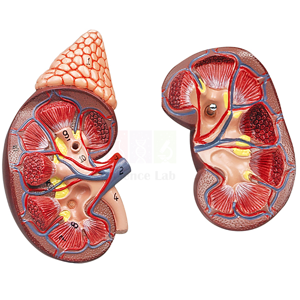
Human Kidney with Adrenal Gland Model
Human Kidney with Adrenal Gland Model (Product Code: SLE/A/85) is a life-size kidney model, sectioned along the frontal plane, used to teach internal renal anatomy in school, nursing and medical classrooms. Dissectible into 2 parts and measuring 23.5 x 11.4 cm, it opens to show the cortex, medulla, pyramids with papillae, partially opened renal pelvis, ureter and blood vessels. It is made by Jain Scientific Equipments Pvt Ltd (ScienceLab), an OEM manufacturer, at its own factory in Ambala, India.
Specifications
| Parameter | Detail |
|---|---|
| Scale | Life size |
| Size | 23.5 x 11.4 cm |
| Construction | Dissectible into 2 parts |
| Section | Sectioned along the frontal plane |
| Internal structures shown | Cortex, medulla, pyramids with papillae, partially opened renal pelvis, ureter and blood vessels |
| Associated organ | Adrenal gland |
| Product code | SLE/A/85 |
| Manufacturer | Jain Scientific Equipments Pvt Ltd (ScienceLab) — OEM |
| Country of origin | India (made in Ambala, Haryana) |
What Is the Kidney with Adrenal Gland Model Used For?
With the two halves opened on the bench, students trace the route urine takes: from the papillae at the tips of the pyramids into the partially opened renal pelvis and on into the ureter. The contrast between the outer cortex and the inner medulla helps explain where filtration and concentration take place, and the blood vessels show how the kidney receives the blood it filters. The adrenal gland gives teachers a natural bridge from the excretory system to the endocrine system in the same lesson.
The model is used in senior school lessons on excretion, in nursing anatomy practicals and in patient-education discussions about kidney structure, and it is a practical addition for institutions building up biology teaching aids in India. Open and close the halves gently on a flat surface, and store the model in a cabinet between sessions to protect the painted detail.
OEM Manufacturer
This kidney model is an own-make product of Jain Scientific Equipments Private Limited, which trades as ScienceLab and, as the OEM (original equipment manufacturer), produces it at its own licensed factory at Saha, Ambala, Haryana, India. Schools, colleges, dealers and importers receive it factory-direct, so quotations, repeat orders and after-sales questions are handled by the manufacturer itself; just ask us. The company profile explains more about the business.
Related Human Models
Other renal models that teachers use to move from organ level to the nephron:
- Human Kidney, Nephron and Renal Corpuscle Model — an enlarged plaque series continuing the lesson down to the filtering unit.
- Human Kidney, Nephron, and Glomerulus Model Set — kidney, nephrons and glomerulus at three magnifications on one base.
- Human Kidney Model — a life-size 2-piece right kidney with a removable anterior portion and laminated key.
- Kidney Stone Model — adds a common clinical condition to a renal anatomy lesson.
- Human Endocrine Organs Model — shows the adrenal gland alongside the pituitary, thyroid and parathyroid glands.
This model is listed with other organ models in our Human Models category.
Frequently Asked Questions
What can be seen when the Human Kidney with Adrenal Gland Model is opened?
Opening the Human Kidney with Adrenal Gland Model along its frontal section reveals the cortex, medulla, pyramids with papillae, a partially opened renal pelvis, the ureter and blood vessels, so the internal organisation of the kidney can be followed step by step.
Is the Human Kidney with Adrenal Gland Model life size?
Yes. The Human Kidney with Adrenal Gland Model shows the kidney at life size and measures 23.5 x 11.4 cm, which lets students relate what they see directly to the real organ.
Does the adrenal gland come off separately?
The Human Kidney with Adrenal Gland Model divides into 2 parts along its frontal section; the adrenal gland is not listed as a separate removable piece. Contact us if a detachable adrenal matters for your lesson plan.
Is a numbered key supplied with the model?
A key is not mentioned in the product details for the Human Kidney with Adrenal Gland Model. Please confirm with us at enquiry stage if you need one, and we will tell you what is packed with the model.
Who manufactures the Human Kidney with Adrenal Gland Model?
Jain Scientific Equipments Pvt Ltd, trading as ScienceLab, is the OEM manufacturer of the Human Kidney with Adrenal Gland Model. It is made at the company's own licensed factory at Saha, Ambala, Haryana, India, and supplied factory-direct.
What details should a purchase enquiry include?
For the Human Kidney with Adrenal Gland Model, quote code SLE/A/85, the quantity and delivery location, and say whether the order is for a school, college or hospital so we can prepare the right paperwork for India or international markets.
Request a Quote
To receive a quotation for the Human Kidney with Adrenal Gland Model direct from the manufacturer, write to us from the contact page or add the model to the enquiry cart along with any other renal models.
Last updated: 24 September 2026
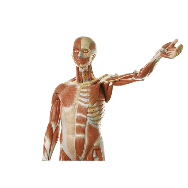
Human Male Muscular Model, Removable Arms
Human Male Muscular Model, Removable Arms (Product Code: SLE/A/86) is a detailed male muscle figure at about 1/2x life size that displays both the surface muscles and the musculature lying beneath them. The model is assembled from parts, and its arms can be taken off so the shoulder girdle and the muscles of the trunk can be seen without obstruction. It is aimed at physical education, sports science, physiotherapy, nursing and anatomy classes, and is made by Jain Scientific Equipments Pvt Ltd (ScienceLab), Ambala.
Specifications
| Parameter | Detail |
|---|---|
| Scale | About 1/2x life size |
| Size | 86 x 49 x 38 cm |
| Construction | Made of parts |
| Removable parts | Arms |
| Anatomy shown | Surface muscles and underlying musculature, in fine detail |
Typical Use
Muscle identification is usually taught region by region, and this figure supports that approach. With the arms attached, students can trace the superficial muscles of the chest, abdomen, back and limbs; once the arms are removed, deeper muscles around the shoulder and the lateral chest wall come into view. Coaches and physiotherapy tutors use it to explain which muscle groups act in a movement, while biology teachers use it to show origin, insertion and the layering of muscles over one another.
Measuring 86 x 49 x 38 cm, it is large enough for a whole class to follow a demonstration without taking over the teaching bench. For sports science departments and school labs looking for human anatomical models, it gives a full-body view of the muscular system that a single limb or torso model cannot.
Manufacturer
This muscle figure is made by Jain Scientific Equipments Private Limited, which operates as ScienceLab from Ambala in Haryana. For tender and vendor registration forms, the company’s identifiers are GSTIN 06AAECJ8618A1ZT and LEI 3358004HRD1JIVXKYX49. Our company profile gives a fuller picture of the business.
Related Human Models
Choices that extend or complement a muscular system lesson:
- Human Muscular Arm Model — closer study of the arm muscles once they are detached from the figure
- Human Muscular Figure Model — another full-body muscle figure to compare before buying
- Miniature Human Muscular Figure — a smaller option for student benches and group work
- Human Sexless Muscular Figure — a gender-neutral alternative for junior classes
- Human Muscular and Ligamentous Skeleton — shows how muscles and ligaments attach to the skeleton
See the complete anatomy models collection for more figures and torsos.
Frequently Asked Questions
What can be removed from the model?
The arms are removable. The model is built from parts; ask us via the contact page if you need a full list of detachable pieces.
What scale and size is it?
It is about 1/2x life size and measures 86 x 49 x 38 cm.
Does it show deep muscles as well as surface muscles?
Yes. It illustrates the surface muscles and the underlying musculature, which is why it suits physiotherapy and sports science teaching.
Is a numbered key supplied?
A key is not mentioned in the product details. Please confirm with us whether identification material can be included.
How should the arms be stored when removed?
Keep them in a padded box or on a soft tray next to the figure, and refit them before moving the model so the joints are not strained.
What warranty applies?
Warranty terms are included in each quotation; our team can confirm them when you enquire.
Do you handle bulk and overseas orders?
Yes. We supply sports colleges, universities and dealers in India and international markets. Share your quantity and destination for a combined quote.
Request a Quote
Contact us through the contact page for a price, or add the figure to the enquiry cart with any other anatomy models you require.
Last updated: 24 September 2026
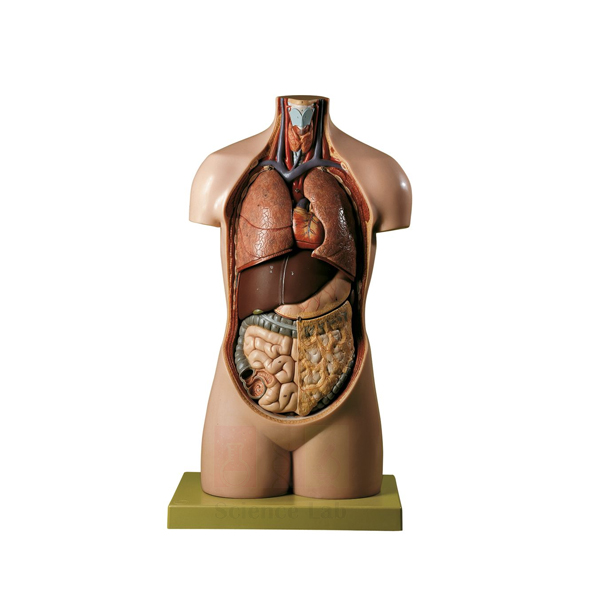
Male Youth Torso without Head
Male Youth Torso without Head (Product Code: SLE/A/87) is a life-size torso of a young male that realistically depicts the anatomy of the neck, thorax and abdomen. Leaving out the head keeps the focus on the internal organs of the trunk, and the model separates into 8 parts so the lungs, heart, liver, stomach and intestines can be lifted out and examined. It is a straightforward choice for secondary school biology, health science and nursing foundation classes, made by Jain Scientific Equipments Pvt Ltd (ScienceLab), Ambala.
Specifications
| Parameter | Detail |
|---|---|
| Scale | Life size |
| Mounting | Mounted on a base |
| Overall size | 39 x 26 x 73 cm H |
| Torso height | 69 cm |
| Regions shown | Neck, thorax and abdomen |
| Separates into | 8 parts |
| Removable organs include | Lungs, 2-piece heart, liver, stomach, small and large intestines |
Typical Use
This torso is often the first model students meet when they study the organs of the body. A teacher can remove the lungs to reveal the 2-piece heart, open the heart to show its interior, and then take out the liver, stomach and intestines to explain the order in which food passes through the digestive tract. Putting the organs back in place is a useful exercise in itself, as it tests whether learners remember where each organ sits.
With 8 parts rather than dozens, it is quick to dismantle and reassemble within a single period, which suits busy school timetables. For schools looking at a basic biology lab setup in India, a headless torso like this covers the thoracic and abdominal organs at a manageable size, leaving head and brain topics to a separate model.
Manufacturer
Made in Ambala, Haryana by Jain Scientific Equipments Private Limited, a company that sells its teaching models and laboratory apparatus under the ScienceLab name. Visit our company profile to learn how the business is organised and what else it produces.
Related Human Models
Alternatives and follow-on models for organ and body-system lessons:
- Human Male and Female Torso Model, 20 Parts — a larger-part torso with head and interchangeable genitalia
- Human Sexless Torso Model — another trunk model to compare for junior classes
- Human Micro Sexless Torso Model — a small torso for student benches and group activities
- Human Digestive System Model — follows the stomach and intestines in greater depth
- Human Heart Model — a larger heart for studying chambers and valves
More torso, organ and system models are available in our anatomy models category.
Frequently Asked Questions
Which organs can be removed?
The model separates into 8 parts, including the lungs, the 2-piece heart, liver, stomach, and small and large intestines.
Why is there no head?
The design concentrates on the neck, thorax and abdomen. If you also need head and brain anatomy, consider a torso with head or a separate brain model.
What are the dimensions?
Overall 39 x 26 x 73 cm H on its base, with the torso itself 69 cm tall.
Is it male or sexless?
It is a male youth torso. For details of the external features shown, please check with us before ordering.
How should it be cleaned?
Remove the organs, wipe each with a soft cloth lightly moistened with mild soapy water, dry thoroughly and refit them.
What should a school include in a quotation request?
The product code SLE/A/87, quantity, delivery address and whether you need a formal proforma invoice. Send these through the contact page.
Request a Quote
For prices and delivery times, reach us via the contact page, or add this torso to your enquiry cart and send one combined request.
Last updated: 24 September 2026
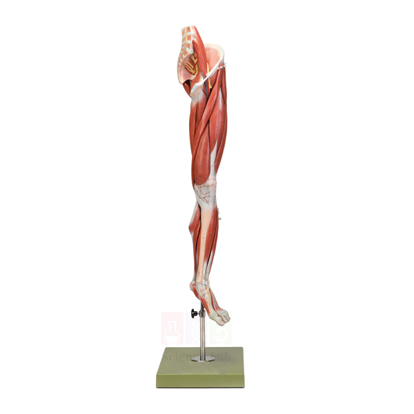
Human Leg and Portion of Pelvis Model
Human Leg and Portion of Pelvis Model (Product Code: SLE/A/88) is a life-size lower limb, attached to part of the pelvis, that stands on a stand with base and can be rotated to view every side. Its surface muscles are removable, exposing the blood vessels and nerves that run through the limb, and the whole model is dissectible into 10 parts. It is intended for anatomy, physiotherapy, orthopaedic, sports medicine and nursing teaching, and is made by Jain Scientific Equipments Pvt Ltd (ScienceLab), Ambala.
Specifications
| Parameter | Detail |
|---|---|
| Scale | Life size |
| Region | Leg with a portion of the pelvis |
| Movement | Rotatable |
| Dissectible into | 10 parts |
| Removable layer | Surface muscles, for study of blood vessels and nerves |
| Mounting | On stand with base |
| Size | 39 x 26 x 108 cm H |
Typical Use
Lower-limb anatomy combines muscles, vessels and nerves in a small space, and a rotatable life-size model is the clearest way to show how they fit together. Teachers typically start with the intact limb to name the thigh, calf and gluteal muscles, then lift the surface muscles away to follow the main arteries, veins and nerves from the pelvis down towards the foot. Rotating the model on its stand lets the class see the front, back and sides without anyone handling the parts.
Physiotherapy and sports medicine tutors use it to point out where nerves are vulnerable to injury and how muscle groups act on the hip and knee. As a piece of biology lab equipment in India for medical and allied health colleges, it complements a skeleton by adding the soft tissue that bones alone cannot show.
Manufacturer
ScienceLab is the brand name used by Jain Scientific Equipments Private Limited, an Ambala, Haryana firm that makes this leg model along with many other anatomy and laboratory products for educational buyers. Our company profile has more about the company.
Related Human Models
Models that fit the same lower-limb and pelvis teaching block:
- 3B Human Left Leg Model — an alternative leg model to compare before ordering
- Human Pelvis with Lower Lumbar and Femur Heads — the bony pelvis and hip joint in more detail
- Human Knee Joint Model, Life Size — focuses on the knee joint within the limb
- Human Foot Model Set — continues the study down to the foot
- Human Femur Bone Model — the thigh bone the muscles attach to
Visit our anatomy models range for more limb, joint and body-system models.
Frequently Asked Questions
How many parts does it come apart into?
It is dissectible into 10 parts, including the removable surface muscles.
Can the model be turned?
Yes. It is rotatable on its stand, so all sides of the limb can be shown to a class.
What does removing the muscles reveal?
The underlying blood vessels and nerves, which is the main teaching purpose of the removable layer.
How tall is it?
The model measures 39 x 26 x 108 cm H, including its stand and base, so check cupboard height before ordering.
Is it a left or right leg?
The side is not stated in the product details. Please confirm with us via the contact page if your syllabus needs a specific side.
How do I look after it?
Handle the removable muscles by their edges, keep the model out of direct sunlight and wipe it with a soft, slightly damp cloth.
Can you supply it to institutions abroad?
Yes. We quote for colleges and distributors in India and international markets; mention the destination and quantity.
Request a Quote
Ask for a quotation on our contact page, or add the model to the enquiry cart to build a full list for your anatomy lab.
Last updated: 24 September 2026
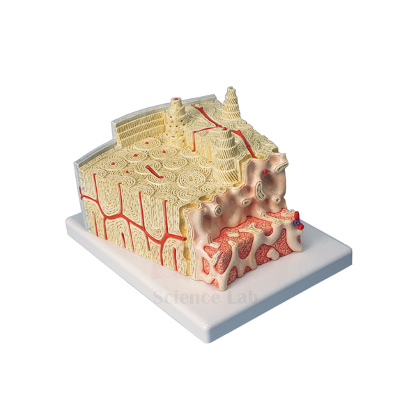
3B Bone Microanatomy Model
3B Bone Microanatomy Model (Product Code: SLE/A/89) enlarges a section of lamellar bone to about 80x life size so that the microscopic architecture of compact bone can be studied without a microscope. The block is cut in both cross and longitudinal section through all layers, down to the bone marrow, with Haversian canals, endosteum, cortical substance and osteocytes clearly visible. It is used in histology, physiology, orthopaedics and senior biology teaching, and is supplied by Jain Scientific Equipments Pvt Ltd (ScienceLab), Ambala, India.
Specifications
| Parameter | Detail |
|---|---|
| Scale | About 80x life size |
| Mounting | Mounted on a base |
| Tissue shown | Section of lamellar bone |
| Sections | Cross and longitudinal, through all layers including bone marrow |
| Visible structures | Haversian canals, endosteum, cortical substance, osteocytes |
Typical Use
Students often find it difficult to turn a flat slide image of compact bone into a mental picture of cylinders of lamellae running along the bone. Because this model shows the same tissue in two planes at once, a teacher can point to a Haversian canal in cross section and then follow it lengthwise in the longitudinal face, explaining how osteocytes sit between the lamellae and how the endosteum lines the inner surface next to the marrow.
It works best alongside the microscope: show the model first, then let learners find the same features on prepared microscope slides of bone. In physiology lessons it also supports discussion of bone remodelling and blood supply, while orthopaedic and physiotherapy students use it to understand how fractures heal at tissue level.
Supplier
This model is supplied by Jain Scientific Equipments Private Limited, which trades as ScienceLab from Ambala, Haryana, and sources selected teaching models to sit alongside its own laboratory products. For more about the company, see our company profile.
Related Human Models
Items that support a complete lesson on bone tissue and structure:
- Microslide Animal Tissue Set 2 Muscle, Bone, Connective — real bone sections to compare with the enlarged model
- Binocular Biological Microscope — for viewing bone slides after the model demonstration
- Human Bone Structures Model — shows bone at the level of the whole bone rather than the tissue
- Human Femur Bone Model — a long bone to relate the microanatomy to gross anatomy
- Skeletal Muscle Fiber Model — another enlarged tissue model for the musculoskeletal unit
Browse our anatomy models category for more microanatomy and organ models.
Frequently Asked Questions
How much is the bone enlarged?
About 80x life size, enough to make individual osteocytes and canals visible from across a classroom.
Which structures can students identify?
Haversian canals, endosteum, cortical substance, osteocytes and the bone marrow, shown in both cross and longitudinal section.
Is this model made by ScienceLab?
No. It is a branded model that we supply; our team can advise on availability and delivery for your order.
What are the dimensions and weight?
These are not given in the product details. Please ask through the contact page if you need them for storage or shipping.
How should it be cleaned?
Use a soft brush to remove dust from the textured surfaces and a slightly damp cloth on the base. Avoid solvents.
Can it be included in a bulk order for a medical college?
Yes. It can be quoted together with our own models for institutions in India and international markets; send your full list and delivery location.
Request a Quote
For availability and pricing, contact us on the contact page or place the model in your enquiry cart with the slides and microscope you need.
Last updated: 24 September 2026
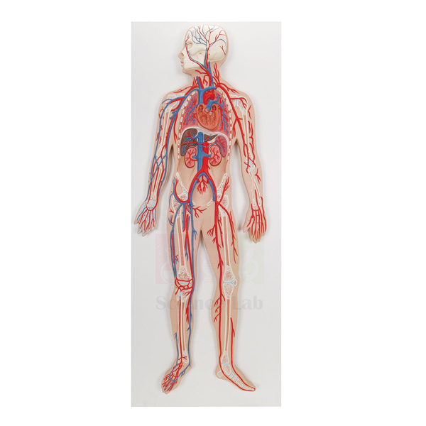
Human Circulatory System Model
Human Circulatory System Model (Product Code: SLE/A/90) is a generalized, about 1/2x life size representation of the human circulatory system, laid out on a durable polymer base with a key. It traces the circuitry of both the pulmonary and systemic pathways and includes a 2-part heart together with the lungs, liver, spleen and kidneys, so students can see how blood reaches and returns from the major organs. It is used in school, nursing and paramedical biology teaching, and is made by Jain Scientific Equipments Pvt Ltd (ScienceLab), Ambala.
Specifications
| Parameter | Detail |
|---|---|
| Scale | About 1/2x life size |
| Size | 32 x 90 x 12 cm |
| Mounting | Durable polymer base |
| Key | Included |
| Organs shown | 2-part heart, lungs, liver, spleen, kidneys |
| Pathways | Pulmonary and systemic circulatory pathways |
| Representation | Generalized schematic of the human circulatory system |
Typical Use
The model is designed to answer the question students always ask: where does the blood actually go? A teacher can start at the heart, open the 2-part heart to show its chambers, and then follow the pulmonary circuit to the lungs and back before tracing the systemic circuit out to the liver, spleen and kidneys. Because it is a generalized layout rather than a crowded full-body figure, the double circulation is easy to follow and to redraw in notebooks.
The key allows the model to be used for self-testing, with students naming the vessels and organs and then checking their answers against it. It is a useful heart model anatomy companion in India’s secondary and nursing curricula, where double circulation, the hepatic portal system and renal blood supply are standard topics.
Manufacturer
The model is built by Jain Scientific Equipments Private Limited in Ambala, Haryana, the company that owns the ScienceLab trading name. Purchase departments that need vendor details can register the company under GSTIN 06AAECJ8618A1ZT, and its Legal Entity Identifier is 3358004HRD1JIVXKYX49. Further background is on our company profile.
Related Human Models
Models and slides that round out a circulation unit:
- Human Heart Model — a larger heart for detailed study of chambers and valves
- Human Cardiac Beat And Blood Circulation — a demonstration aid for the heartbeat and blood flow
- Human Lymphatic System Model — the second transport system that works alongside the blood vessels
- Human Fetal Circulation Model — shows how circulation differs before birth
- Prepared Microscope Slide Set – Circulatory System — vessel and blood tissue sections for the microscope practical
Explore further organ system models in our anatomy models range.
Frequently Asked Questions
Which organs are included?
A 2-part heart, the lungs, liver, spleen and kidneys, connected by the pulmonary and systemic pathways.
Is it a realistic full-body model?
No. It is a generalized representation designed for clarity, which makes the circulatory routes easier to follow than on a whole-body figure.
What size is it?
About 1/2x life size, measuring 32 x 90 x 12 cm on its polymer base.
Does it come with a key?
Yes, a key is included for identifying the structures shown.
Can it be hung on a wall?
Wall-mounting fittings are not mentioned in the product details. Please check with us through the contact page.
How should the model be cleaned?
Dust regularly with a soft cloth and wipe gently with mild soap solution when needed; keep it away from heaters and strong sunlight.
Is a warranty included?
Warranty terms are set out in each quotation. Our team will confirm them when you enquire.
Request a Quote
Send your enquiry through the contact page or add the model to your enquiry cart; we quote for single schools, institutions and distributors in India and international markets.
Last updated: 24 September 2026
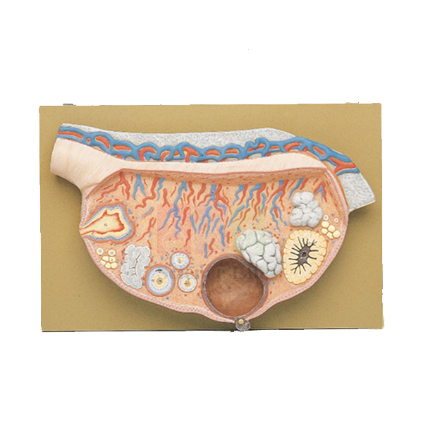
Human Female Ovary Model
Human Female Ovary Model (Product Code: SLE/A/91) is a 10x life size model of the ovary that presents the follicle at different phases of maturation together with the corpus rubrum, corpus luteum and corpus albicans. By showing the whole ovarian cycle on one enlarged organ, it helps students link the stages of follicle development to ovulation and hormone production. It is used in senior biology, physiology, gynaecology, nursing and midwifery teaching, and is made by Jain Scientific Equipments Pvt Ltd (ScienceLab), Ambala.
Specifications
| Parameter | Detail |
|---|---|
| Scale | 10x life size |
| Size | 28 x 40 x 8 cm |
| Weight | 1.8 kg |
| Follicles | Shown in different maturation phases |
| Other structures | Corpus rubrum, corpus luteum, corpus albicans |
Typical Use
The ovarian cycle is usually taught from diagrams that show one stage at a time. On this model a teacher can walk around the ovary in order: from early follicles through maturing stages, to the ruptured follicle as the corpus rubrum, on to the corpus luteum and finally the corpus albicans left after it regresses. Seeing all these structures at once makes it easier to explain the timing of ovulation and the link between the corpus luteum and progesterone.
Nursing and midwifery tutors use it when discussing fertility, contraception and early pregnancy, and it pairs naturally with ovary sections viewed under the microscope. For colleges adding to their human biology model set in India, the 10x enlargement at a manageable 1.8 kg means it can be passed around a small group without difficulty.
Manufacturer
Jain Scientific Equipments Private Limited produces this ovary model in Ambala, Haryana, and markets it under its trading name, ScienceLab, to schools, colleges and training institutes. The company profile page explains more about who we are.
Related Human Models
Items used alongside this model in reproduction and ovarian cycle lessons:
- Human Ovary Model — another ovary model to compare before ordering
- Human Female Genital Organs Model — places the ovary within the female reproductive organs
- Ts Of Ovary (Mammalian) — a transverse section of mammalian ovary to relate the model to real tissue
- Human Embryo Development Set — follows on from ovulation to fertilisation and development
- Human Adult Female Pelvis Structural Model — the bony pelvis that houses the ovaries
Find more reproductive and organ models in our anatomy models category.
Frequently Asked Questions
Which stages of the ovarian cycle are shown?
Follicles at different maturation phases, plus the corpus rubrum, corpus luteum and corpus albicans.
How large is the model?
It is 10x life size and measures 28 x 40 x 8 cm.
How heavy is it?
About 1.8 kg, so it is easy to move between classrooms.
Does it include a base or a key?
The product details do not specify a base or key. Please confirm what is included with us through the contact page.
Is it suitable for school students?
Yes. It works for senior secondary biology as well as nursing and medical courses, as the structures are presented clearly at a large scale.
How do I clean it?
Wipe with a soft cloth, slightly moistened if necessary, and avoid alcohol or solvent cleaners that could affect the colours.
Do you supply outside India?
Yes. We serve institutions and distributors in India and international markets; include your country and quantity when you enquire.
Request a Quote
Request pricing on our contact page or add this ovary model to the enquiry cart together with other reproductive system models.
Last updated: 24 September 2026
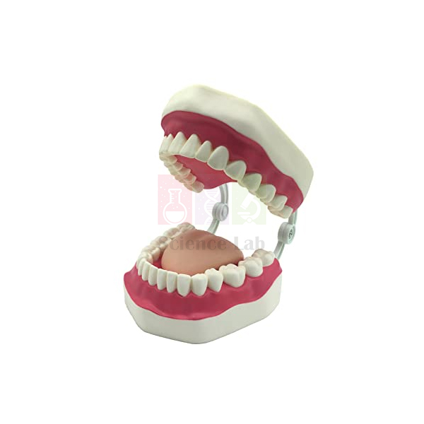
Human Oral Hygiene Model
Human Oral Hygiene Model (Product Code: SLE/A/92) is a 3x life size set of teeth mounted on a base and supplied with a giant toothbrush, built for one clear purpose: teaching the correct way to brush teeth. The enlarged scale lets a presenter show brush angle, stroke direction and hard-to-reach surfaces so that a whole room of children or adults can follow along. It is widely useful in primary schools, dental and nursing colleges, community health programmes and clinic waiting areas, and is made by Jain Scientific Equipments Pvt Ltd (ScienceLab), Ambala.
Specifications
| Parameter | Detail |
|---|---|
| Scale | 3x life size |
| Mounting | Mounted on base |
| Included accessory | Giant toothbrush |
| Purpose | Teaching the correct way to brush teeth |
Typical Use
Good brushing habits are easier to demonstrate than to describe. Using the giant toothbrush on the enlarged teeth, a teacher or dental hygienist can show how to hold the brush at an angle to the gumline, how to cover the outer, inner and chewing surfaces, and why the back teeth need special attention. Children can then take turns with the brush themselves, which turns a health talk into an active lesson they are more likely to remember.
The model is a natural fit for health education in schools across India, school dental camps and oral health awareness days. In dental hygiene and nursing training it is used to rehearse patient instruction, and in clinics it gives staff a quick visual aid when advising parents on their children’s brushing routine.
Manufacturer
This model is manufactured by Jain Scientific Equipments Private Limited, whose products carry the ScienceLab brand. The company is headquartered in Ambala, Haryana, and supplies teaching models and lab equipment to schools and training institutions. More information is on our company profile.
Related Human Models
Teaching aids that complement an oral health or dental anatomy session:
- Dental Caries Model — shows what tooth decay looks like and why brushing matters
- Human Teeth (Denture) — for naming the different teeth before the brushing demonstration
- Human Teeth — an additional teeth model for group work
- Human Tongue Model — extends the lesson to the tongue and the rest of the mouth
- Mini Science Kit – Teeth and Eating — a hands-on activity kit for younger pupils
Our anatomy models range includes further health education and organ models.
Frequently Asked Questions
Is a toothbrush included?
Yes. A giant toothbrush sized for the enlarged teeth comes with the model.
How large are the teeth?
The model is 3x life size. Overall dimensions are not listed, so ask us via the contact page if you need them for storage.
Who is this model for?
Anyone teaching brushing technique — primary teachers, school nurses, dental hygiene students and community health workers.
Can children handle it?
Yes, under supervision. It stands on a base, so pupils can practise brushing while the teacher watches their technique.
How should it be cleaned after a session?
Wipe the teeth and base with a damp cloth and mild soap, rinse the brush in plain water, and let both dry before storing.
Can we order several for a school health programme?
Yes. Multiple units can be quoted for programmes, schools and distributors in India and international markets; send the quantity and delivery details.
Request a Quote
To get a price, use our contact page, or add the model to your enquiry cart with any other health education aids you need.
Last updated: 24 September 2026
Human Spinal Cord Section Model
Human Spinal Cord Section Model (Product Code: SLE/A/93) is a cross section of the spinal cord at the level of the 5th cervical vertebra, enlarged to about 7x life size and set on a base. It shows the spinal nerves, the spinal ganglion and the blood vessels in their positions around the cord, giving students a clear three-dimensional view of a region they otherwise see only in diagrams and stained slides. It is used in neuroanatomy, physiology, physiotherapy and senior biology courses, and is made by Jain Scientific Equipments Pvt Ltd (ScienceLab), Ambala.
Specifications
| Parameter | Detail |
|---|---|
| Scale | About 7x life size |
| Level | 5th cervical vertebra |
| Type | Cross section |
| Structures shown | Spinal nerves, spinal ganglion, blood vessels |
| Mounting | On base |
| Size | 40 x 10 x 28 cm |
Typical Use
This section model is ideal for explaining how a spinal nerve is formed and where sensory and motor information enters and leaves the cord. An instructor can point to the spinal ganglion to show where sensory cell bodies lie, follow the spinal nerves outwards, and use the blood vessels to discuss how the cord is supplied. Because the section is taken at a cervical level, it also helps when teaching why injuries in the neck region can affect the arms as well as the body below.
Physiotherapy and nursing students use it to understand reflex arcs and the effects of spinal cord injury, while biology students use it to connect the reflex arc diagram in their textbook to real anatomy. For departments building up biology lab equipment for a neuroscience unit, it bridges the gap between a whole spine model and microscope slides of nerve tissue.
Manufacturer
Our company, Jain Scientific Equipments Private Limited, makes this spinal cord section in Ambala, Haryana and trades under the ScienceLab name. To see what else we make and how we work with institutions, visit the company profile.
Related Human Models
Models and slides used in the same nervous system teaching sequence:
- Human Vertebral And Partial Spinal Cord Model — shows the cord inside the vertebral column
- Human Cervical Spinal Column Model — the neck vertebrae that surround this level of the cord
- Human Nervous System Model — the wider network the spinal nerves belong to
- Enlarged Human Brain Stem Model — continues the pathway upwards into the brain stem
- Microslide Nerve & Muscle Action — prepared slides for the microscope part of the lesson
See more neuroanatomy and body-system models in our anatomy models section.
Frequently Asked Questions
Which level of the spinal cord is shown?
The cross section is taken at the 5th cervical vertebra.
What structures can be identified?
Spinal nerves, the spinal ganglion and blood vessels. If you need a list of any further labelled features, please ask us before ordering.
What is the enlargement and size?
About 7x life size, measuring 40 x 10 x 28 cm on its base.
Does it come with a key or labels?
A key is not mentioned in the product details. Contact us through the contact page to confirm what is supplied.
How should it be cared for?
Keep it on its base in a closed cabinet, dust with a soft brush and avoid wet cleaning around the fine nerve roots.
What warranty applies?
Warranty terms are provided with our quotation; ask the team for the current conditions.
What should we include in a tender or bulk enquiry?
The product code SLE/A/93, quantity, delivery location in India or abroad, and any documents your procurement process needs.
Request a Quote
Reach us on the contact page for pricing, or add the model to the enquiry cart alongside other nervous system models.
Last updated: 24 September 2026
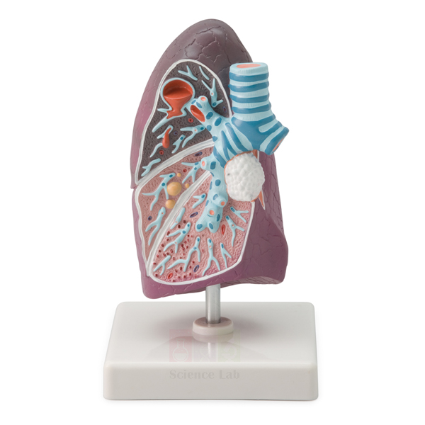
Pathology of the Human Lung Model
Pathology of the Human Lung Model (Product Code: SLE/A/94) is a 1-part, 2/3x life-size model used to teach how microbial infections damage the lung in medical, nursing and paramedical colleges. Lung abscess, pneumonia and tuberculosis are each shown separately with a realistic representation of their morphology on a compact 11 x 11 x 11 cm model with stand and base. It is made by Jain Scientific Equipments Pvt Ltd (ScienceLab), an OEM manufacturer, at its own factory in Ambala, India.
Specifications
| Parameter | Detail |
|---|---|
| Scale | 2/3x life size |
| Size | 11 x 11 x 11 cm |
| Construction | 1-part model |
| Mounting | On stand with base |
| Pathologies shown | Lung abscess, pneumonia and tuberculosis (caused by microbial infections) |
| Presentation | Each pathology shown separately with a realistic representation of its morphology |
| Product code | SLE/A/94 |
| Manufacturer | Jain Scientific Equipments Pvt Ltd (ScienceLab) — OEM |
| Country of origin | India (made in Ambala, Haryana) |
What Is the Lung Pathology Model Used For?
In a pathology or microbiology lecture, the model lets the tutor move from the organism to the damage it causes: a localised lung abscess, the consolidated tissue of pneumonia and the lesions associated with tuberculosis. Seeing all three on one piece helps students recognise how different infections look and why they are managed differently. It is equally useful in community-health sessions where tuberculosis awareness is part of the programme.
Its small footprint makes it easy to pass round a tutorial group or to place at a practical station beside a healthy lung model, which is the most effective way to teach disease against normal anatomy. Colleges adding pathology pieces to their biology lab equipment in India often start with a respiratory model like this one. Handle it by the base and clean it with a dry, soft cloth.
OEM Manufacturer
ScienceLab is the trading name of Jain Scientific Equipments Private Limited, the OEM (original equipment manufacturer) that makes this pathology model at its own licensed factory at Saha, Ambala, Haryana, India. For vendor files, the company's MSME registration is Udyam UDYAM-HR-01-0003714 and its DGFT Importer-Exporter Code is AAECJ8618A. Medical and nursing colleges, dealers and importers buy it factory-direct, and repeat orders are handled by the manufacturer itself; ask us. Learn more on the company profile page.
Related Human Models
Models that pair with this one for normal-versus-disease teaching:
- Human Segmented Lungs Model — healthy life-size lung anatomy for side-by-side comparison.
- Somso Human Lung Model — enlarged normal lobule structure to contrast with infected tissue.
- Pathology of the Human Trachea and Bronchi Model — continues respiratory pathology with bronchitis and bronchiectasis.
- Human Liver Pathology Model — another pathology model for a broader disease-teaching set.
You can find more anatomical and pathology models in our Human Models category.
Frequently Asked Questions
Which lung diseases does the Pathology of the Human Lung Model show?
The Pathology of the Human Lung Model shows lung abscess, pneumonia and tuberculosis, all pathologies caused by microbial infections. Each is represented separately with a realistic depiction of its morphology, so students can compare the three disease patterns directly.
Does the Pathology of the Human Lung Model come apart?
No. The Pathology of the Human Lung Model is a 1-part model, which keeps it robust for frequent handling when it is passed around a tutorial group or used at a practical station.
How big is the Pathology of the Human Lung Model?
The Pathology of the Human Lung Model is 2/3x life size and measures 11 x 11 x 11 cm, mounted on a stand with base, so it needs very little bench or cabinet space.
Is a key or explanatory card included?
A key or explanatory card is not listed for the Pathology of the Human Lung Model. Contact us to confirm what is supplied with the model before ordering, especially if you need labelled material for self-study.
Who manufactures the Pathology of the Human Lung Model?
Jain Scientific Equipments Pvt Ltd (ScienceLab) is the OEM manufacturer of the Pathology of the Human Lung Model, producing it at its own licensed factory at Saha, Ambala, Haryana, India, and supplying colleges and dealers directly.
Is the model suitable for TB awareness programmes?
Yes. Because tuberculosis appears alongside pneumonia and lung abscess, the Pathology of the Human Lung Model helps non-specialist audiences see how TB differs from other chest infections in community-health and patient-education sessions.
Do you supply training institutes outside India?
Yes. The Pathology of the Human Lung Model is quoted for institutions and distributors in India and international markets; include product code SLE/A/94, quantity and destination so packing and shipping are set out in the quotation.
Request a Quote
For a manufacturer-direct quotation on the Pathology of the Human Lung Model, use our contact page or add it to the enquiry cart with any healthy-anatomy models you want to compare it with.
Last updated: 24 September 2026
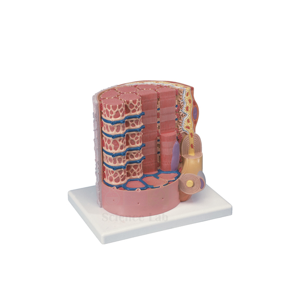
Skeletal Muscle Fiber Model
Skeletal Muscle Fiber Model (Product Code: SLE/A/95) represents the microstructure of a single skeletal muscle fiber in great detail, enlarged to 10,000x life size and mounted on a reinforced polymer base. It includes the neuromuscular junction, sarcoplasmic reticulum, T tubules and myofibrils, the structures students need to understand how a nerve signal becomes a muscle contraction. The model is used in physiology, histology, sports science and senior biology teaching, and is made by Jain Scientific Equipments Pvt Ltd (ScienceLab), Ambala.
Specifications
| Parameter | Detail |
|---|---|
| Scale | 10,000x life size |
| Size | 25 x 23 x 23 cm |
| Mounting | Reinforced polymer base |
| Subject | Microstructure of a skeletal muscle fiber |
| Structures included | Neuromuscular junction, sarcoplasmic reticulum, T tubules, myofibrils |
Typical Use
Excitation–contraction coupling is one of the most abstract topics in physiology, and this model makes it concrete. A lecturer can begin at the neuromuscular junction where the motor nerve meets the fiber, show how the T tubules carry the signal deep into the cell, point to the sarcoplasmic reticulum that releases calcium, and finish at the myofibrils where contraction actually takes place. Seeing these structures wrapped around one another explains why the signal reaches the whole fiber almost at once.
Sports science and physiotherapy tutors use it when discussing fatigue, cramps and training adaptations, and histology classes pair it with stained muscle sections so students can relate the striations they see under the microscope to the myofibrils on the model. It is a compact addition to cell biology equipment for any department teaching muscle physiology.
Manufacturer
Manufacturing of this microstructure model is carried out by Jain Scientific Equipments Private Limited of Ambala, Haryana, a company better known to schools and colleges as ScienceLab. Our company profile describes the business and its product lines.
Related Human Models
Models and slides that complete a unit on muscle tissue:
- Microstructure of Smooth Muscle Model — contrasts skeletal fibers with smooth muscle cells
- Skeletal Muscle, Longitudinal and Transverse Incisions — real muscle sections to compare with the enlarged fiber
- Microslide Animal Tissue Set 2 Muscle, Bone, Connective — prepared tissue slides for the microscope practical
- Human Male Muscular Model, Removable Arms — shows whole muscles made up of fibers like this one
- 3B Bone Microanatomy Model — the matching tissue-level model for bone in a musculoskeletal unit
More microanatomy and organ models are listed in our anatomy models range.
Frequently Asked Questions
How much is the muscle fiber enlarged?
The model is 10,000x life size, which brings sub-cellular structures up to a size that can be pointed out in class.
Which structures does it include?
The neuromuscular junction, sarcoplasmic reticulum, T tubules and myofibrils.
What are the dimensions?
25 x 23 x 23 cm on a reinforced polymer base, small enough for a desk or display shelf.
Is there an identification key?
The product details do not mention a key. Please ask us through the contact page about identification material.
How do I clean it?
Remove dust with a soft brush and wipe the base with a lightly damp cloth; avoid solvents on the painted detail.
Can it be supplied with other microanatomy models in one order?
Yes. We can quote a combined order for colleges and distributors in India and international markets; list all items in your enquiry.
Request a Quote
Ask for a price via the contact page, or add this model to your enquiry cart together with the muscle slides and related models you need.
Last updated: 24 September 2026
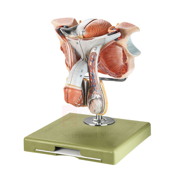
Human Male Genital Organs Model
Human Male Genital Organs Model (Product Code: SLE/A/96) is a life-size anatomical model of the male reproductive organs that separates into 5 parts, letting students see how the bladder, prostate, urethra, testes and spermatic cord connect. It is used for reproduction and urogenital anatomy lessons in senior-secondary biology classes, nursing schools and medical colleges, and is manufactured by Jain Scientific Equipments Pvt Ltd (ScienceLab), Ambala, as part of its range of human anatomical models made in India.
Specifications
| Parameter | Detail |
|---|---|
| Type | Anatomical model of the male genital organs |
| Scale | Life size |
| Dissection | Dissectible into 5 parts |
| Size | 18 × 20 × 21 cm (H) |
| Mounting | On stand with base |
| Structures shown in fine detail | Penis, prostate, bladder, testes, spermatic cord, urethra and scrotum |
Typical Use
Teachers use this model to trace the pathway shared by the urinary and reproductive systems in the male: with the parts separated, a class can follow the urethra from the bladder through the prostate and along the penis, then locate each testis within the scrotum and the spermatic cord running upward from it. It supports textbook chapters on human reproduction, nursing anatomy practicals and health-education talks where a three-dimensional object explains far more than a diagram.
Because it is life size, learners get a realistic sense of scale that flat charts cannot give. Many schools place it alongside a female counterpart as part of a human biology model set, so both systems can be compared in the same lesson. A practical tip: lay the 5 parts out in order on a tray while teaching and count them back onto the stand before the model goes into storage.
Manufacturer
This model comes from Jain Scientific Equipments Private Limited, a company based in Ambala, Haryana, that sells its laboratory and anatomy products under the ScienceLab name to schools, colleges and distributors in India and international markets. Read the company profile for background on the business and its product lines.
Related Human Models
Models often ordered with this one for reproduction and urogenital teaching:
- Human Male Reproductive System Model Set — a wider set for extended lessons on the male reproductive system
- Human Male and Female Urogenital Systems Model — places the bladder and urethra within the complete urinary tract of both sexes
- Human Female Genital Organs Model — the female counterpart for side-by-side comparison
- Male Pelvis Section Model — shows these organs in position inside a sectioned pelvis
- Male Skeletal Pelvis — the bony framework that surrounds and protects them
See the complete Human Models range for more anatomy teaching models.
Frequently Asked Questions
How many parts does the model come apart into?
Five. The model is dissectible into 5 parts so internal structures such as the prostate and urethra can be exposed; if you need the exact part-by-part breakdown before ordering, please contact us.
Is it life size, and how much space does it need?
Yes, it is life size and measures 18 × 20 × 21 cm in height on its stand with base, so it fits easily on a demonstration bench or display shelf.
Which structures can students identify on it?
The penis, prostate, bladder, testes, spermatic cord, urethra and scrotum are all modelled in fine detail.
Does it come with a key or labelling guide?
A key is not listed in our product details for this model. Contact us to confirm what documentation is packed with it.
How should it be cleaned between classes?
Wipe it with a soft, slightly damp cloth and let it dry before reassembling. Avoid solvents and abrasive cleaners, which can damage the surface finish.
What warranty applies?
Warranty terms are stated in the quotation. Ask us for the current terms before you place an order.
Can you supply several units for a department or distributor?
Yes. Multiple units can be supplied to schools, colleges and distributors across India and international markets; share the quantity and delivery location via the contact page or the enquiry cart.
Request a Quote
To receive pricing for the Human Male Genital Organs Model, contact us or add it to your enquiry cart together with any other anatomy models you need.
Last updated: 24 September 2026
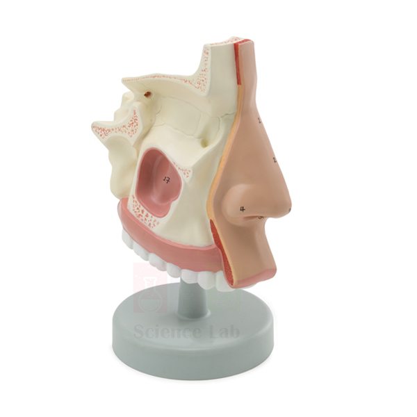
Human Nasal Cavity Model
Human Nasal Cavity Model (Product Code: SLE/A/97) is a compact, life-size sectional model that opens up the inside of the nose, showing a section of the nasal cavity together with the sinus cavities around it. Biology teachers, nursing tutors and ENT-focused trainers use it to explain where inhaled air first enters the respiratory tract. It is manufactured by Jain Scientific Equipments Pvt Ltd (ScienceLab), Ambala, and is a space-saving piece of biology lab equipment in India's schools and colleges.
Specifications
| Parameter | Detail |
|---|---|
| Type | Sectional model of the human nasal cavity |
| Scale | Life size |
| Size (with base) | 16.5 x 4.5 x 14.5 cm |
| Mounting | On a base |
| Structures shown | Section of the nasal cavity, with the sinus cavities also shown |
Typical Use
The model is most often used at the start of a respiratory-system unit. Holding the section up to the class, a teacher can point out the passage air follows after the nostrils, discuss how the nasal lining warms, moistens and filters incoming air, and then show where the sinus cavities sit in relation to it. That link makes it useful in health-education lessons that explain why a blocked nose and sinus congestion so often occur together.
With a depth of only 4.5 cm, it stands neatly on a narrow shelf or in a display cabinet and can be passed around a small group without difficulty. It pairs well with an external nose model and an airway model so learners can follow the respiratory route from face to lungs, which is why it is a common choice among school biology lab equipment in India for middle and senior classes.
Manufacturer
The Human Nasal Cavity Model is manufactured by Jain Scientific Equipments Private Limited, whose products carry the ScienceLab trading name. The company operates from Ambala, Haryana, a long-established centre for scientific and educational equipment in India. Its company profile describes the business and the categories it covers.
Related Human Models
Useful companions for a lesson on the upper respiratory tract:
- Human Nose Model — shows the nose from outside, complementing this internal section
- Anatomical Human Nasal Cavity Model — an alternative nasal cavity model for comparison or larger classes
- Head Section with Larynx Model — places the nasal cavity in context with the throat and larynx
- Human Larynx, Trachea, and Bronchi Model — continues the airway below the throat
- Human Respiratory System Model, 2-parts — an overview of the whole breathing system
Explore all Human Models in our anatomy range.
Frequently Asked Questions
What exactly does the model show?
A section of the nasal cavity with the sinus cavities also shown. For the outside of the nose, see the separate Human Nose Model listed above.
How big is it?
It measures 16.5 x 4.5 x 14.5 cm including its base and is modelled at life size.
Does it have removable parts?
Our listing describes a section on a base and does not mention removable parts. If you need a dissectible model, contact us and we will suggest options.
Is a numbered key supplied?
A key is not listed for this model. Please check with us about the documentation included before ordering.
Is it suitable for nursing or paramedical training?
Yes. It is used wherever the anatomy of the nasal passages and sinuses is taught, from school biology to introductory nursing and paramedical courses.
What should we include in a request for quotation?
Tell us the number of units, the delivery city or country, and whether you need other anatomy models in the same shipment. Send this through our contact form or the enquiry cart.
How are warranty questions handled?
Warranty terms are provided with each quotation; contact us for the terms that apply to your order.
Request a Quote
For prices and delivery details, get in touch or place the Human Nasal Cavity Model in your enquiry cart.
Last updated: 24 September 2026
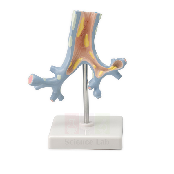
Pathology of the Human Trachea and Bronchi Model
Pathology of the Human Trachea and Bronchi Model (Product Code: SLE/A/98) is a 10x enlarged teaching model that shows two diseases of the bronchi, bronchitis and bronchiectasis, so students can see how each one changes the airway. It is aimed at pathology and respiratory lessons in medical, nursing and paramedical courses and is a practical addition to pharmacy college lab equipment where disease states are taught. The model is manufactured by Jain Scientific Equipments Pvt Ltd (ScienceLab), Ambala.
Specifications
| Parameter | Detail |
|---|---|
| Type | Pathology model of the human trachea and bronchi |
| Scale | 10x life size |
| Size | 11 x 11 x 20 cm |
| Mounting | On stand with base |
| Pathologies shown | 2 — bronchitis and bronchiectasis |
| Bronchitis | Inflammation of the bronchi |
| Bronchiectasis | Abnormal chronic dilatation of 1 or more of the bronchi |
Typical Use
Lecturers use the model to contrast two conditions that are easy to confuse on paper. Bronchitis is an inflammation of the airway lining, while bronchiectasis is a lasting, abnormal widening of one or more bronchi; seeing both on one enlarged model helps learners connect each diagnosis with the structural change behind it and with the cough and mucus problems patients describe. The 10x scale means details remain visible from the back of a tutorial room.
It fits naturally into sessions on smoking, recurrent chest infections and chronic lung disease, and works well next to a normal airway model so students first learn healthy anatomy and then the disease. Colleges building a set of biology teaching aids for pathology often combine it with prepared slides of diseased tissue, moving from the naked-eye view to the microscopic one.
Manufacturer
This pathology model is manufactured by Jain Scientific Equipments Private Limited, an Ambala, Haryana company that trades as ScienceLab and supplies teaching models to institutions in India and international markets. Vendor registration details needed for tender files can be requested along with your quotation. More about the company is available on our company profile page.
Related Human Models
Items that complete a respiratory pathology lesson:
- Human Larynx, Trachea, and Bronchi Model — a normal airway to compare against the diseased bronchi
- Pathology of the Human Lung Model — extends the lesson from the bronchi to diseases of the lung itself
- Human Respiratory System Model, 2-parts — shows where the trachea and bronchi sit in the whole system
- Microslide Smoking & Health — prepared slides for the microscopic side of the smoking-and-health topic
- Microslide Human Diseases — slides of diseased tissue to follow the model demonstration
The full Human Models category lists further anatomy and pathology models.
Frequently Asked Questions
Which conditions are shown on this model?
Two pathologies of the bronchi: bronchitis, an inflammation, and bronchiectasis, an abnormal chronic dilatation of one or more bronchi.
How large is the model?
It is enlarged to 10x life size and measures 11 x 11 x 20 cm on its stand with base, compact enough for a tutorial table.
Does it also show a healthy airway for comparison?
Our product details list only the two pathologies. For a normal reference, pair it with the Human Larynx, Trachea, and Bronchi Model linked above, or ask us to confirm the model's full appearance.
Which courses is it suited to?
It suits pathology, respiratory medicine and pharmacology teaching in medical, nursing, pharmacy and paramedical programmes, as well as advanced school biology.
Is a descriptive key included?
A key is not listed for this item. Contact us to confirm what accompanies the model.
What do we need to send for a tender or institutional quote?
Send the quantity, delivery address and any tender reference, and mention if you need our vendor registration details. Use the contact page or add the model to the enquiry cart.
Do you ship outside India?
Yes, we supply institutions in India and international markets; delivery terms are confirmed in the quotation.
Request a Quote
Request pricing for the Pathology of the Human Trachea and Bronchi Model through our contact page, or collect it with other items in the enquiry cart.
Last updated: 24 September 2026
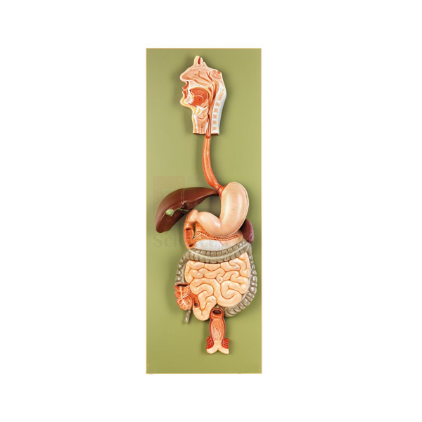
Human Digestive Tract Model
Human Digestive Tract Model (Product Code: SLE/A/99) presents the human digestive tract at life size on a single base, with a stomach that can be opened and a design that dissects into 3 pieces. It gives biology classes, nursing students and nutrition trainers a full-scale reference for how food travels through the body. The model is manufactured by Jain Scientific Equipments Pvt Ltd (ScienceLab), Ambala, a biology lab equipment supplier in India serving schools, colleges and training institutes.
Specifications
| Parameter | Detail |
|---|---|
| Type | Model of the human digestive tract |
| Scale | Life size |
| Dissection | Dissectible into 3 pieces |
| Stomach | Can be opened to view the interior |
| Size | 32 × 12 × 91 cm |
| Mounting | On base |
Typical Use
In a digestion lesson the teacher can start at the top of the model and move downward, naming each stage of the journey and explaining what happens to food there. Opening the stomach turns an abstract idea into something visible: students can discuss how the stomach stores and churns food and why its lining matters, then close it again and continue along the tract. Because the model is life size, learners come away with a realistic sense of how long the digestive route is.
The model suits nutrition and digestion chapters in senior school, introductory anatomy for nursing and allied-health courses, and community health talks. It is a staple of high school biology lab equipment for this reason. With a longest dimension of 91 cm, it needs a stable table or a dedicated display space; move it by the base rather than by the organs, and keep the 3 pieces together between classes.
Manufacturer
ScienceLab is the brand name of Jain Scientific Equipments Private Limited, which manufactures this digestive tract model and runs its business from Ambala in the state of Haryana. The company equips laboratories in India and international markets; the company profile gives an overview of its work.
Related Human Models
Pieces that extend a digestive system lesson:
- Human Stomach Model — a closer look at the organ that opens on this model
- Human Liver and Gallbladder Model — the accessory organs that supply bile to the tract
- Human Spleen, Duodenum, and Pancreas Model — detail of the region just beyond the stomach
- Human Digestive System Model — an alternative model of the digestive system for comparison
- Microslide The Digestive System — prepared slides for the microscopic view of digestive tissues
For more anatomy teaching models, browse the Human Models category.
Frequently Asked Questions
How many pieces does it separate into?
Three. The model is dissectible into 3 pieces, and the stomach can also be opened.
How much space does it need?
It measures 32 × 12 × 91 cm on its base, so plan for a sturdy table or cabinet shelf that can take the 91 cm dimension.
Which organs are represented?
It represents the human digestive tract at life size, including an opening stomach. For a structure-by-structure list, please contact us before ordering.
Can it be combined with microscope work?
Yes. Many teachers follow the model with prepared digestive-system slides so students see the same tract at tissue level; see the slide set in the related list.
How do we keep it in good condition?
Dust it regularly, wipe marks with a slightly damp soft cloth, and store it away from direct sunlight and heat. Reassemble the pieces fully before moving it.
What warranty is offered?
Warranty terms are confirmed at the quotation stage; contact us for details.
Can you supply it in quantity for several labs?
Yes. We supply schools, colleges and distributors in India and international markets; include the quantity and delivery location in your enquiry cart request.
Request a Quote
Ask for a price on the Human Digestive Tract Model via our contact page, or add it to the enquiry cart with related digestive system models.
Last updated: 24 September 2026
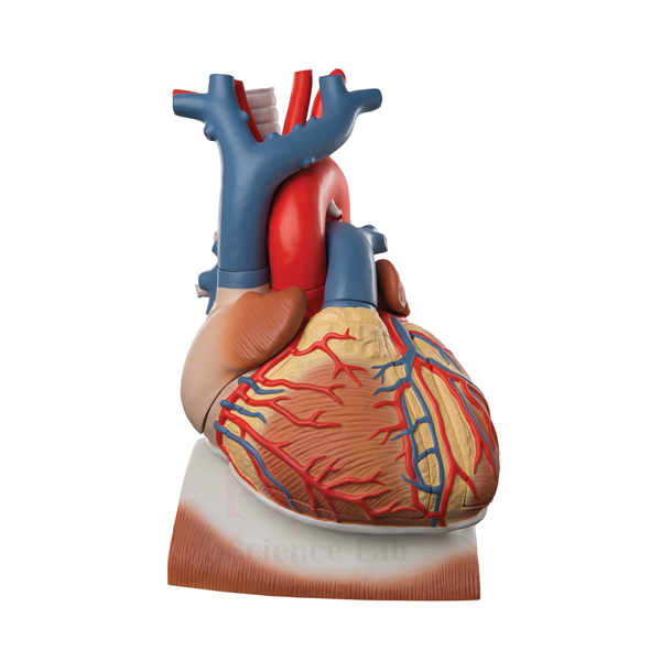
Human Heart on Diaphragm Model
Human Heart on Diaphragm Model (Product Code: SLE/A/100) shows a life-size human heart resting on the diaphragm, the way it sits in the chest, and dissects into 8 parts that include the diaphragm with a section of pericardium, the thymus gland and the ventricles. It helps students connect cardiac anatomy with the heart's position and surroundings, making it a strong heart model for anatomy teaching in India's schools, nursing colleges and medical institutes. It is manufactured by Jain Scientific Equipments Pvt Ltd (ScienceLab), Ambala.
Specifications
| Parameter | Detail |
|---|---|
| Type | Heart model mounted on the diaphragm |
| Scale | Life size |
| Dissection | Dissects into 8 parts |
| Parts include | Diaphragm with section of pericardium, thymus gland, apex of the heart, lower part of the ventricles, and ventricles |
| Size | 29 x 18 x 19 cm |
| Mounting | On a green base |
Typical Use
Most heart models show the organ on its own; this one keeps it on the diaphragm so a lesson can begin with position. Students see that the heart rests on the muscular floor of the chest, wrapped by the pericardium, with the thymus gland in front of it. The teacher can then lift away the thymus and open the lower heart by removing the apex and ventricle sections, moving from surface anatomy to the internal chambers in one sequence.
This makes it useful when teaching breathing and circulation together, since the diaphragm's role in respiration can be discussed right beside the heart it supports. It is a sensible centrepiece for a school biology lab setup in India and for first-year anatomy practicals. After each class, check that all 8 parts are back in place so none are lost between sessions.
Manufacturer
Made by Jain Scientific Equipments Private Limited, the Ambala, Haryana company behind the ScienceLab brand, this model belongs to a wider line of anatomical teaching models supplied to institutions in India and international markets. See the company profile for more about the manufacturer.
Related Human Models
Other models that support cardiovascular and thoracic lessons:
- Human Heart Model — a stand-alone heart for studying external features
- Heart 3 times Natural Size Model — an enlarged heart for viewing detail from a distance
- Human Lungs with Heart and Larynx Model — shows the heart together with the lungs and airway
- Human Thoracic Organs Model — the wider chest cavity in which heart and diaphragm sit
- Microslide The Circulatory System — prepared slides that take circulation lessons to tissue level
Browse the complete Human Models range for further options.
Frequently Asked Questions
Which parts come off the model?
It dissects into 8 parts, including the diaphragm with a section of pericardium, the thymus gland, the apex of the heart, the lower part of the ventricles and the ventricles. For the complete part list, contact us.
Why is the heart shown on the diaphragm?
It keeps the heart in its natural relationship with the diaphragm and pericardium, so students learn position and surroundings as well as the organ itself.
What are its size and scale?
The model is life size and measures 29 x 18 x 19 cm on a green base.
Is there a numbered key?
A key is not mentioned in our product details for this model; please ask us to confirm what is supplied.
Which classes is it best for?
It suits senior school biology, nursing and paramedical anatomy, and introductory medical anatomy where heart position and structure are taught together.
How should it be looked after?
Handle the removable parts over a table, wipe with a soft, lightly damp cloth, and store the assembled model covered to keep dust off.
How do we order several units?
Send your quantity and delivery location through the contact page or enquiry cart; we supply institutions and distributors in India and international markets.
Request a Quote
For a quotation on the Human Heart on Diaphragm Model, contact our team or add the model to your enquiry cart.
Last updated: 24 September 2026
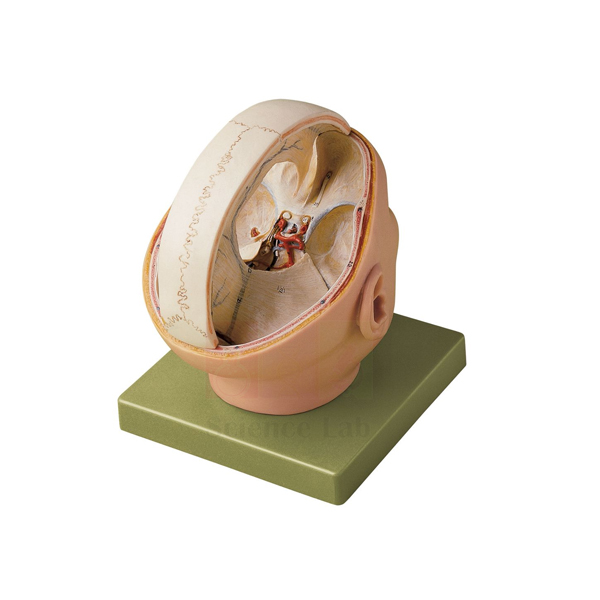
Human Dura Mater Model
Human Dura Mater Model (Product Code: SLE/A/101) is a life-size neuroanatomy model that shows the dura mater with its sinuses, the 12 pairs of cranial nerves and the arteries at the base of the brain, and separates into 2 parts for closer study. Anatomy lecturers, nursing tutors and senior biology teachers use it to explain how the brain is protected, supplied with blood and connected to the head and neck. It is manufactured by Jain Scientific Equipments Pvt Ltd (ScienceLab), an anatomical models manufacturer in Ambala.
Specifications
| Parameter | Detail |
|---|---|
| Type | Neuroanatomy model of the dura mater |
| Scale | Life size |
| Dissection | Dissectable into 2 parts |
| Size | 18 × 21 × 23 cm |
| Mounting | On base |
| Structures shown | Dura mater with its sinuses in proportion, the 12 pairs of cranial nerves, and the arteries at the base of the brain |
Typical Use
The model is built for lessons on the meninges and the base of the brain. A lecturer can first show the dura mater as the tough outer covering and point out its sinuses, which collect blood leaving the brain, then turn the model over to trace the 12 pairs of cranial nerves and the arteries that run along the underside. Students revising the cranial nerves often find it easier to learn their order and position on a three-dimensional model than from a textbook drawing.
It is used in human anatomy and physiology practicals at medical, dental, nursing and physiotherapy colleges, and in advanced school biology when the nervous system is covered in depth. Institutions expanding their educational laboratory equipment in India often add it alongside a brain model and a skull, so the brain, its coverings and the bony case can be taught as one topic. Set it on a table at student eye level when discussing the base-of-brain structures.
Manufacturer
Jain Scientific Equipments Private Limited manufactures the Human Dura Mater Model and sells it under its trading name, ScienceLab. The company is headquartered in Ambala, Haryana, and supplies anatomy and laboratory products to customers across India and international markets. Visit the company profile to learn more.
Related Laboratory Equipment
Models that complete a lesson on the brain, its coverings and its nerves:
- Human Brain Model with Arteries — follows the arterial supply of the brain in more detail
- Human Brain Stem Model — shows the region from which most cranial nerves emerge
- Human Brain Model, 15 Parts — a dissectible brain for the structures beneath the dura
- Human Skull Model — the bony case that the dura mater lines
- Human Nervous System Model — carries the nerve pathways beyond the skull
Find more options in our Human Models category.
Frequently Asked Questions
What anatomy does the model display?
The dura mater with its sinuses, the 12 pairs of cranial nerves, and the arteries at the base of the brain, all at life size.
Into how many parts does it separate?
Two. It is dissectable into 2 parts so both the covering and the underlying structures can be examined.
What are the dimensions?
It measures 18 × 21 × 23 cm and is supplied on a base.
Are the cranial nerves labelled or numbered?
All 12 pairs are shown, but a numbered key is not listed in our product details. Please contact us to confirm labelling before you order.
Is it suitable for school use as well as colleges?
Yes, although it is most often chosen for college-level anatomy; senior school classes use it when studying the nervous system in depth.
How are warranty and after-sales questions handled?
Warranty terms are set out in the quotation. Reach out with any after-sales query.
What should an institutional enquiry include?
Quantity, delivery location and any companion models you need. Submit it via the contact form or the enquiry cart.
Request a Quote
Prices and availability for the Human Dura Mater Model are available on request: contact us or use the enquiry cart.
Last updated: 24 September 2026
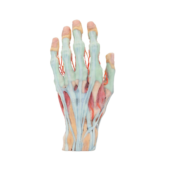
3-D Printed Human Superficial Hand Dissection Model
3-D Printed Human Superficial Hand Dissection Model (Product Code: SLE/A/102) reproduces a superficial dissection of the human hand at about life size, showing the vasculature, musculature and tendons in intricate detail. Because it is 3-D printed, students can study a specimen that is as realistic as possible without the need for human cadavers. It is used in anatomy, physiotherapy and occupational therapy teaching, and is one of the human anatomical models manufactured in India by Jain Scientific Equipments Pvt Ltd (ScienceLab), Ambala.
Specifications
| Parameter | Detail |
|---|---|
| Type | 3-D printed superficial dissection model of the human hand |
| Scale | About life size |
| Size | About 6-7/10" W x 5-9/10" H x 11-2/5" L |
| Structures shown | Vasculature, musculature and tendons of the hand, in intricate detail |
| Purpose | Realistic specimen study without the need for human cadavers |
Typical Use
The hand packs a great deal of anatomy into a small space, and this model lets learners examine the layer just beneath the skin: the blood vessels that supply the fingers and palm, the small muscles that give the hand its precision, and the tendons that transmit movement from the forearm. Instructors use it to explain grip, pinch and finger movement, and to show why injuries to a single tendon or vessel can affect function so noticeably.
For courses where cadaver access is limited, or where the biology dissection set is used only on animal specimens, the model gives a realistic human reference that can be handled in every session without preservation or special storage. Physiotherapy, occupational therapy, nursing and medical students use it for hand-anatomy practicals, and art and design departments use it to study the hand's form. Pairing it with a skeletal hand helps students relate each tendon to the bones it moves.
Manufacturer
The model is produced by Jain Scientific Equipments Private Limited, which trades as ScienceLab from its base in Ambala, Haryana, and serves teaching institutions in India and international markets. Buyers who need company registration details for procurement can request them with their quotation. The company profile has further background.
Related Human Models
Models that help build a complete picture of the hand and upper limb:
- Human Skeletal Hand Model — the bones beneath the muscles and tendons shown here
- Human Hand Joint Model with Ligaments — adds the joints and ligaments of the hand
- Human Arm and Hand model — follows the tendons back to the forearm muscles
- 3-D Printed Internal Heart Structures Model — another 3-D printed model for cadaver-free study
- 3D Printed Human Flexed Knee Joint — a 3-D printed joint for lower-limb lessons
See all Human Models for more anatomy teaching options.
Frequently Asked Questions
What does "superficial dissection" mean on this model?
It shows the structures that lie just beneath the skin of the hand: the vasculature, musculature and tendons.
How large is it?
About life size, measuring about 6-7/10" W x 5-9/10" H x 11-2/5" L.
Why choose a 3-D printed model over a cadaveric specimen?
It lets students study a realistic specimen without the need for human cadavers, so it can be used in ordinary classrooms and handled repeatedly.
Are the bones of the hand shown?
Our product details describe the vasculature, musculature and tendons. For bone anatomy, add the Human Skeletal Hand Model, or ask us about the model's deeper layers.
Does it come on a stand or base?
A stand is not listed for this model. Please contact us to confirm how it is supplied.
How should it be cleaned?
Dust it with a soft dry brush or cloth, and use only a lightly damp cloth for marks; avoid solvents and hot water.
What information speeds up a quotation?
The number of units, your delivery location and any registration documents your purchasing office requires. Send these via the contact page or the enquiry cart.
Request a Quote
To order the 3-D Printed Human Superficial Hand Dissection Model, contact us for pricing or add it to your enquiry cart.
Last updated: 24 September 2026
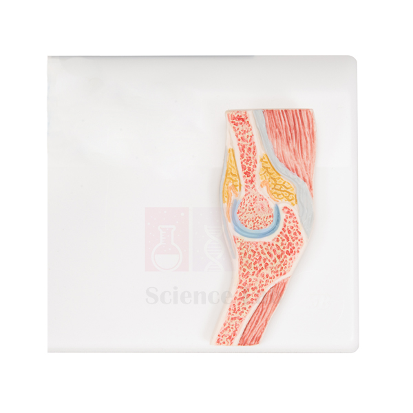
Human Elbow Cross-Section Model
Human Elbow Cross-Section Model (Product Code: SLE/A/104) is an intricately crafted, life-size section through the human elbow in which the muscles, ligaments and bones are clearly shown. Important structures are numbered against a key printed on the green base, and a removable transparent plastic cover protects the section. Anatomy, physiotherapy and sports-science teachers use it to explain how the elbow is built in layers. It is manufactured by Jain Scientific Equipments Pvt Ltd (ScienceLab), a laboratory equipment supplier in India based in Ambala.
Specifications
| Parameter | Detail |
|---|---|
| Type | Cross-section model of the human elbow |
| Scale | Life size |
| Size | 32 x 26 x 4 cm |
| Structures shown | Muscles, ligaments and bones |
| Identification | Important structures numbered; key printed on the green base |
| Protection | Removable, transparent plastic cover |
Typical Use
A cross-section answers a question that whole-limb models leave open: what lies next to what at one level of the arm. With this model, students can see how muscle groups wrap around the bones of the elbow and where the ligaments hold the joint together, then use the numbered key to name each structure. It works well for self-study, because learners can write down the numbers on a worksheet and check their answers against the printed key.
Used next to a human skeleton model, it helps students relate the flat section back to the whole arm and understand how injuries around the elbow affect nearby tissues. The slim 4 cm depth and protective cover make it practical for display cabinets, anatomy museums and corridor showcases as well as bench teaching; keep the cover in place whenever the model is not being examined.
Manufacturer
Behind the ScienceLab name is Jain Scientific Equipments Private Limited, an Ambala, Haryana company that manufactures this elbow section among its many anatomical and laboratory products for buyers in India and international markets. Learn more on the company profile page.
Related Human Models
Models that pair with this section for upper-limb teaching:
- Human Elbow Joint Model — the complete joint for comparing with the sectional view
- Functional Elbow Joint — demonstrates how the joint moves, which a section cannot show
- Human Muscular Arm Model — follows the muscles seen here along the full arm
- Shoulder Girdle with Musculature Model — continues the upper limb up to the shoulder
- Human Skeleton, Rod-Supported — a full skeleton for placing the elbow in context
More anatomy models are listed in the Human Models category.
Frequently Asked Questions
Does it come with a protective cover?
Yes. A removable, transparent plastic cover is included to protect the section.
How are the structures identified?
Important structures are numbered on the model, and the key is printed on the green base.
What are its dimensions?
The model is life size and measures 32 x 26 x 4 cm.
Can it demonstrate elbow movement?
No. It is a fixed cross-section for studying structure; for flexion and extension, see the Functional Elbow Joint in the related list.
How should the model and cover be cleaned?
Wipe the cover with a soft, lint-free cloth, and dust the model only when the cover is off. Avoid solvent-based cleaners on the transparent plastic.
What is the warranty?
Warranty terms are confirmed in the quotation; contact us for current terms.
Can it be supplied in multiples or exported?
Yes. Multiple units can be supplied to institutions and distributors in India and international markets; send your requirement through the enquiry cart or contact page.
Request a Quote
For a price on the Human Elbow Cross-Section Model, send us an enquiry or add it to the enquiry cart.
Last updated: 24 September 2026
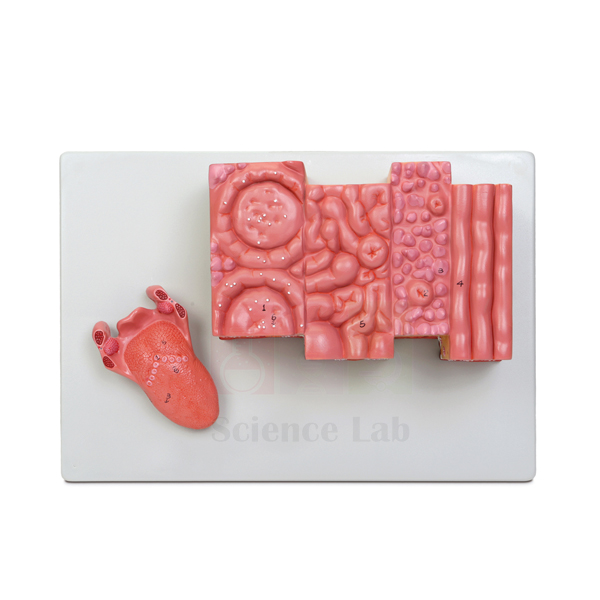
Microstructure of the Human Tongue Model
Microstructure of the Human Tongue Model (Product Code: SLE/A/105) enlarges a block of tongue tissue about 20x so that taste buds, lingual and mucous glands and the fine structure of the musculature can be seen without a microscope. A separate life-sized model of the intact tongue, with the palatine and lingual tonsils and the epiglottis, is included for orientation. Histology lecturers and biology teachers use the pair to link tissue structure with the whole organ. It is manufactured by Jain Scientific Equipments Pvt Ltd (ScienceLab), a biology lab instruments supplier in Ambala.
Specifications
| Parameter | Detail |
|---|---|
| Type | Enlarged microstructure model of the human tongue |
| Scale | About 20x life size |
| Size | 33 × 23 × 11 cm |
| Mounting | Durable polymer base |
| Histological features | Taste buds, lingual and mucous glands, fine structure of the musculature |
| Key | Numbered key included |
| Also included | Separate life-sized intact tongue model featuring the palatine and lingual tonsils and the epiglottis |
Typical Use
This model is designed for teaching at two scales. Start with the life-sized tongue to orient the class: where the tonsils sit, how the epiglottis relates to the back of the tongue, and why this region matters for swallowing. Then move to the enlarged block, where students can find the taste buds, follow the glands that keep the mouth moist, and see how muscle fibres run in different directions to give the tongue its flexibility.
It is especially helpful just before a practical with prepared microscope slides, because students who have already seen the structures in three dimensions recognise them far more quickly under the lens. Dental, medical, nursing and food-science courses use it in oral histology and taste physiology, and senior school classes use it in lessons on the senses. The numbered key makes it easy to set identification exercises.
Manufacturer
Jain Scientific Equipments Private Limited, trading as ScienceLab, manufactures this tongue microstructure model. The company is located in Ambala, Haryana, and provides histology, anatomy and general laboratory products to education buyers in India and international markets. Its background is summarised on the company profile.
Related Laboratory Equipment
Items that support an oral histology or taste lesson:
- Human Tongue Model — an additional whole-tongue model for group work
- Prepared Microscope Slide Set – Digestion (Liver, Pancreas And Tongue) — real tongue tissue to examine after the model
- Microslide Basic Human Histology — broader histology slides for comparing tissue types
- Compound Microscope — for viewing the slides that follow the model demonstration
- Larynx Human Model — continues from the epiglottis into the larynx
Explore more anatomy models in the Human Models category.
Frequently Asked Questions
What is included in the set?
The enlarged microstructure model on its polymer base, a numbered key, and a separate life-sized model of the intact tongue.
How much is the tissue enlarged?
About 20x life size; the enlarged model measures 33 × 23 × 11 cm.
Which microscopic features can students identify?
Taste buds, lingual and mucous glands, and the fine structure of the tongue musculature.
What does the life-sized tongue show?
The intact tongue with the palatine and lingual tonsils and the epiglottis, helping students place the enlarged tissue in context.
Can it replace microscope slides in a practical?
No, it complements them. The model prepares students to recognise structures, and slides then show real tissue.
How should it be cleaned?
Wipe the model and its polymer base with a soft, slightly damp cloth, then dry. Keep the life-sized tongue with the main model so it is not misplaced.
How can we get a quotation for several departments?
List the quantities and delivery addresses in the enquiry cart or on our contact page; warranty terms are included with the quotation.
Request a Quote
Pricing for the Microstructure of the Human Tongue Model is available on request — contact us or use the enquiry cart.
Last updated: 24 September 2026
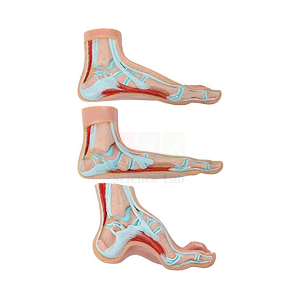
Human Foot Model Set
Human Foot Model Set (Product Code: SLE/A/107) contains 3 life-size models representing cross sections of a normal, a flat and an arched human foot, supplied with a key. Unlike a general human body model kit, this set focuses on one clear comparison: how the shape of the foot's arch changes the structure of the foot. It is used in physiotherapy, podiatry, sports science and biology teaching, and is manufactured by Jain Scientific Equipments Pvt Ltd (ScienceLab), Ambala.
Specifications
| Parameter | Detail |
|---|---|
| Type | Set of cross-section models of the human foot |
| Models in set | 3 — normal foot, flat foot and arched foot |
| Scale | Life size |
| Size | 22 x 12 x 8 cm (each model) |
| Key | Included |
Typical Use
Lined up side by side in the same orientation, the three sections let students see at once how a flat foot and an arched foot differ from a normal one. Teachers use the comparison to explain what the arch does, spreading body weight and absorbing impact during walking and running, and why a collapsed or exaggerated arch can lead to discomfort in the feet, knees or back.
Physiotherapy, podiatry and orthotics students use the set when learning to assess foot posture, sports-science courses use it in lessons on gait and footwear, and schools use it in health education sessions on posture and choosing suitable shoes. Because each model is life size, learners can compare the sections directly with their own feet. Keep the key with the set so every structure can be named during practicals.
Manufacturer
This foot set is manufactured by Jain Scientific Equipments Private Limited. Operating as ScienceLab out of Ambala, Haryana, the company builds and supplies anatomy models and laboratory equipment for education buyers throughout India and international markets. Details are on the company profile.
Related Human Models
Models that extend a lesson on the foot and lower limb:
- Human Skeletal Foot Model — shows the bones that form the arches
- Human Foot Set Model — an alternative foot set for comparison
- Human Leg and Portion of Pelvis Model — places the foot within the whole lower limb
- Human Knee Joint Model, Life Size — for discussing how foot posture affects the knee
- Human Elbow Cross-Section Model — another life-size section for a sectional anatomy series
The Human Models category lists our complete range.
Frequently Asked Questions
What does the set contain?
Three life-size cross-section models, one each of a normal, flat and arched human foot, plus a key.
How big is each model?
Each model measures 22 x 12 x 8 cm.
What is the teaching point of having three feet?
Comparing them side by side shows how arch shape alters the structure of the foot, which is the basis for understanding flat feet and high arches.
Can the models be purchased separately?
The item is listed as a set of 3. If you need individual models, please contact us to check availability.
Is the set suitable for physiotherapy and podiatry courses?
Yes, it is well suited to foot-posture and gait teaching in these courses, as well as sports science and school health education.
How should the set be stored?
Store the three models together in a closed cabinet, away from heat and direct sunlight, and wipe them with a soft damp cloth when needed.
Can you deliver outside India?
Yes, we supply institutions in India and international markets. Send the quantity and destination via the enquiry cart; warranty and delivery terms come with the quotation.
Request a Quote
To receive a quotation for the Human Foot Model Set, contact us or add it to your enquiry cart.
Last updated: 24 September 2026
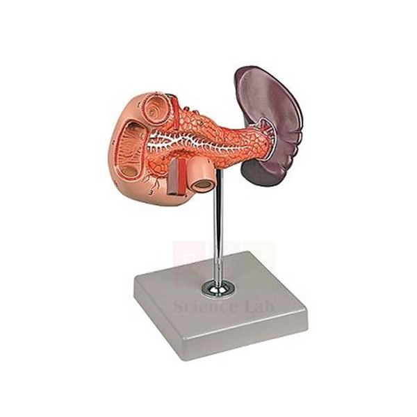
Human Spleen, Duodenum, and Pancreas Model
Human Spleen, Duodenum, and Pancreas Model (Product Code: SLE/A/108) is a life-size model of three neighbouring upper-abdominal organs, mounted on a stand with base. It shows the pancreatic duct, the blood vessels and a partially opened duodenum, helping students understand how the pancreas delivers its secretions into the intestine. The model is used in anatomy, physiology and nursing teaching and is made by Jain Scientific Equipments Pvt Ltd (ScienceLab), Ambala, one of the human anatomical model manufacturers in India.
Specifications
| Parameter | Detail |
|---|---|
| Type | Model of the spleen, duodenum and pancreas |
| Scale | Life size |
| Size | 12 × 22 × 23 cm (H) |
| Mounting | On stand with base |
| Structures shown | Pancreatic duct, blood vessels and partially opened duodenum |
Typical Use
This region is hard to picture from diagrams because the organs sit deep in the abdomen, tucked behind the stomach. The model brings them out where a class can see them: students follow the pancreatic duct through the pancreas towards the duodenum, then look into the opened section of the duodenum to understand where digestive juices meet food leaving the stomach. The blood vessels give a starting point for discussing how these organs are supplied.
It also prompts a useful discussion of function, since the pancreas works in both digestion and blood-sugar control while the spleen, despite its position, belongs to the immune and blood-filtering systems. Zoology and physiology departments add it to their zoology lab equipment for these lessons, and nursing courses use it when introducing conditions such as pancreatitis and diabetes. Its compact size makes it easy to pass between small groups.
Manufacturer
ScienceLab is the name under which Jain Scientific Equipments Private Limited manufactures and sells its products, including this abdominal organ model. The company works from Ambala, Haryana, and serves schools, colleges and dealers in India and international markets. More information is on the company profile.
Related Human Models
Models used alongside this one in digestive and abdominal anatomy:
- Human Pancreas, Spleen and Duodenum Model — an alternative model of the same organ group
- Pathology of the Human Pancreas, Duodenum, and Gallbladder Model — adds disease states for clinical courses
- Human Liver and Gallbladder Model — completes the story of secretions reaching the duodenum
- Human Digestive Tract Model — shows where the duodenum fits in the whole tract
- Rear Organs of the Upper Abdomen — a regional view of the organs behind the stomach
Browse all Human Models for more.
Frequently Asked Questions
Which structures are shown?
The spleen, duodenum and pancreas, with the pancreatic duct, blood vessels and a partially opened duodenum.
Why is the duodenum partly opened?
So students can see inside the part of the intestine that receives the pancreatic duct, rather than only its outer surface.
What size is it?
It is life size and measures 12 × 22 × 23 cm in height on its stand with base.
Does the model come apart?
Our listing does not describe it as dissectible. Contact us if you need to confirm this before ordering.
Are the liver and gallbladder included?
No, they are not listed as part of this model. The Human Liver and Gallbladder Model in the related list covers them.
What care does it need?
Wipe it gently with a soft cloth, lift it by the stand or base, and store it covered when not in use.
How do we request a formal quotation?
Send the quantity, delivery location and any companion models through the contact form or enquiry cart; warranty terms are given in the quotation.
Request a Quote
For pricing on the Human Spleen, Duodenum, and Pancreas Model, contact us or add it to the enquiry cart.
Last updated: 24 September 2026
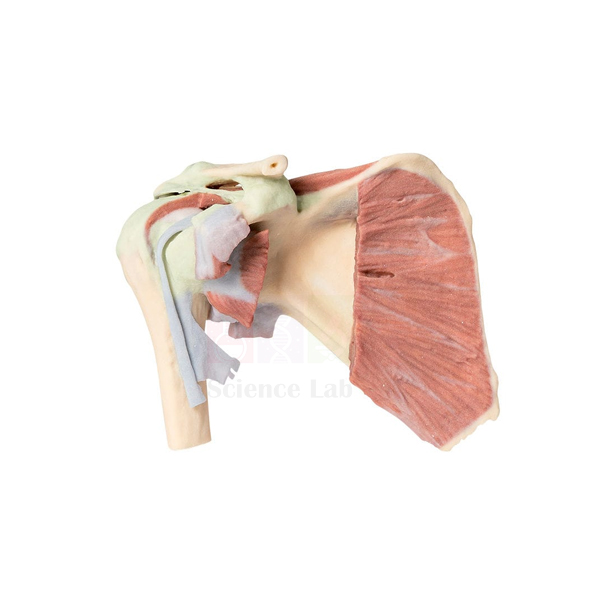
Shoulder Girdle with Musculature Model
Shoulder Girdle with Musculature Model (Product Code: SLE/A/109) is a one-piece, about life-size model that gives a 360° view of the muscular and bony elements of the human shoulder girdle and upper arm. Rod-mounted on a durable polymer base and supplied with a numbered key, it is a practical piece of educational lab equipment in India's physiotherapy, sports-science, nursing and medical colleges. It is manufactured by Jain Scientific Equipments Pvt Ltd (ScienceLab), Ambala.
Specifications
| Parameter | Detail |
|---|---|
| Type | Muscular model of the shoulder girdle and upper arm |
| Scale | About life size |
| Size (without base) | 17 × 21 × 10 cm |
| Construction | One-piece model |
| Mounting | Rod-mounted on durable polymer base |
| View | 360° view of muscular and bony elements |
| Key | Numbered key included |
Typical Use
The shoulder is the most mobile joint in the body, and that mobility depends on muscles as much as bones. Mounted on a rod above its base, this model can be studied from the front, back and sides, so students can see how muscles wrap over and around the shoulder girdle and continue down the upper arm. Using the numbered key, they can identify each muscle and bone and then discuss which muscles lift, rotate and stabilise the arm.
Physiotherapy and sports-science tutors use it when explaining common shoulder injuries and rehabilitation exercises, while nursing and medical students use it for upper-limb anatomy practicals and injection-site orientation. Because it is a single piece with no removable parts, it withstands frequent handling in busy practical classes, a useful quality in shared biology lab equipment. Place it in the centre of a group table so everyone gets a different viewing angle.
Manufacturer
Ambala-based Jain Scientific Equipments Private Limited manufactures this shoulder model and markets it, like the rest of its range, as ScienceLab. The company is registered in Haryana and supplies educational institutions in India and international markets. For more about who we are, see the company profile.
Related Human Models
Models that complete an upper-limb and shoulder lesson:
- Human Shoulder Joint Model — the joint itself, for studying bones and articulation
- Shoulder Joint Model — an alternative shoulder joint model for group work
- Human Muscular Arm Model — continues the musculature down to the forearm and hand
- Human Elbow Cross-Section Model — a sectional view of the next joint along the arm
- Human Muscular Torso with Head and Open Back Model — shows the back and chest muscles that attach to the shoulder girdle
Find further models in the Human Models category.
Frequently Asked Questions
Does the model come apart?
No. It is a one-piece model, so there are no parts to lose or reassemble.
How large is it?
It is about life size and measures 17 × 21 × 10 cm without the base.
What anatomy can be studied?
The muscular and bony elements of the shoulder girdle and upper arm, visible from all sides thanks to the 360° view.
Is a key supplied?
Yes, a numbered key is included for identifying the structures.
How is it mounted?
It is rod-mounted on a durable polymer base, which holds the model clear of the table for viewing.
How should it be cleaned?
Wipe with a soft cloth lightly dampened with water, dry it, and avoid harsh chemicals on the model and base.
Can you quote for export or large orders?
Yes. We quote for institutions and distributors in India and international markets; provide quantity and destination via the contact page or enquiry cart. Warranty terms are confirmed in the quotation.
Request a Quote
Get a price for the Shoulder Girdle with Musculature Model by contacting us or adding it to your enquiry cart.
Last updated: 24 September 2026
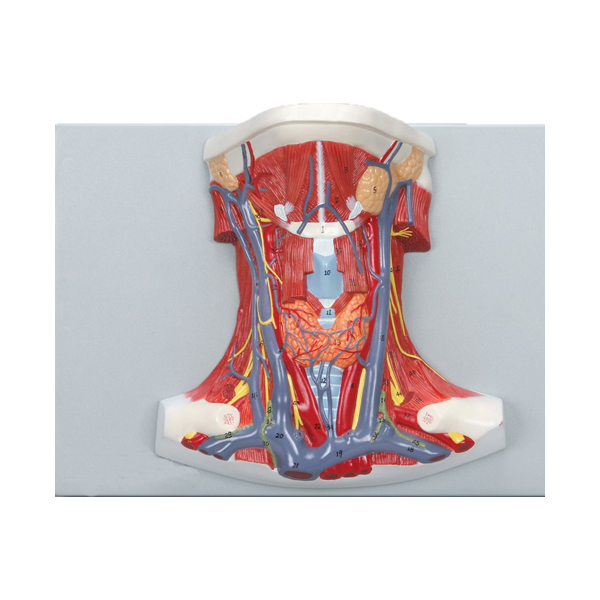
Anatomy of the Human Neck Model
Anatomy of the Human Neck Model (Product Code: SLE/A/124) is a life-size ventral dissection of the human neck that gives clear views of its muscles, vascular structures, nerves and glands, with a printed key identifying 47 anatomical features. Anatomy departments, dental and nursing colleges and paramedical training centres use it to teach one of the most crowded regions of the body. It is manufactured by Jain Scientific Equipments Pvt Ltd (ScienceLab), a laboratory equipment manufacturer in India based in Ambala.
Specifications
| Parameter | Detail |
|---|---|
| Type | Ventral dissection model of the human neck |
| Scale | Life size |
| Size (without base) | 23 x 14 x 32 cm |
| Mounting | Durable polymer base |
| Structures shown | Muscles, vascular structures, nerves and glands |
| Key | Printed key identifying 47 anatomical features |
Typical Use
In a short stretch of the body the neck carries blood to and from the head, air to the lungs, food to the stomach and nerve signals in both directions, all wrapped in layers of muscle and alongside important glands. Viewed from the front, this dissection lets a lecturer work through those layers in order, using the printed key to name each of the 47 features and to explain how the vessels, nerves and glands lie in relation to the neck muscles.
The model supports head-and-neck anatomy courses and clinical skills teaching, where students need to picture what lies beneath the skin before learning procedures or examinations in this area. It is a long-lasting addition to biology teaching aids for colleges because the key allows independent revision: students can cover the key, name the numbered features themselves and then check their answers.
Manufacturer
The Anatomy of the Human Neck Model is manufactured by Jain Scientific Equipments Private Limited, known to customers as ScienceLab. From Ambala in Haryana, the company provides anatomical models and laboratory equipment to educational and training institutions in India and international markets. Read more on the company profile.
Related Laboratory Equipment
Models that build on a neck anatomy lesson:
- Head and Neck Model — follows the neck structures up into the head
- 3-D Printed Head and Neck Section Model — a sectional view to complement the front dissection
- Larynx Human Model — a closer look at the airway structures in the neck
- Human Cervical Spinal Column Model — the vertebrae that support the neck from behind
- Human Endocrine Organs Model — places the neck glands within the endocrine system
See the full Human Models range for more.
Frequently Asked Questions
How many features does the key identify?
The printed key identifies 47 anatomical features.
From which direction is the neck dissected?
It is a ventral dissection, so the neck is shown from the front.
What are the dimensions?
It is life size and measures 23 x 14 x 32 cm without its base, which is made of durable polymer.
Which body systems are represented?
Muscles, vascular structures, nerves and glands are all shown.
Can parts be removed?
Removable parts are not listed for this model. Contact us to confirm before ordering if you need a dissectible version.
Which courses is it designed for?
Human anatomy, dental and ENT teaching, nursing and paramedical training, and any course covering the anatomy of the head and neck.
What should be included in a quotation request?
The required quantity, delivery location and preferred delivery date; warranty terms will be stated in our reply. Use the contact form or enquiry cart.
Request a Quote
To receive pricing for the Anatomy of the Human Neck Model, write to us or add it to the enquiry cart.
Last updated: 24 September 2026
Enlarged Human Cerebellum Model
Enlarged Human Cerebellum Model (Product Code: SLE/A/131) magnifies the human cerebellum to 4x life size and is dissected to show details of its internal organization. It separates into 2 parts and comes with a numbered key, giving neuroanatomy lecturers a large, clear model for teaching the part of the brain that coordinates movement and balance. As school and college biology lab equipment in India, it suits advanced biology, psychology and health-science courses. It is manufactured by Jain Scientific Equipments Pvt Ltd (ScienceLab), Ambala.
Specifications
| Parameter | Detail |
|---|---|
| Type | Enlarged, dissected model of the human cerebellum |
| Scale | 4x life size |
| Size | 40 x 19 x 18 cm |
| Dissection | Dissectible into 2 parts |
| Internal detail | Dissected to show details of internal organization |
| Key | Numbered key included |
| Mounting | Not on base |
Typical Use
At life size the cerebellum is small and tucked beneath the back of the brain, so its structure is easy to overlook. Enlarged four times, it becomes a centrepiece for lessons on how the brain controls posture, balance and smooth, coordinated movement. Separating the 2 parts exposes the internal organization, and the numbered key lets students name what they see while the teacher explains how grey and white matter are arranged inside.
Physiotherapy and sports-science courses use it when discussing balance disorders and motor learning, psychology and neuroscience students use it in brain-structure modules, and senior biology classes use it to go beyond a whole-brain overview. It works well as part of a college biology lab setup alongside a brain stem model. Since it is not supplied on a base, rest it on a flat, padded surface during lessons and handle it with both hands.
Manufacturer
This cerebellum model is made by Jain Scientific Equipments Private Limited, an Ambala, Haryana manufacturer whose products are sold under the ScienceLab brand to education buyers in India and international markets. The company profile explains more about the business.
Related Laboratory Equipment
Models that fit alongside it in a neuroanatomy course:
- Enlarged Human Brain Stem Model — an enlarged companion for the structure the cerebellum connects to
- Human Brain Stem Model — a smaller brain stem model for group work
- Human Brain Model, 15 Parts — shows where the cerebellum sits in the whole brain
- Human Regional Brain Model — for teaching the functional regions of the brain
- Human Nervous System Model — links the brain to the rest of the nervous system
More models are available in the Human Models category.
Frequently Asked Questions
How large is the model?
It is 4x life size and measures 40 x 19 x 18 cm.
Is it supplied on a base or stand?
No, it is not on a base. It rests directly on a table, so a padded mat or tray is advisable.
How many parts does it have?
It is dissectible into 2 parts, which open to show the internal organization of the cerebellum.
Does it include a key?
Yes, a numbered key is included.
Which courses benefit most from it?
Neuroanatomy, psychology, physiotherapy and sports science, as well as senior school biology units on the brain.
How should it be stored?
Keep the two parts together in a closed cabinet or padded box, away from heat and sunlight, and wipe with a soft damp cloth when needed.
How do we get a price and warranty terms?
Send the quantity and delivery location via our contact page or enquiry cart; pricing and warranty terms are provided in the quotation.
Request a Quote
For a quotation on the Enlarged Human Cerebellum Model, contact us or add it to the enquiry cart.
Last updated: 24 September 2026
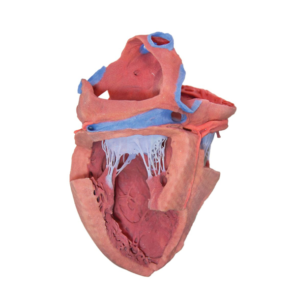
3-D Printed Internal Heart Structures Model
3-D Printed Internal Heart Structures Model (Product Code: SLE/A/132) is an about life-size, 3-D printed heart that has been dissected to display the internal structures of its chambers. It lets students study a specimen that is as realistic as possible without the need for human cadavers, which makes it valuable for cardiovascular teaching in medical, nursing and paramedical programmes. It is manufactured by Jain Scientific Equipments Pvt Ltd (ScienceLab), Ambala, a biology lab equipment supplier to institutions in India and abroad.
Specifications
| Parameter | Detail |
|---|---|
| Type | 3-D printed dissected model of the internal heart structures |
| Scale | About life size |
| Size | 11-4/5" W x 9-4/5" H x 9-4/5" L |
| Structures shown | Internal structures of the chambers of the heart |
| Purpose | Realistic specimen study without the need for human cadavers |
Typical Use
Understanding the heart means understanding what happens inside it. With the chambers opened, a lecturer can trace the route of blood from the body into the right side of the heart, out to the lungs, back into the left side and onward to the body, using the model's interior to explain how valves and the dividing wall keep that flow moving in one direction. Students who have only seen a heart model from the outside often find this the point at which the double circulation finally makes sense.
Because it is 3-D printed rather than preserved tissue, the model can be handled freely in ordinary classrooms and skills labs, with no specimen storage or chemical handling. It is used in anatomy practicals, cardiac nursing modules and paramedic training, and it complements a conventional heart model in anatomy classes across India: the external model for surface features, this one for the chambers within.
Manufacturer
Jain Scientific Equipments Private Limited manufactures the 3-D Printed Internal Heart Structures Model and trades under the ScienceLab name from Ambala, Haryana. Purchasing departments that require the company's registration documents for vendor onboarding can ask for them when requesting a quotation. See our company profile for more background.
Related Human Models
Models that round out a cardiovascular lesson:
- Human Heart Model — for the external features that complement this internal view
- Heart 3 times Natural Size Model — an enlarged heart for lecture-hall demonstrations
- Human Heart on Diaphragm Model — shows the heart in position with the diaphragm and pericardium
- 3-D Printed Human Superficial Hand Dissection Model — another cadaver-free 3-D printed dissection
- Microslide The Circulatory System — prepared slides for studying circulatory tissues under the microscope
Browse the Human Models category for our full anatomy range.
Frequently Asked Questions
What does the model show?
The internal structures of the chambers of the heart, exposed by dissection.
What are its dimensions?
It is listed as about life size, measuring 11-4/5" W x 9-4/5" H x 9-4/5" L. If exact dimensions matter for display or storage, contact us to confirm.
What is the advantage of a 3-D printed heart?
It gives students a realistic specimen to study without the need for human cadavers, so it can be used in any classroom.
Do sections come apart?
The model is dissected to reveal the chambers; removable parts are not listed in our product details. Please ask us if you need to confirm this.
Is a key or labelling guide included?
A key is not listed for this model. Contact us to check what is supplied with it.
How should it be cleaned?
Remove dust with a soft brush or dry cloth and use only a lightly dampened cloth for marks; keep it away from heat and solvents.
How do we arrange a quotation or export order?
Tell us the quantity, destination and any vendor documents you need through the contact page or enquiry cart. We supply India and international markets, and warranty terms are included in the quotation.
Request a Quote
For pricing on the 3-D Printed Internal Heart Structures Model, contact our team or add it to your enquiry cart.
Last updated: 24 September 2026
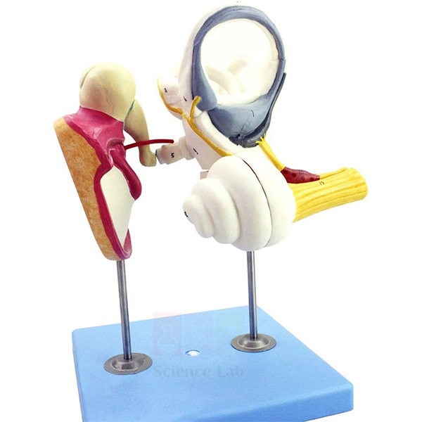
Human Inner Ear Model, 4x
The Human Inner Ear Model, 4x (Product Code: SLE/A/133) enlarges the organs of hearing that sit hidden inside the skull to about four times life size, so a class can follow sound from the eardrum, across the ear bones and into the cochlea. Biology teachers, nursing and paramedical tutors and ENT or audiology instructors use it as a compact ear model for anatomy lab teaching in India and abroad. It is made by Jain Scientific Equipments Pvt Ltd (ScienceLab), Ambala.
Specifications
| Parameter | Detail |
|---|---|
| Scale | About 4× life size |
| Size | 18 × 18 × 20 cm (H) |
| Mounting | On stand with base |
| Cochlea | Dissectible into 3 parts |
| Structures included | Cochlea, ear bones and tympanic membrane |
Typical Use
In a lesson on hearing, the teacher can start at the tympanic membrane, show how the ear bones pass vibration inward, and then lift the cochlea apart into its 3 parts to reveal the coiled interior where sound is turned into nerve signals. Because the structures are held on a stand, the model can be turned to face the class or passed along a bench for close inspection without anyone handling loose pieces.
The model also supports revision of common hearing problems, such as why damage to the eardrum or the ear bones reduces conduction while cochlear damage affects sensing. Among biology teaching aids for secondary school, nursing and allied-health courses, a bench-sized inner ear like this is easy to store and quick to set out for practical examinations.
Manufacturer
This inner ear model comes from Jain Scientific Equipments Private Limited, a company in Ambala, Haryana that sells its educational and laboratory range under the ScienceLab name. Schools, colleges and distributors can read about the company’s background on our company profile page.
Related Human Models
Models and apparatus that fit the same lesson on the ear and hearing:
- Human Inner Ear Model, 18x — a much larger view of the inner ear for detailed study of the labyrinth
- Human Ear Model, 5 Parts — puts the inner ear in context with the outer and middle ear
- Human Ear Model — a one-piece ear with blood vessels for quick overview teaching
- Tuning Fork Set, Steel — for simple sound and hearing demonstrations alongside the model
See the complete Human Models range for more anatomy teaching models.
Frequently Asked Questions
Which parts of the ear does this model include?
It includes the cochlea, the ear bones and the tympanic membrane (eardrum), mounted together on a stand with base.
Can the cochlea be opened?
Yes. The cochlea is dissectible into 3 parts so students can see its internal structure.
How does it compare with the 18x inner ear model?
This 4x version keeps the eardrum, ear bones and cochlea together at a bench-friendly 18 × 18 × 20 cm. Choose the 18x model if you need much greater magnification of the inner ear alone.
Is a numbered key or teaching guide supplied?
A key is not listed in our current documentation for this model. Please contact us to confirm what accompanies it before you order.
How should the model be cleaned?
Wipe it with a soft, dry or slightly damp cloth and avoid solvents or abrasive cleaners. Keep the small cochlea parts together on the stand after each class.
Can you supply several units for a school or an overseas order?
Yes. We supply schools, colleges and distributors across India and international markets; share the quantity and delivery location for a quotation.
Request a Quote
To order the Human Inner Ear Model, 4x, contact us with your quantity and delivery details, or add it to the enquiry cart together with other models for one combined quotation.
Last updated: 24 September 2026
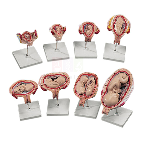
3B Human Development Set
The 3B Human Development Set (Product Code: SLE/A/134) is a series of uterus models that follows pregnancy from the early embryo to a well-formed fetus, with the embryo or fetus lifting out of each uterus for closer study. It is used in embryology, reproductive biology, midwifery and nursing teaching, and works well as a human biology model set for colleges in India. The set is supplied by Jain Scientific Equipments Pvt Ltd (ScienceLab), Ambala.
Specifications
| Parameter | Detail |
|---|---|
| Uterus models | Eight models of the uterus |
| Embryo stages | Removable embryo in the 1st, 2nd and 3rd months |
| Fetus stages | Removable fetus in the 5th and 7th months |
| 5th-month models | Four models showing twins as well as a fetus in various positions |
| Mounting | Each model mounted on a base |
Typical Use
Laid out in order on a bench, the models let students compare how the embryo grows through the first three months and how the fetus fills the uterus by the 5th and 7th months. Lifting each embryo or fetus out of its uterus makes the relationship between the developing baby and the uterine wall easy to discuss, and the 5th-month models open up topics such as twin pregnancy and fetal position that are central to obstetric and midwifery training.
Tutors often rotate small groups through the set during practical sessions, asking each group to identify the stage and describe the changes they see. Institutions equipping several teaching rooms can combine it with other pregnancy and pelvis models in a single biology model kit bulk order.
Supplier
The set reaches customers through Jain Scientific Equipments Private Limited, the Ambala, Haryana company behind the ScienceLab brand, which supplies anatomy models to schools, medical and nursing colleges and distributors. More about the company is on our company profile.
Related Human Models
Models that are commonly taught alongside this development set:
- Human Placenta Model — shows how the placenta and umbilical cord support the fetus
- Human Birth Demonstration Model — continues the story from late pregnancy to delivery through the pelvis
- Human Adult Female Pelvis Structural Model — explains the bony birth canal the fetus must pass through
- Human Embryo Development Set — an alternative gestation series for comparison
- Human Embryo Set, PVC — another uterus-and-fetus series covering the gestation period
Browse all Human Models for further reproductive and developmental anatomy aids.
Frequently Asked Questions
Which stages of pregnancy does the set cover?
It shows a removable embryo in the 1st, 2nd and 3rd months and a removable fetus in the 5th and 7th months.
What do the 5th-month models show?
Four models in the 5th month show twins as well as a fetus in various positions, which helps when teaching fetal lie and multiple pregnancy.
How many pieces will I receive?
Our listing describes eight uterus models plus the four 5th-month models, each on its own base. Please contact us to confirm the exact piece count and packing before ordering.
Can I confirm the brand and origin before buying?
Yes. The product name carries the 3B brand; ask us for brand, origin and packing details as part of your quotation.
How should the models be stored between classes?
Return each embryo or fetus to its own uterus, keep the models upright on their bases and store them away from direct sunlight and heat. Clean with a soft cloth only.
What should an institutional enquiry include?
Tell us the quantity, the delivery address, and whether you need a formal quotation or tender format. We supply buyers across India and international markets.
Request a Quote
For pricing and availability of the 3B Human Development Set, contact us or place it in the enquiry cart with any other models you need.
Last updated: 24 September 2026
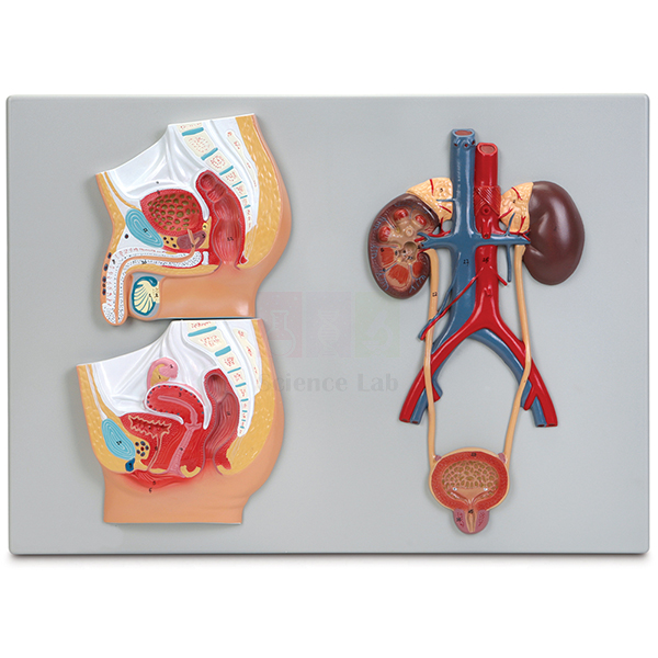
Human Male and Female Urogenital Systems Model
The Human Male and Female Urogenital Systems Model (Product Code: SLE/A/135) places a male pelvis, a female pelvis and a generalized urinary system side by side on one plaque, so students can compare how the urinary and reproductive tracts are arranged in each sex. It is designed for school biology lab equipment in India as well as nursing, paramedical and pre-medical anatomy teaching, and is made by Jain Scientific Equipments Pvt Ltd (ScienceLab), Ambala.
Specifications
| Parameter | Detail |
|---|---|
| Scale | About 3/4× life size |
| Overall size | 53 × 38 × 3 cm |
| Format | 3-part plaque mount |
| Base | Durable polymer base |
| Pelvic sections | Detailed sagittal sections of the male and female pelvis, highlighting the major urogenital structures of each |
| Urinary system | Renal artery and vein, kidneys (right side showing interior anatomy), ureters, and urinary bladder with urethra |
Typical Use
Teachers usually begin with the generalized urinary system, following blood into the kidney through the renal artery, looking at the opened right kidney, and tracing urine down the ureters to the bladder and urethra. The two sagittal pelvic sections then show where the bladder sits relative to the reproductive organs in men and women, which makes differences such as urethral length and the position of the uterus or prostate easy to point out.
Because everything is on one flat plaque, the model is convenient to prop up at the front of a class or to lay on a table for small-group revision, and it stores flat in a cupboard. It is a useful addition to any human body model kit used for reproduction, excretion and health-education topics.
Manufacturer
Manufacture is by Jain Scientific Equipments Private Limited, better known to schools and colleges by its trading name ScienceLab, and based in Ambala in the state of Haryana. You can find out more about the company on its company profile.
Related Human Models
Items that extend the same urinary and reproductive system lesson:
- Human Male Reproductive System Model Set — more detail on the male tract than the pelvic section can give
- Human Female Urogenital Model — a three-dimensional companion for the female side
- Human Kidney, Nephron, and Glomerulus Model Set — zooms into how the kidney filters blood
- Human Excretory System — a laminated wall chart to reinforce the same content
View the full Human Models category for other organ-system models.
Frequently Asked Questions
Does the model show both male and female anatomy?
Yes. It carries sagittal sections of both the male and the female pelvis, plus a generalized urinary system on the same plaque.
Can students see inside the kidney?
Yes. The right kidney is shown with its interior anatomy exposed, while the renal artery and vein, ureters and bladder with urethra complete the urinary tract.
How large is it?
The overall size is 53 × 38 × 3 cm at about 3/4× life size, so it fits on a standard classroom table.
Can it be hung on a wall?
It is a plaque mount on a polymer base. Wall-hanging fittings are not listed in our documentation, so please contact us if you plan to wall-mount it.
Is a numbered key included?
A key is not mentioned in our current product details. Ask us to confirm the teaching material supplied with the model.
What warranty applies?
Warranty terms are confirmed at the time of quotation; contact us for details.
Request a Quote
Schools, colleges and distributors in India and international markets can request pricing through our contact page or by adding this model to the enquiry cart.
Last updated: 24 September 2026
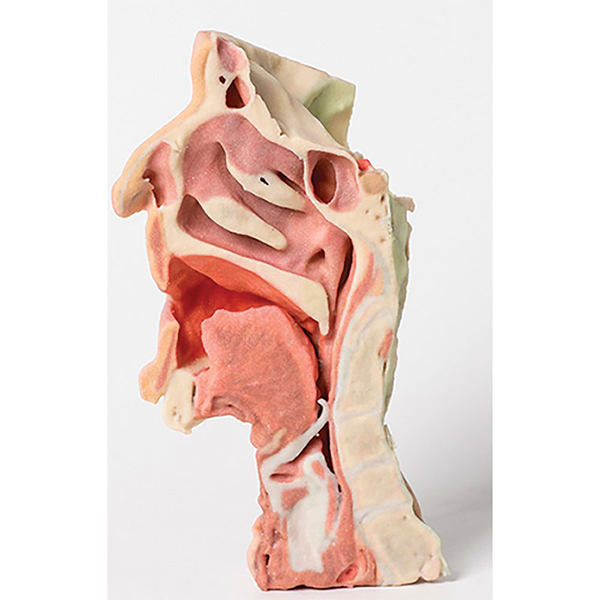
3-D Printed Head and Neck Section Model
The 3-D Printed Head and Neck Section Model (Product Code: SLE/A/136) is a midsagittally sectioned right face and neck, produced by 3-D printing to look as close as possible to a real specimen so students can study head and neck anatomy without a cadaver. Medical, dental, nursing and allied-health colleges use it for regional anatomy, and it comes from Jain Scientific Equipments Pvt Ltd (ScienceLab), Ambala, a manufacturer of human anatomical models in India.
Specifications
| Parameter | Detail |
|---|---|
| Scale | Approximately life size |
| Size | 26 × 48 × 32 cm |
| Production method | 3-D printed |
| Section shown | Midsagittal section of the right face and neck |
| Included | Laminated scientific manual |
Typical Use
A midline section is one of the most useful views in head and neck anatomy because it lets learners follow the air and food passages as a continuous route and see how deeper structures relate to the features of the face. Students can use the model to trace these pathways, identify the regions they will meet again in clinical work, and connect surface landmarks with what lies beneath them.
The laminated manual lets the model be used in self-directed study stations as well as in lectures, and because it does not need the storage conditions of a preserved specimen it can stay out in the dissection hall or skills room. It is a practical piece of lab equipment for colleges in India that want cadaver-like realism for routine teaching.
Manufacturer
The model is produced by Jain Scientific Equipments Private Limited, the Haryana company that trades as ScienceLab from Ambala and serves teaching institutions with anatomy and laboratory equipment. Read more about the company on its company profile.
Related Human Models
Other models that complement this head and neck section:
- Anatomy of the Human Neck Model — a front-on dissection of neck muscles, vessels, nerves and glands to pair with the midline view
- Head Section with Larynx Model — focuses on the upper airway and swallowing passage
- 3-D Printed Human Superficial Hand Dissection Model — another realistic 3-D printed specimen for the same teaching collection
- 3-D Printed Internal Heart Structures Model — extends 3-D printed realism to cardiac anatomy
Explore more in our Human Models range.
Frequently Asked Questions
Which side of the head does the model show?
It shows the right face and neck, sectioned along the midsagittal plane so the internal midline structures are exposed.
Why choose a 3-D printed model over a standard one?
It is made to be as realistic as possible, giving students a specimen-like appearance without the handling and storage demands of a cadaver.
What is supplied with it?
A laminated scientific manual is included to help students identify what they see.
How big is the model?
It is approximately life size and measures 26 × 48 × 32 cm.
How should it be cleaned?
Dust with a soft brush or dry cloth and avoid solvents, alcohol wipes or scrubbing, which can dull painted detail on printed surfaces.
Can you provide company documents for our purchase file?
Yes. Tell us which vendor registration details your institution needs through our contact page and we will include them with the quotation.
Request a Quote
To get a quotation for the 3-D Printed Head and Neck Section Model, contact us or add it to the enquiry cart; we supply institutions across India and international markets.
Last updated: 24 September 2026
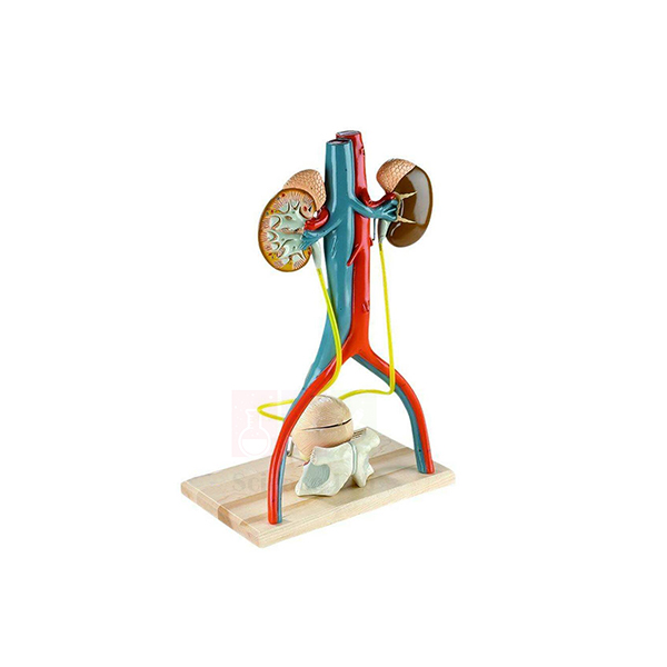
Denoyer-Geppert Free-Standing Human Urinary System Model
The Denoyer-Geppert Free-Standing Human Urinary System Model (Product Code: SLE/A/138) is a fully three-dimensional, larger-than-life replica of the male urinary apparatus and its blood supply. Unlike flat plaque models that present only half the picture, it stands on its own so students can examine the kidneys, bladder and prostate from every side. Anatomy departments, nursing schools and pharmacy colleges use it for renal and pelvic teaching, and it is supplied by Jain Scientific Equipments Pvt Ltd (ScienceLab), Ambala, a biology lab equipment supplier in India.
Specifications
| Parameter | Detail |
|---|---|
| Size | 12 × 19 × 8 in (30 × 48 × 20 cm) |
| Scale | Larger than life |
| Construction | Free-standing, fully 3-dimensional; molded entirely of non-breakable vinyl polymer |
| Part 1 | Kidneys, adrenals and blood vessels |
| Part 2 | Section of the pelvic bones with pubic symphysis, lower portion of the bladder and prostate |
| Part 3 | Upper portion of the bladder and prostate |
| Kidney section | Right kidney sagittally sectioned, revealing cortex, medulla, pyramids, calyces, pelvis and the ramifications of the renal artery and vein |
| Bladder interior | Opening the bladder exposes the folded mucosa, ureteric orifices, trigone, prostatic urethra, seminal vesicles, ejaculatory ducts and vas deferens |
| Ureters | Flexible plastic tubing |
| Key | Accompanying key identifying fifty hand-numbered features |
Typical Use
Lecturers can follow the whole urinary pathway in three dimensions: blood reaching the kidneys through the renal vessels, filtration explained on the sectioned right kidney, urine carried down the flexible ureters, and storage and outflow at the bladder. Opening the bladder brings the trigone and ureteric openings into view, while the pelvic bone section shows exactly where the bladder and prostate sit behind the pubic symphysis, a relationship that is hard to grasp from a flat diagram.
Because the model is free-standing and made of non-breakable vinyl, students can pick it up and turn it during practicals, and the key to fifty numbered features makes it suitable for spotting tests. It is a durable choice among pharmacy college lab equipment and anatomy teaching aids where renal structure underpins lessons on drug excretion and urinary disorders.
Supplier
This model is offered by Jain Scientific Equipments Private Limited, which operates under the ScienceLab name from Ambala, Haryana and sources and supplies teaching models for institutions and distributors. Our company profile gives more background.
Related Human Models
Models and charts used in the same renal and urinary teaching sequence:
- Human Kidney, Nephron, and Glomerulus Model Set — takes the lesson down to nephron and glomerulus level
- Human Kidney with Adrenal Gland Model — a closer look at a single kidney and its adrenal gland
- Human Male and Female Urogenital Systems Model — adds the female pelvis for comparison
- Male Pelvis Section Model — a sectional view of the male pelvic organs
- Human Excretory System — a laminated chart for classroom display and revision
More options are listed in our Human Models section.
Frequently Asked Questions
How is this different from a plaque-mounted urinary model?
It is fully three-dimensional and free-standing, so the urinary organs and their blood supply can be studied from all aspects rather than from one face only.
Does it show the female urinary system?
No. It represents the male urinary apparatus, including the prostate and seminal vesicles. For a male-female comparison, see the Human Male and Female Urogenital Systems Model.
How many parts does it separate into?
Three: the kidneys with adrenals and blood vessels; the pelvic bone section with the lower bladder and prostate; and the upper bladder and prostate.
Is it robust enough for student handling?
It is molded entirely of non-breakable vinyl polymer, with flexible tubing for the ureters, which suits hands-on practical sessions.
Is a key provided?
Yes. An accompanying key identifies fifty hand-numbered features on the model.
Can I confirm the brand and country of origin before ordering?
Yes. The name carries the Denoyer-Geppert brand; contact us for brand, origin and delivery details with your quotation.
Request a Quote
Send your quantity and delivery location through our contact page, or use the enquiry cart to request this model with other items. We supply India and international markets.
Last updated: 24 September 2026
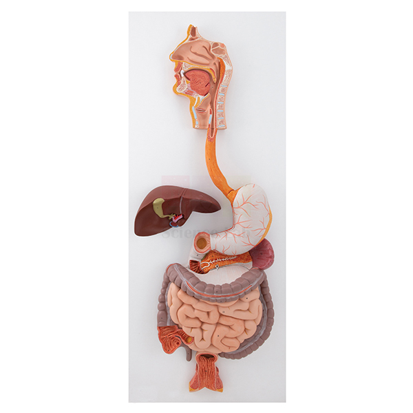
Human Digestive System Model
The Human Digestive System Model (Product Code: SLE/A/139) presents the entire digestive tract from mouth to rectum at about life size, together with the liver and pancreas, so a class can follow food through every stage of digestion on one model. A separate head section adds the chewing muscles and salivary glands. It is made by Jain Scientific Equipments Pvt Ltd (ScienceLab), Ambala, for secondary schools, colleges and nursing institutes planning a biology lab setup in India or overseas.
Specifications
| Parameter | Detail |
|---|---|
| Scale | About life size |
| Size (with base) | 92 × 32 cm |
| Second listed size | 15 × 20 cm (the part this refers to is not specified; please confirm with us) |
| Coverage | Entire digestive tract from mouth to rectum, including the liver and pancreas |
| Dissected views | Open dissection of the stomach; interior views of the small and large intestines |
| Head section | Separate section showing the musculature involved in mastication and the salivary glands |
| Key | All structures numbered and referenced on the accompanying key |
Typical Use
A digestion lesson can start with the separate head section, where students see how the chewing muscles and salivary glands begin mechanical and chemical breakdown, and then move down the tract. The opened stomach and the interior views of the small and large intestines show how the lining changes along the way, while the liver and pancreas explain where bile and digestive enzymes come from.
With every structure numbered and listed on the key, teachers can set identification exercises and practical-exam stations without extra labelling. The model suits high school biology lab equipment lists as well as nursing and nutrition courses that need a complete overview before moving on to individual organ models.
Manufacturer
Jain Scientific Equipments Private Limited makes this model and markets its educational range as ScienceLab; the company is located in Ambala, Haryana, India. Visit the company profile to learn more about us.
Related Human Models
Organ models and slides that deepen the same digestion topic:
- Human Stomach Model — a dissectible stomach for closer study of its wall and interior
- Human Liver With Gall Bladder Model — the liver lobes, gall bladder and bile ducts in detail
- Pathology of the Human Pancreas, Duodenum, and Gallbladder Model — shows common diseases of the organs seen here in healthy form
- Human Digestive Tract Model — an alternative whole-tract model on a single base
- Prepared Microscope Slide Set – Digestion (Liver, Pancreas And Tongue) — tissue sections to connect the model with microscopy
Browse the whole Human Models range for more system models.
Frequently Asked Questions
Does the model cover the whole digestive system?
Yes. It shows the entire tract from mouth to rectum, including the liver and pancreas, plus a separate head section for chewing and saliva.
Which organs are opened up?
The stomach is shown as an open dissection and the small and large intestines have interior views.
Is there a key for the numbered structures?
Yes. All structures are numbered and referenced on the accompanying key.
What are the dimensions?
The size with base is listed as 92 × 32 cm. A second size of 15 × 20 cm also appears in our records without saying which part it refers to, so please contact us to confirm before planning display space.
How should it be looked after?
Wipe with a soft, lightly damp cloth, keep it out of direct sunlight, and store the separate head section with the main model so it is not misplaced.
What information do you need for a school or college quotation?
The number of units, the delivery address and any tender or purchase-order format you must follow. We deliver across India and to international markets.
Request a Quote
Ask for pricing on the Human Digestive System Model through our contact page, or collect it with other items in the enquiry cart and send one request.
Last updated: 24 September 2026
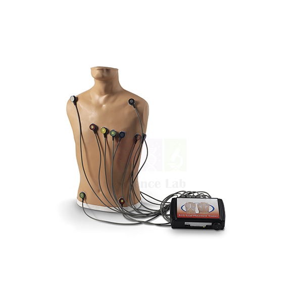
15-Lead ECG Placement Trainer
The 15-Lead ECG Placement Trainer (Product Code: SLE/A/141) is an adult trainer for learning where ECG electrodes belong and how accurately they have been placed, for recordings of up to 15 leads. Nursing, paramedical, cardiac-technology and medical students practise on its anatomical landmarks before working with patients, making it a practical aid for electrocardiograph (ECG) teaching in India lab and skills-centre settings. It is made by Jain Scientific Equipments Pvt Ltd (ScienceLab), Ambala.
Specifications
| Parameter | Detail |
|---|---|
| Type | Adult ECG electrode placement trainer |
| Lead capacity | Teaches placement for up to 15-lead ECG |
| Connection sites | 4 limb leads and V1 through V9 |
| Chest lead side | Right- or left-sided electrodes can be attached |
| Anatomical landmarks | Intercostal spaces, midclavicular line, anterior axillary line, midaxillary line and scapula |
| Signal output | Does not provide ECG output signals |
| Rhythm simulation | Rhythm and hands-free defibrillation simulations possible by adding any ECG arrhythmia simulator directly to an ECG monitor |
Typical Use
Misplaced chest electrodes are a common source of misleading ECG tracings, so this trainer focuses on the skill that comes before interpretation: finding the right spot. Learners palpate and count intercostal spaces, locate the midclavicular, anterior axillary and midaxillary lines, and then position the standard chest leads and the extra posterior and right-sided leads that make up an extended recording. The scapula landmark supports practice of the posterior positions such as V7 to V9.
Instructors can check each student’s placement against the connection sites and repeat the exercise as often as needed without a volunteer. When rhythm recognition or defibrillation drills are required, an ECG arrhythmia simulator can be connected directly to the ECG monitor, keeping this unit focused on placement. It fits well in vocational training lab equipment for paramedical and emergency-care courses.
Manufacturer
This trainer is a product of Jain Scientific Equipments Private Limited, an Ambala, Haryana manufacturer that trades as ScienceLab and supplies teaching equipment to medical, nursing and technical institutions. See the company profile for more information.
Related Human Models
Equipment often set up in the same clinical-skills room:
- Advanced Adult CPR Training Manikin — for chest-compression and rescue-breathing practice alongside ECG skills
- Human Heart Model — explains the heart anatomy that each lead views
- Stethoscope — for pairing heart-sound auscultation with electrode placement practice
- Blood Pressure Monitor — completes a basic vital-signs training station
Find more trainers and models in our Human Models category.
Frequently Asked Questions
Does the trainer produce an ECG trace?
No. It does not provide ECG output signals. Rhythm and hands-free defibrillation simulations can be run by adding an ECG arrhythmia simulator directly to an ECG monitor.
Is an arrhythmia simulator or ECG monitor included?
Neither is listed as part of this trainer. Contact us if you need advice on what to pair with it.
Which electrode positions can be practised?
There are connection sites for 4 limb leads and V1 through V9, and electrodes can be attached on the right or left side.
Is it an adult or a paediatric trainer?
It is an adult trainer, with landmarks such as the intercostal spaces, midclavicular line, axillary lines and scapula.
Are spare parts and after-sales support available?
Please ask us about spares and warranty terms when you request a quotation, as these are confirmed per order.
Can you supply several trainers for a nursing or paramedical college?
Yes. We supply institutions in India and international markets; include the quantity and delivery location in your enquiry.
Request a Quote
For a quotation on the 15-Lead ECG Placement Trainer, contact us or add it to the enquiry cart with any other skills-lab items you need.
Last updated: 24 September 2026
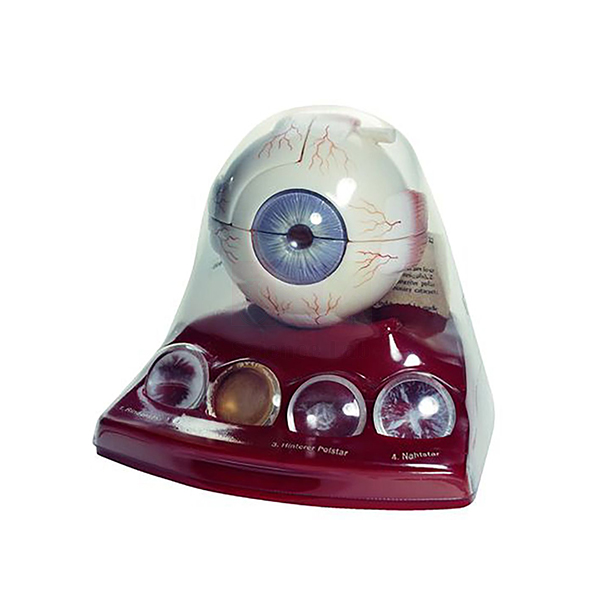
Cataract Eye Model with 4 Forms of Cataract
The Cataract Eye Model with 4 Forms of Cataract (Product Code: SLE/A/142) is an enlarged eye on a base, supplied with four examples of lens clouding (cortical, nuclear, posterior and coronary cataract) that sit in labeled compartments in the base. Optometry and ophthalmic-assistant courses, nursing schools and patient-education desks use it to explain how each type of cataract affects vision, and it serves as a focused eye model for anatomy and pathology teaching in India. It is made by Jain Scientific Equipments Pvt Ltd (ScienceLab), Ambala.
Specifications
| Parameter | Detail |
|---|---|
| Scale | About 3× life size |
| Size | 13 × 16 × 15 cm |
| Cataract forms included | 4: cortical, nuclear, posterior and coronary |
| Mounting | Eye mounted on base |
| Storage | Cataract forms rest in labeled compartments at the bottom of the base |
Typical Use
Cataracts are among the most common causes of reduced vision, and they are easier to understand when learners can see where the opacity forms within the lens. With the four forms laid out beside the eye, a tutor can contrast clouding at the centre of the lens (nuclear), in its outer layers (cortical), at the back (posterior) and in a crown-like ring (coronary), and discuss how the position of the opacity changes symptoms such as glare or blurred near vision.
The labeled compartments keep each example identified and stored with the model, which makes it practical for counselling patients before surgery as well as for classroom teaching. Schools can also include it in a health education kit on eye care and ageing.
Manufacturer
ScienceLab is the brand under which Jain Scientific Equipments Private Limited of Ambala, Haryana manufactures and sells its anatomy and laboratory products, and it produces this cataract model. The company profile has further details.
Related Human Models
Eye models that pair well with this cataract model:
- Human Eye Model — a bisected eyeball for teaching the healthy lens and focusing first
- Eye Microanatomy Model — magnified retinal layers for the back of the eye
- Human Eye in Orbit Model — shows the eye and optic nerve in the bony orbit
- Human Eye Model with Eyelids — adds eyelids, eye muscles and tear apparatus
For other sense-organ and anatomy models, see the Human Models range.
Frequently Asked Questions
Which types of cataract does the model show?
Four: cortical, nuclear, posterior and coronary cataract.
Where are the cataract examples kept?
They rest in labeled compartments at the bottom of the base, so each form stays identified and stored with the eye.
Can each cataract form be fitted into the eye?
Our documentation lists them as separate pieces stored in the base but does not describe how they are exchanged. Please contact us to confirm before ordering.
How large is the model?
It is about 3× life size and measures 13 × 16 × 15 cm, compact enough for a consulting desk.
How do I keep the model clean?
Dust with a soft cloth, avoid solvents, and return the four cataract pieces to their compartments after use so none are lost.
Do you supply eye clinics and training institutes outside India?
Yes. We serve buyers in India and international markets; send your quantity and destination for a quotation.
Request a Quote
To request pricing, contact us or add the Cataract Eye Model with 4 Forms of Cataract to the enquiry cart.
Last updated: 24 September 2026
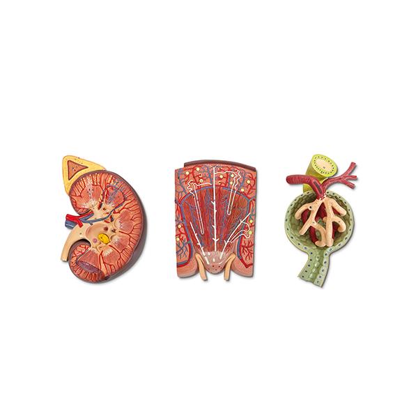
Human Kidney, Nephron and Renal Corpuscle Model
Human Kidney, Nephron and Renal Corpuscle Model (Product Code: SLE/A/143) is a plaque-mounted series of 3 greatly enlarged models used to teach renal physiology from organ to filtering unit in medical, nursing, pharmacy and senior school programmes. It combines a 2 1/2× life-size dissection of the kidney and adrenal gland, a schematic nephron and a renal corpuscle on a plaque measuring 69 × 29 × 10 cm overall. It is made by Jain Scientific Equipments Pvt Ltd (ScienceLab), an OEM manufacturer, at its own factory in Ambala, India.
Specifications
| Parameter | Detail |
|---|---|
| Format | Plaque-mounted series of 3 models |
| Overall size | 69 × 29 × 10 cm |
| Scale | Greatly enlarged |
| Model 1 | 2 1/2× life-size dissection of the kidney and adrenal gland |
| Model 2 | Schematic enlargement showing anatomical details of an individual nephron |
| Model 3 | Renal corpuscle with Bowman's capsule, glomerulus and juxtaglomerular apparatus |
| Product code | SLE/A/143 |
| Manufacturer | Jain Scientific Equipments Pvt Ltd (ScienceLab) — OEM |
| Country of origin | India (made in Ambala, Haryana) |
What Is the Kidney, Nephron and Renal Corpuscle Model Used For?
The three models on one plaque follow the order in which urine formation is usually taught. Lecturers begin with the dissected kidney, move to the nephron to explain how fluid is modified as it passes along the tubule, and finish at the renal corpuscle, where blood is filtered from the glomerulus into Bowman's capsule. The juxtaglomerular apparatus then gives a starting point for discussing how the kidney helps regulate blood pressure.
Because everything is fixed to one plaque, the full sequence stays together for display in a physiology laboratory or museum room and is quick to set up for a lecture. It is a strong addition to biology teaching aids in India for courses that examine the nephron in detail. Dust the plaque regularly and avoid propping it where it could slip.
OEM Manufacturer
Jain Scientific Equipments Private Limited, known by its trading name ScienceLab, is the OEM (original equipment manufacturer) of this renal plaque series and makes it at its own licensed factory at Saha, Ambala, Haryana, India. Medical colleges, schools, dealers and importers buy it factory-direct instead of through a trader, and repeat orders or questions about the plaque are answered by the manufacturer itself; ask us. Visit our company profile for more.
Related Human Models
Kidney models that complement this enlarged series:
- Human Kidney with Adrenal Gland Model — a life-size, two-part frontal section for hands-on study at real scale.
- Human Kidney, Nephron, and Glomerulus Model Set — an alternative set with the glomerulus enlarged 700×.
- Human Kidney Model — a life-size single-organ model with laminated key for introductory classes.
- Kidney Stone Model — connects renal anatomy to a common clinical problem.
It is catalogued in our Human Models category alongside other organ models.
Frequently Asked Questions
What are the three models in the Human Kidney, Nephron and Renal Corpuscle Model?
The Human Kidney, Nephron and Renal Corpuscle Model combines a 2 1/2× life-size dissection of the kidney and adrenal gland, a schematic enlargement of an individual nephron, and a renal corpuscle with Bowman's capsule, glomerulus and juxtaglomerular apparatus.
How much space does the model need?
The Human Kidney, Nephron and Renal Corpuscle Model measures 69 × 29 × 10 cm overall, so it suits a wall display area, a museum cabinet or a large lecture bench rather than a small student table.
Is the nephron shown to scale?
No. In the Human Kidney, Nephron and Renal Corpuscle Model the nephron is a schematic enlargement, simplified to make its anatomical details clear rather than to reproduce the exact proportions of a real nephron.
Can the models be removed from the plaque?
The Human Kidney, Nephron and Renal Corpuscle Model is supplied plaque-mounted, and the product details do not say whether the models detach. Contact us if you need that confirmed before ordering.
Who manufactures the Human Kidney, Nephron and Renal Corpuscle Model?
Jain Scientific Equipments Pvt Ltd, trading as ScienceLab, is the OEM manufacturer of the Human Kidney, Nephron and Renal Corpuscle Model, making it at its own licensed factory at Saha, Ambala, Haryana, India, and supplying it factory-direct.
What do you need to prepare a quotation for a college?
For the Human Kidney, Nephron and Renal Corpuscle Model, send product code SLE/A/143, the number of sets, the delivery address in India or abroad and your purchase procedure, and the manufacturer will prepare a matching quotation.
Request a Quote
Get a manufacturer-direct price for this kidney, nephron and renal corpuscle series by messaging us through the contact page, or save it in your enquiry cart while you browse other renal models.
Last updated: 24 September 2026
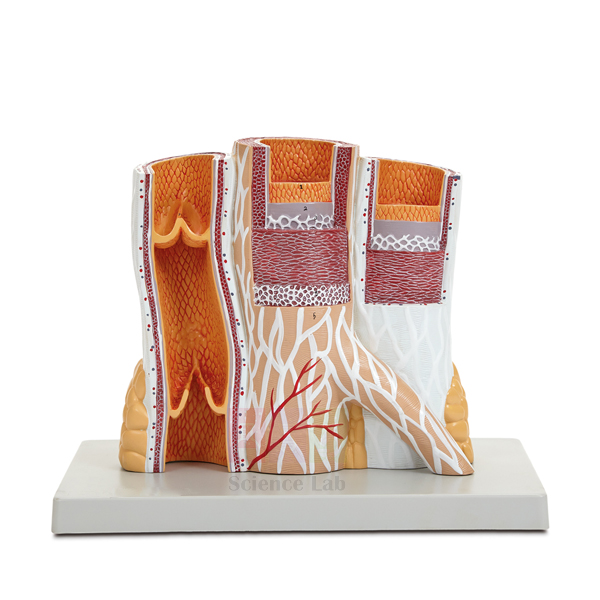
Microstructure of Artery and Vein Model
Microstructure of Artery and Vein Model (Product Code: SLE/A/144) is an enlarged block model used to teach the layered wall structure of arteries and veins in histology, physiology, nursing and school biology classes. Serial sectioning exposes the sequential muscular and membranous layers and shows the difference between arterial and venous wall thickness, while a cut section of vein with venous valves and a numbered key are included. It is made by Jain Scientific Equipments Pvt Ltd (ScienceLab), an OEM manufacturer, at its own factory in Ambala, India.
Specifications
| Parameter | Detail |
|---|---|
| Type | Enlarged block model of artery and vein microstructure |
| Size (without base) | 25 × 10 × 23 cm |
| Sectioning | Serial sectioning exposes sequential muscular and membranous layers of the vessel walls |
| Comparison shown | Differences between arterial and venous wall thickness |
| Additional piece | Cut section of vein with venous valves |
| Mounting | Durable polymer base |
| Supplied with | Numbered key |
| Product code | SLE/A/144 |
| Manufacturer | Jain Scientific Equipments Pvt Ltd (ScienceLab) — OEM |
| Country of origin | India (made in Ambala, Haryana) |
What Is the Artery and Vein Model Used For?
Under a microscope, students often find it hard to tell an artery from a vein on a prepared slide. This enlarged block gives them a three-dimensional reference first: the stepped serial sections show each layer of the wall in turn, and placing the two vessels next to each other makes it obvious that the artery has the thicker, more muscular wall needed to cope with higher pressure. The teacher can then move to the microscope and ask the class to apply what they have just seen.
The cut vein with its valves is ideal for explaining how blood returns to the heart against gravity and why faulty valves lead to varicose veins. With the numbered key, the model also works for self-directed revision in the lab, and it sits naturally beside heart model anatomy lessons in India's school and college curricula on the circulatory system.
OEM Manufacturer
This vessel model comes from ScienceLab, the trading name of Jain Scientific Equipments Private Limited, which is the OEM (original equipment manufacturer) and makes it at its own licensed factory at Saha, Ambala, Haryana, India. The company is a registered MSME manufacturer (Udyam UDYAM-HR-01-0003714), and its export consignments move under Importer-Exporter Code AAECJ8618A issued by the DGFT.
Colleges, schools, dealers and importers buy the model factory-direct, so quotations, repeat orders for additional teaching rooms and any question about the numbered key are answered by the maker itself. Read the company profile for more.
Related Human Models
These models and slides support the same circulatory-system lessons:
- Microstructure of Smooth Muscle Model — explains the muscle tissue that forms the vessel wall layers.
- Human Heart Model — shows where the arteries leave and the veins return.
- Human Heart, Pumping Model — demonstrates the pumping action that drives blood through these vessels.
- Human Circulatory System Model — places arteries and veins in the context of the whole circulation.
- Prepared Microscope Slide Set – Circulatory System — real tissue for the microscope follow-up to the model.
Browse the Human Models category for more anatomy and histology teaching models.
Frequently Asked Questions
What does the serial sectioning on the artery and vein model show?
On the Microstructure of Artery and Vein Model, serial sectioning exposes the sequential muscular and membranous layers of each vessel wall and highlights how much thicker the arterial wall is than the venous wall, the key difference students must recognise.
Are venous valves included with the Microstructure of Artery and Vein Model?
Yes. The Microstructure of Artery and Vein Model includes a cut section of vein with venous valves, so teachers can show how valves stop blood flowing backwards as it returns to the heart.
What size is the Microstructure of Artery and Vein Model?
The Microstructure of Artery and Vein Model measures 25 × 10 × 23 cm without its base and is mounted on a durable polymer base, a size that is clearly visible from the front rows of a classroom.
Does the artery and vein model come with a key?
Yes. The Microstructure of Artery and Vein Model is supplied with a numbered key, so students can identify each labelled layer and structure on their own during revision or practical sessions.
Is the Microstructure of Artery and Vein Model suitable for schools as well as colleges?
Yes. The enlarged block and simple artery-versus-vein comparison of the Microstructure of Artery and Vein Model suit school biology, while its layer-by-layer detail supports histology, physiology and nursing courses at college level.
Who manufactures the Microstructure of Artery and Vein Model?
The Microstructure of Artery and Vein Model is manufactured by Jain Scientific Equipments Pvt Ltd (ScienceLab), the OEM, at its own licensed factory at Saha, Ambala, Haryana, India, and is supplied factory-direct to institutions and dealers in India and international markets.
Are calibration or test documents available for the artery and vein model?
The Microstructure of Artery and Vein Model is a teaching model, not a measuring device, so calibration does not apply. Any documents your procurement process needs can be discussed through our contact page.
Request a Quote
Ask for a quotation on SLE/A/144 through the contact page, or add it to the enquiry cart along with heart and circulation models. The quotation comes direct from the manufacturer.
Last updated: 24 September 2026
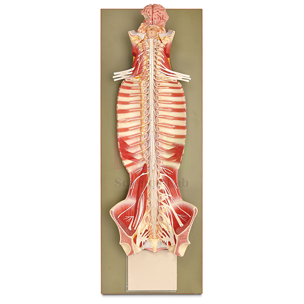
Human Nervous System Model
The Human Nervous System Model (Product Code: SLE/A/145) is a life-size, base-mounted model that shows the brain, brain stem, spinal cord and spinal nerves in their natural position within the vertebral column, with the sympathetic trunk added on the left side. It gives school, college and nursing students a single view of how the central and peripheral parts of the nervous system connect, and is part of the biology lab equipment in India made by Jain Scientific Equipments Pvt Ltd (ScienceLab), Ambala.
Specifications
| Parameter | Detail |
|---|---|
| Scale | Life size |
| Size | 32 × 19 × 90 cm |
| Mounting | On base |
| Structures shown | Brain, brain stem, spinal cord and spinal nerves, in position in the vertebral column |
| Autonomic detail | Sympathetic trunk shown on the left side |
Typical Use
Because the brain and cord are shown inside the vertebral column, students can see how the spinal nerves leave between the vertebrae and why injuries at different levels of the spine affect different parts of the body. The sympathetic trunk running alongside the column on the left side introduces the autonomic nervous system and its chain of ganglia, a topic that is often hard to picture from textbook diagrams alone.
Being life size, the model reads well from the back of a classroom and can stand beside a human skeleton model so learners relate the nerves to the bones that protect them. It is used in secondary biology, physiotherapy, nursing and first-year medical anatomy for lectures, practical identification and exam revision.
Manufacturer
Made by Jain Scientific Equipments Private Limited, a private limited company in Ambala, Haryana whose teaching and laboratory products carry the ScienceLab brand. Further company information is on our company profile.
Related Human Models
Models that break the nervous system down into its parts:
- Human Brain Model, 4 Parts — a hands-on brain for studying lobes and the brain stem in detail
- Human Spinal Cord Section Model — an enlarged cross section of the cord and its nerve roots
- Human Vertebral And Partial Spinal Cord Model — how a cord segment sits within the vertebrae
- Neuron Model — the single nerve cell that makes up every pathway on this model
- Nervous System Model — a markable model with activity guide for younger classes
Our Human Models category lists further system models.
Frequently Asked Questions
What parts of the nervous system are shown?
The brain, brain stem, spinal cord and spinal nerves in position in the vertebral column, plus the sympathetic trunk on the left side.
How much space does it need?
It measures 32 × 19 × 90 cm on its base, so check your shelf or cupboard dimensions before it arrives.
Does it show the nerves of the arms and legs?
Our documentation lists the spinal nerves and sympathetic trunk only; for limb nerve detail, please contact us before ordering.
Can the brain be removed from the model?
Removable parts are not mentioned in our product details. Ask us to confirm if you need a dissectible version.
Is a numbered key supplied?
A key is not listed for this model; we can confirm the accompanying teaching material with your quotation.
Can it be delivered outside India?
Yes. We supply customers across India and international markets; tell us the destination so we can quote packing and delivery.
Request a Quote
Use our contact page or the enquiry cart to ask for a quotation on the Human Nervous System Model.
Last updated: 24 September 2026
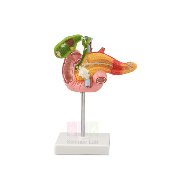
Pathology of the Human Pancreas, Duodenum, and Gallbladder Model
The Pathology of the Human Pancreas, Duodenum, and Gallbladder Model (Product Code: SLE/A/146) is a life-size model on a base that brings together the most important diseases of three closely linked digestive organs: gallstones, inflammation, polyps and cancer of the gallbladder, pancreatitis and pancreatic cancer, and a duodenal ulcer. Pathology and medical teaching departments, nursing colleges and gastroenterology clinics use it to explain disease to students and patients. It is made by Jain Scientific Equipments Pvt Ltd (ScienceLab), Ambala, an anatomical models manufacturer.
Specifications
| Parameter | Detail |
|---|---|
| Scale | Life size |
| Size (with base) | 11 × 11 × 24 cm |
| Mounting | On a base |
| Gallbladder | Partially opened; shows stones in various locations, cholecystitis, polyposis and carcinoma |
| Pancreas | Pancreatitis in the pancreas tail and cancer in the pancreas head |
| Duodenum | Section of the duodenum with a duodenal ulcer |
Typical Use
The gallbladder, pancreas and duodenum share ducts and blood supply, so disease in one often affects the others. This model lets a lecturer show, on one compact piece, how gallstones lodged in different places can cause pain or obstruction, why an inflamed or polypoid gallbladder needs follow-up, and how a tumour in the head of the pancreas differs in position and consequences from inflammation in its tail. The duodenal ulcer section adds a common condition that students need to recognise.
Clinicians use the same model at the desk to show patients what a diagnosis means before treatment or surgery. In pharmacology lab equipment collections it helps link drug classes, such as those used for acid-related ulcers or pancreatic enzyme support, to the organs they act on.
Manufacturer
Our company, Jain Scientific Equipments Private Limited, produces this pathology model in Ambala, Haryana and sells it under the ScienceLab trade name. Learn more on the company profile page.
Related Human Models
Normal-anatomy and pathology items that complement this model:
- Human Spleen, Duodenum, and Pancreas Model — the healthy organs for side-by-side comparison
- Human Liver and Gallbladder Model — normal liver and gallbladder anatomy before discussing gallstones
- Human Liver Pathology Model — extends the pathology lesson to the liver
- Prepared Microscope Slide Set – Digestion (Liver, Pancreas And Tongue) — normal tissue sections of the pancreas and related organs
See the wider Human Models range for more pathology and organ models.
Frequently Asked Questions
Which conditions are shown on the model?
Gallstones in various locations, cholecystitis, polyposis and carcinoma of the gallbladder; pancreatitis in the pancreas tail and cancer in the pancreas head; and a duodenal ulcer.
Is healthy anatomy shown as well?
The model concentrates on disease. For a normal comparison, pair it with the Human Spleen, Duodenum, and Pancreas Model.
How big is it?
It is life size and measures 11 × 11 × 24 cm including the base, small enough for a clinic desk.
Does it come with a description card or key?
A key is not listed in our documentation for this model. Please contact us to check what accompanies it.
How should it be cleaned in a clinical setting?
Wipe with a soft cloth dampened with mild soapy water and dry it; avoid solvents and harsh disinfectants that may damage painted surfaces.
What do you need to prepare a quotation for a hospital or college?
The quantity, delivery location, and any purchase or tender format. We supply India and international markets.
Request a Quote
To price this pathology model, contact us or add it to the enquiry cart together with any normal-anatomy models you need.
Last updated: 24 September 2026
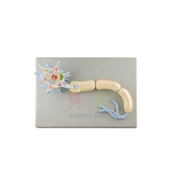
Neuron Model
The Neuron Model (Product Code: SLE/A/147) enlarges a typical myelinated nerve cell about 2,500 times, from the dendrites and cell body to the axon wrapped in its myelin sheath, and shows the organelles inside the cell body. Part of the myelin can be removed to reveal the interior of the axon. Biology teachers, physiology lecturers and psychology departments use it as cell biology equipment in India and elsewhere, and it is made by Jain Scientific Equipments Pvt Ltd (ScienceLab), Ambala.
Specifications
| Parameter | Detail |
|---|---|
| Magnification | About 2,500× life size |
| Size | 53 × 38 × 17 cm |
| Base | Mounted on a durable polymer base |
| Cell body | Nucleus and nucleolus, Golgi complex, mitochondria and endoplasmic reticulum |
| Processes | Dendrites and axon |
| Myelin sheath | With nodes of Ranvier and Schwann cells; a portion can be removed to view the interior of the axon |
| Key | Numbered key included |
Typical Use
The model is ideal for explaining how structure supports function in nerve cells: dendrites receive signals, the cell body with its nucleus, Golgi complex, mitochondria and endoplasmic reticulum keeps the cell supplied, and the axon carries impulses away. Lifting off part of the myelin sheath shows what lies underneath, while the nodes of Ranvier and Schwann cells set up a clear explanation of why myelinated fibres conduct faster.
Teachers often use it straight after an animal cell model lesson, so students can spot familiar organelles in a specialised cell. The numbered key supports labelling worksheets, practical exams and self-study, from senior secondary biology to undergraduate neuroscience.
Manufacturer
Jain Scientific Equipments Private Limited, trading as ScienceLab, builds this neuron model and runs its business from Ambala in Haryana, India. The company profile explains more about who we are.
Related Human Models
Resources for the same lesson on nerve cells and impulses:
- Human Nerve Ending and Synapse Model — follows the axon to the point where the signal passes to the next cell
- Dissectible Human Neuron — a simpler schematic neuron for introductory classes
- Denoyer-Geppert 3-D Human Motor Neuron Model — a motor neuron shown ending on skeletal muscle
- Microslide Nerve & Muscle Action — real microscope images of nerve and muscle to compare with the model
- Micro Slide Viewer — needed to view Microslide image strips
More models are available in our Human Models range.
Frequently Asked Questions
Which cell structures are shown?
The cell body with nucleus and nucleolus, dendrites and axon, Golgi complex, mitochondria, endoplasmic reticulum, and the myelin sheath with nodes of Ranvier and Schwann cells.
Can any part be removed?
Yes. A portion of the myelin sheath can be removed to show the interior of the axon.
Is a key included?
Yes, the model comes with a numbered key.
How is it different from the Dissectible Human Neuron?
This model shows organelle-level detail of a myelinated neuron at about 2,500× on a polymer base, whereas the Dissectible Human Neuron is a schematic nerve cell that separates into parts on a stand.
How large is it?
It measures 53 × 38 × 17 cm, large enough for whole-class demonstration.
Can you supply it for several classrooms or for export?
Yes. We supply schools, universities and distributors in India and international markets; include the quantity with your enquiry.
Request a Quote
Request pricing through our contact page, or add the Neuron Model to the enquiry cart along with any other items you need.
Last updated: 24 September 2026
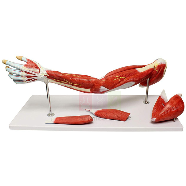
Human Muscular Arm Model
The Human Muscular Arm Model (Product Code: SLE/A/148) is a life-size upper limb that shows the muscles of the shoulder, arm and hand in great detail, along with the significant nerves and blood vessels, and comes apart into 7 parts so deeper layers can be studied. Physiotherapy, sports science, nursing and medical students use it for limb anatomy, and it is among the anatomy lab equipment for colleges in India made by Jain Scientific Equipments Pvt Ltd (ScienceLab), Ambala.
Specifications
| Parameter | Detail |
|---|---|
| Scale | Life size |
| Dissection | Dissectible into 7 parts |
| Size | 74 × 18 × 11 cm |
| Regions covered | Shoulder, arm and hand |
| Structures shown | Muscles in great detail, with significant nerves and blood vessels |
Typical Use
Upper-limb anatomy is taught layer by layer, and a dissectible model lets students do the same: start with the superficial muscles around the shoulder and arm, remove parts to reach deeper muscles, and follow the main nerves and vessels as they run towards the hand. It helps explain muscle actions such as flexing the elbow or gripping, and why an injury to a particular nerve weakens a specific group of muscles.
Physiotherapy and sports-science tutors use it when discussing common strains and rehabilitation exercises, while biology teachers can place it next to a human skeleton model to show how muscles attach across joints. Being life size, it also makes a clear reference for surface-anatomy and palpation practice.
Manufacturer
This arm model is manufactured by Jain Scientific Equipments Private Limited, which has built its ScienceLab brand in Ambala, Haryana around educational models and lab apparatus. Visit our company profile for more about the business.
Related Human Models
Models that support the same upper-limb lesson:
- Human Arm and Hand model — the skeleton of the same limb, from shoulder girdle to fingers
- Human Elbow Joint Model — shows the joint that many arm muscles act across
- Human Skeletal Hand Model — the small bones of the wrist and hand where forearm tendons insert
- Skeletal Muscle Fiber Model — explains how the muscles on this model contract at fibre level
Our Human Models range includes further muscular and limb models.
Frequently Asked Questions
What does the model show?
The muscles of the shoulder, arm and hand in great detail, together with the significant nerves and blood vessels of the limb.
How many parts does it separate into?
It is dissectible into 7 parts. The exact breakdown of parts is not detailed in our documentation, so contact us if you need it for a tender specification.
Is it a left or a right arm?
The side is not stated in our current product details; please ask us to confirm before ordering.
What are its dimensions?
It is life size and measures 74 × 18 × 11 cm.
Any tips for looking after a dissectible model?
Reassemble all parts after each class, store the model where it will not be knocked over, and clean it only with a soft, slightly damp cloth.
Can you supply physiotherapy and medical colleges in bulk?
Yes. We supply institutions in India and international markets; share your quantities and delivery details for a quotation.
Request a Quote
For price and availability of the Human Muscular Arm Model, contact us or add it to the enquiry cart.
Last updated: 24 September 2026
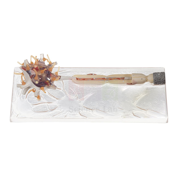
Dissectible Human Neuron
The Dissectible Human Neuron (Product Code: SLE/A/150) is a schematic model of a nerve cell and its axon, showing the medullary (myelin) sheath and Schwann’s sheath, that separates into 3 parts and stands on its own base. Its simplified, uncluttered design suits secondary school and pre-university classes that need to learn the basic parts of a neuron before moving to more detailed cell biology. It is one of the biology teaching aids in India made by Jain Scientific Equipments Pvt Ltd (ScienceLab), Ambala.
Specifications
| Parameter | Detail |
|---|---|
| Representation | Schematic nerve cell and axon |
| Sheaths shown | Medullary sheath and Schwann’s sheath |
| Dissection | Dissectible into 3 parts |
| Mounting | On stand with base |
| Size | 53 × 17 × 22 cm |
Typical Use
A schematic neuron strips away microscopic clutter so beginners can concentrate on the essentials: the cell body, the long axon that carries the impulse, and the two coverings around it. Teachers can separate the 3 parts to show how the axon sits inside its medullary sheath and how Schwann’s sheath lies outside, then reassemble the model on its stand to summarise the whole cell.
It is a practical piece of science lab equipment for class 12 and junior college biology chapters on neural control and coordination, and also works for introductory nursing and psychology courses. Because it is mounted on a stand, it can be left on display at the front of the class throughout a unit on the nervous system.
Manufacturer
The neuron is made in India by Jain Scientific Equipments Private Limited, an Ambala, Haryana enterprise that trades as ScienceLab and supplies schools and colleges with teaching models. For more on the company, open the company profile.
Related Human Models
Useful next steps after this introductory neuron:
- Neuron Model — a highly magnified neuron with organelles for advanced classes
- Human Nerve Ending and Synapse Model — shows how one neuron passes its signal to the next
- Human Nervous System Model — places neurons in the context of the brain, cord and spinal nerves
- Human Spinal Cord Section Model — where nerve cell bodies and fibres meet in the cord
Browse more in the Human Models category.
Frequently Asked Questions
What does “schematic” mean for this model?
It is a simplified representation of a nerve cell and axon, designed to show the main parts and sheaths clearly rather than every microscopic detail.
Which coverings of the axon are shown?
The medullary sheath and Schwann’s sheath.
Into how many parts does it separate?
Three. When assembled, it stands on its stand with base for display.
Is it suitable for school students?
Yes. The schematic style matches secondary and pre-university syllabi; for organelle-level detail, see the Neuron Model.
How big is it?
It measures 53 × 17 × 22 cm.
What should a school include in its purchase enquiry?
The number of models, the delivery address and any quotation format required by your school or department. We supply across India and to international markets.
Request a Quote
To receive a quotation for the Dissectible Human Neuron, contact us or add it to the enquiry cart.
Last updated: 24 September 2026
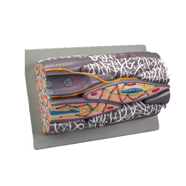
Microstructure of Smooth Muscle Model
The Microstructure of Smooth Muscle Model (Product Code: SLE/A/151) is a block model of a greatly enlarged bundle of smooth muscle fibers, revealing the structures inside and between the cells, from the nucleus, mitochondria and sarcoplasmic reticulum to the dense bodies and contractile myofilaments that produce contraction. Histology, physiology and zoology lab equipment buyers in India choose it to make an invisible tissue visible to a whole class. It is made by Jain Scientific Equipments Pvt Ltd (ScienceLab), Ambala.
Specifications
| Parameter | Detail |
|---|---|
| Type | Block model of a greatly enlarged bundle of smooth muscle fibers |
| Size | 28 × 16 × 11 cm |
| Base | Mounted on a durable polymer base |
| Structures shown | Significant extra- and intracellular structures, including sarcoplasmic reticulum, nucleus, mitochondria, dense bodies and contractile myofilaments |
Typical Use
Smooth muscle lines the walls of hollow organs and blood vessels, and its contraction works differently from the striated muscle students already know. On this block model, the nucleus, the mitochondria and the dense bodies that anchor the contractile myofilaments give the teacher a clear basis to explain why smooth muscle has no visible striations and how it can contract slowly and steadily.
Lecturers typically use it alongside prepared microscope slides of muscle tissue, so students first see the enlarged three-dimensional structure and then recognise the same tissue under the microscope. It suits undergraduate zoology, physiology, pharmacy and medical histology practicals.
Manufacturer
The model is manufactured by Jain Scientific Equipments Private Limited, an Indian company in Ambala, Haryana; its products for schools, colleges and laboratories are sold under the name ScienceLab. Details are on our company profile.
Related Human Models
Items for a complete muscle-tissue lesson:
- Skeletal Muscle Fiber Model — the striated counterpart, for a side-by-side comparison
- Microstructure of Artery and Vein Model — shows where smooth muscle layers sit in vessel walls
- Microslide Animal Tissue Set 2 Muscle, Bone, Connective — photographed microscope views of muscle and related tissues
- Micro Slide Viewer — for viewing the Microslide image strips without a microscope
For other microstructure models, see the Human Models range.
Frequently Asked Questions
Which cell structures can students identify?
The sarcoplasmic reticulum, nucleus, mitochondria, dense bodies and contractile myofilaments, plus significant structures between the fibers.
What magnification is the model?
It is described as greatly enlarged; an exact magnification is not stated in our documentation. Contact us if you need this figure.
How does it differ from a skeletal muscle model?
Smooth muscle has no sarcomere striations; its myofilaments are anchored to dense bodies, which this model shows clearly. Pair it with the Skeletal Muscle Fiber Model for comparison.
Where is smooth muscle found in the body?
In the walls of the digestive tract, blood vessels, airways, bladder and uterus, where it works involuntarily.
What size is it?
The block measures 28 × 16 × 11 cm and sits on a durable polymer base.
Can overseas universities order it?
Yes. We supply institutions and distributors in India and international markets; send the quantity and destination for a quotation.
Request a Quote
Ask for a quotation via our contact page, or place the Microstructure of Smooth Muscle Model in the enquiry cart with other histology items.
Last updated: 24 September 2026
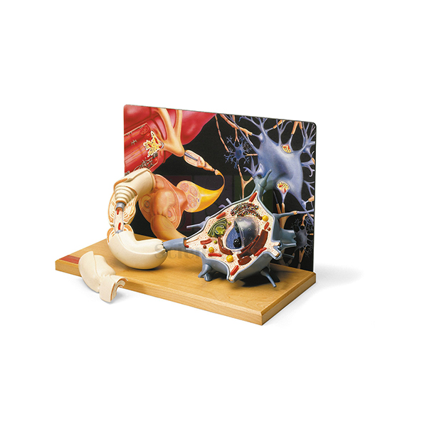
Denoyer-Geppert 3-D Human Motor Neuron Model
The Denoyer-Geppert 3-D Human Motor Neuron Model (Product Code: SLE/A/152) is a diorama that reproduces a motor nerve cell at more than 2,500 times life size against a colour backdrop of associated neurons, and follows its axon all the way to the neuromuscular junction on a skeletal muscle. Physiology, neuroscience, pharmacology and sports-science departments use it to connect nerve-cell structure with how muscles are made to contract. It is supplied by Jain Scientific Equipments Pvt Ltd (ScienceLab), Ambala, a biology lab instruments supplier.
Specifications
| Parameter | Detail |
|---|---|
| Magnification | More than 2,500× life size |
| Size | 43 × 20 × 28 cm |
| Base | Mounted on a hardwood base |
| Format | Diorama: detailed motor nerve cell with a colour backdrop showing a network of associated neurons |
| Removable section | A section of the axon lifts off to show the layers of the myelin sheath, the neurolemma and the Schwann cell that formed them |
| Neuromuscular detail | Convergence of multiple axons into a motor nerve fiber, ending at the neuromuscular junction of a skeletal muscle, with synaptic vesicles carrying neurotransmitters |
| Other highlights | Cellular organelles, branching dendrites, communicating synapses, and a myelin-wrapped axon with node of Ranvier |
| Key | Illustrated key with 54 identified features |
Typical Use
Most neuron models stop at the cell itself; this diorama carries the story through to its effect. Students can start with the dendrites and synapses receiving input, look at the organelles in the cell body, follow the myelinated axon past a node of Ranvier, and then see several axons converge into a motor nerve fiber that ends on skeletal muscle. Lifting away the axon section shows how the Schwann cell wraps the myelin layers and neurolemma around the fibre.
The synaptic vesicles at the neuromuscular junction make the model particularly useful in pharmacology lab equipment sets, where lecturers explain how neuromuscular blockers and related drugs act on transmission. With the 54-feature illustrated key, it also serves well for identification exercises in physiology practicals.
Supplier
This diorama is supplied through Jain Scientific Equipments Private Limited, the Ambala, Haryana company known to its customers as ScienceLab, which provides teaching models and lab apparatus to universities, colleges and distributors. Visit the company profile for background on the company.
Related Human Models
Complementary models and media for teaching nerve-to-muscle signalling:
- Skeletal Muscle Fiber Model — the muscle fibre that the motor neuron stimulates, at the microstructure level
- Human Nerve Ending and Synapse Model — a closer look at a single synapse
- Neuron Model — a general myelinated neuron for comparison with the motor neuron
- Microslide Nerve & Muscle Action — microscope images of nerve and muscle to match the model
- Micro Slide Viewer — the viewer needed for Microslide image sets
Explore further models in our Human Models category.
Frequently Asked Questions
What makes this a diorama model?
The motor neuron is reproduced in three dimensions in front of a colour backdrop that shows a network of associated neurons, so the cell is seen in context.
Does it show the neuromuscular junction?
Yes. Multiple axons converge into a motor nerve fiber that ends at the neuromuscular junction of a skeletal muscle, complete with synaptic vesicles carrying neurotransmitters.
Can the myelin sheath be examined in layers?
Yes. A section of the axon lifts off to show the layers of the myelin sheath, the neurolemma and the Schwann cell.
How are the features identified?
An illustrated key identifies 54 features on the model.
What are the size and base?
It measures 43 × 20 × 28 cm and is mounted on a hardwood base.
Can I confirm the brand and origin before ordering?
Yes. The name carries the Denoyer-Geppert brand; ask us for brand, origin and delivery details through our contact page.
Request a Quote
Universities and distributors in India and international markets can contact us for pricing or add this model to the enquiry cart for a combined quotation.
Last updated: 24 September 2026
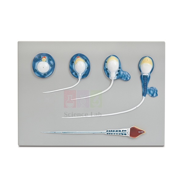
Spermatogenesis Model
The Spermatogenesis Model (Product Code: SLE/A/154) is an enlarged teaching model that follows the final phase of sperm formation, from the spermatid to the mature spermatozoon. Magnified about 10,000 times, it turns a microscopic change in cell shape into something a whole class can see from their seats, making it a practical choice among biology teaching aids for reproduction and cell biology units in senior secondary schools, colleges and nursing or paramedical institutes. It is manufactured by Jain Scientific Equipments Pvt Ltd (ScienceLab), Ambala, India.
Specifications
| Parameter | Detail |
|---|---|
| Process shown | Spermatogenesis from the spermatid to the mature spermatozoon |
| Magnification | Enlarged about 10,000x |
| Size | 53 x 38 x 5 cm |
| Mounting | Durable polymer base |
| Identification | Supplied with key |
Typical Use
In lessons on gametogenesis, the model gives students a fixed reference while the teacher explains how a rounded spermatid is remodelled into a streamlined sperm cell: the nucleus becomes compact, a cap forms over the head and a tail develops for movement. Because the whole sequence is presented on one mounted model, learners compare changes in shape and size directly instead of piecing them together from separate textbook drawings, and the key lets them check their own identifications during revision.
For a stronger practical link, pair the model with a prepared slide of mammalian testis: students first locate developing cells in real tissue under the microscope, then return to the enlarged model to see what those cells become. Used this way, it sits naturally within a human biology model set built around the reproductive system.
Manufacturer
ScienceLab is the brand name under which Jain Scientific Equipments Private Limited, a company based in Ambala, Haryana, makes anatomical models and laboratory apparatus for teaching institutions. The company profile describes its background and product range in more detail.
Related Laboratory Equipment
Items that help teach the same topic, from tissue level to the finished gamete:
- Ts Of Testis (Mammalian) — prepared slide of the tissue in which sperm are produced
- Mitosis & Meiosis Set, Microscope Slides — covers the cell divisions that come before the spermatid stage
- Human Sperm Model — a base-mounted model of the finished male gamete for introductory lessons
- Human Male Reproductive System Model Set — places sperm production in its organ-level context
See the complete Human Models range for other reproduction and anatomy teaching models.
Frequently Asked Questions
Which part of sperm development does this model show?
It shows spermatogenesis from the spermatid to the mature spermatozoon. If you need earlier stages such as spermatogonia or spermatocytes represented, please contact us before ordering, as these are not listed in the product documentation.
What are the dimensions?
The model measures 53 x 38 x 5 cm and is mounted on a durable polymer base.
How do students identify the stages?
A key is supplied with the model, so each stage can be matched to its name during lessons or self-study.
Why is the model enlarged about 10,000 times?
Sperm cells and the cells they develop from are microscopic; the enlargement makes their changes visible to a full class without a microscope. It complements slide work rather than replacing it.
How should the model be cleaned?
Wipe it with a soft, slightly damp cloth and let it dry; avoid solvents and abrasive cleaners, which can damage the finish. Store it away from direct sunlight.
Is a warranty offered?
Warranty terms are not stated in the product documentation, so please ask us for the terms that apply to your order.
Can you supply several units or ship abroad?
Yes. We supply schools, colleges and distributors in India and international markets; send the quantity and delivery location through our contact page for a quotation.
Request a Quote
To request pricing or a formal quotation for the Spermatogenesis Model, contact our sales team or add the item to your enquiry cart together with any related models you need.
Last updated: 24 September 2026
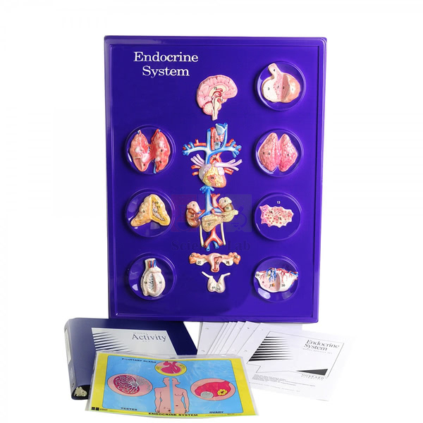
Endocrine System Model Activity Set
The Endocrine System Model Activity Set (Product Code: SLE/A/155) represents seven endocrine glands, each depicted whole and again as a magnified section in detail, together with closely related organs. Covering the pituitary, thyroid, parathyroid and adrenal glands as well as the pancreas, ovaries and testes, it gives biology teachers one human biology model set for chapters on hormones and chemical coordination in schools, colleges and nursing programmes. The set is manufactured by Jain Scientific Equipments Pvt Ltd (ScienceLab) in Ambala, India.
Specifications
| Parameter | Detail |
|---|---|
| Glands depicted | Seven: pituitary, thyroid, parathyroid and adrenal glands, pancreas, ovaries and testes |
| Views | Each gland shown whole and as a magnified section in detail |
| Additional structures | Closely related organs are also depicted |
| Format | Model activity set |
Typical Use
A useful approach is to begin with the whole-gland views so students can place each gland in the body, then move to the magnified sections to discuss what happens inside the tissue that produces hormones. Because both ovaries and testes are included, the set supports a side-by-side comparison of male and female reproductive hormones, while the pancreas opens a link to blood-sugar regulation and digestion.
For group work, give each team one or two glands and ask them to build a short profile — location, neighbouring organs, the hormones released and the tissues those hormones act on — then present it to the class using the model. Activities like this turn the set into working school biology lab equipment rather than a display piece.
Manufacturer
Jain Scientific Equipments Private Limited produces this activity set under its trading name, ScienceLab, from its base in Ambala, Haryana, where it builds biology models and science laboratory apparatus for education buyers. Read the company profile for an overview of the business.
Related Human Models
Models teachers often use alongside this set when covering hormones and the organs they affect:
- Human Endocrine Organs Model — an organ-level model for showing where the glands sit in the body
- Kidney with Adrenal Gland Model — a closer look at the adrenal gland and its vessels
- Pathology of the Human Thyroid Model — extends thyroid lessons into disorders of the gland
- Human Spleen, Duodenum, and Pancreas Model — context for the pancreas and its neighbouring organs
- Circulatory System Model Activity Set — a companion activity set, since hormones travel in the blood
Explore more in our Human Models category.
Frequently Asked Questions
Which glands are in the set?
Seven: the pituitary, thyroid, parathyroid and adrenal glands, the pancreas, the ovaries and the testes, along with closely related organs.
Are the glands shown at actual size?
Each gland appears whole and also as a magnified section. The scale of the whole-gland views is not stated in our documentation; contact us if you need it.
What activity materials come with the set?
The product documentation describes the glands and their sections. For a full list of components, such as any printed guide or worksheets, please confirm with us before ordering.
Which courses is it suited to?
It matches the endocrine topics taught in secondary and higher secondary biology and in introductory college and nursing courses; the depth of study is set by the teacher.
Can I order it together with other activity sets?
Yes. Add it to the enquiry cart with items such as the Circulatory System Model Activity Set and we will quote them together.
What information do you need to quote a school or college?
Tell us the number of sets, the delivery address, the billing name and any documents your purchase procedure requires, and we will prepare the quotation accordingly.
Request a Quote
For a quotation on the Endocrine System Model Activity Set, send your requirement through our contact page or place it in the enquiry cart. We supply schools and colleges in India and international markets.
Last updated: 24 September 2026
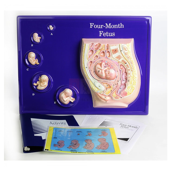
Foetus Model, Four Month
The Foetus Model, Four Month (Product Code: SLE/A/156) presents a 16 week foetus within a model of the womb, with realistic life-sized models of the embryo/foetus at 6, 8, 10, 12 and 14 weeks arranged in sequence beside it. Seeing the stages together at true size helps students grasp how much the developing baby grows during the first four months of pregnancy. Manufactured by Jain Scientific Equipments Pvt Ltd (ScienceLab), Ambala, it is suited to school biology lab equipment collections in India as well as nursing, midwifery and health education classrooms.
Specifications
| Parameter | Detail |
|---|---|
| Main model | 16 week foetus within a model of the womb |
| Earlier stages | Embryo/foetus at 6, 8, 10, 12 and 14 weeks |
| Scale | Realistic, life-sized |
| Arrangement | Earlier stages sequenced next to the 16 week foetus |
| Illustrates | Size, physical features and positioning of the embryo/foetus |
Typical Use
Teachers use the sequence to show the transition from embryo to foetus: learners compare the 6 week stage with the 14 and 16 week stages and describe what changes in body proportions, limbs and facial features. The womb model adds a second lesson on positioning, allowing a discussion of how the foetus lies inside the uterus and how the available space changes as development continues.
Since every stage is life-sized, the model is also effective in health education and antenatal awareness sessions, where real dimensions carry more meaning than enlarged diagrams. Keep it on a stable bench at eye level and let small groups examine it in turn; it then works as hands-on biology lab equipment for reproduction topics rather than a cupboard display.
Manufacturer
This model comes from Jain Scientific Equipments Private Limited, the Ambala, Haryana company known by its trading name ScienceLab, which manufactures teaching models and laboratory equipment for educational institutions. Our company profile gives further background.
Related Human Models
Useful companions for a complete lesson on pregnancy and development:
- Period of Gestation Model — a set of stand-mounted stages that extends the developmental timeline
- Human Embryo Development Set — additional models of early development
- Human Placenta Model — explains how the foetus is nourished during pregnancy
- Human Foetus Model — focuses on the foetus, its membrane and the uterus
- Human Birth Demonstration Model — carries the lesson through to birth
Browse our full Human Models collection for more reproduction and anatomy models.
Frequently Asked Questions
What does “Four Month” refer to?
The main foetus is at 16 weeks, roughly four months of pregnancy, and is shown within a model of the womb. Earlier stages at 6, 8, 10, 12 and 14 weeks are placed alongside it.
Are the models life-sized?
Yes. The embryo and foetus models are described as realistic and life-sized, so students see the real size at each stage.
Does it show how the foetus is positioned?
Yes. Placing the 16 week foetus within the womb model illustrates its positioning as well as its size and physical features.
Can the individual stages be lifted out?
This is not stated in our product documentation. Please contact us if you need the stages to be removable.
Who typically uses this model?
Biology teachers covering human reproduction, and instructors in nursing, midwifery and health education courses who need an accurate sense of early foetal size.
How long does delivery take?
Lead time depends on quantity and destination; share your requirement with us and we will confirm it in the quotation.
Do you export this model?
Yes. We supply institutions and distributors in India and international markets.
Request a Quote
Ask for a quotation on the Foetus Model, Four Month through our contact form, or add it to the enquiry cart with other reproduction models for a combined price.
Last updated: 24 September 2026
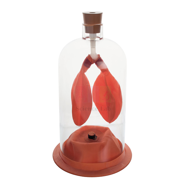
Lung Demonstration Model
The Lung Demonstration Model (Product Code: SLE/A/157) gives students a first-hand view of how the lungs work. Two sacs stand in for the lungs, a rubber membrane acts as the diaphragm and a clear plastic enclosure represents the chest cavity; when the diaphragm is made to relax and contract, the sacs visibly empty and fill with air. Manufactured by Jain Scientific Equipments Pvt Ltd (ScienceLab), Ambala, this simple working model is practical biology lab equipment for respiration lessons in schools across India.
Specifications
| Parameter | Detail |
|---|---|
| Lungs | Two sacs representing the lungs |
| Diaphragm | Rubber membrane |
| Chest cavity | Clear plastic enclosure |
| Size | Height 30 cm x Width 14.5 cm |
| Operation | Diaphragm is made to relax and contract so the lungs empty and fill with air |
Typical Use
When the rubber diaphragm is drawn away from the enclosure, the space around the sacs increases, the pressure inside falls and air flows into the “lungs”, just as it does when the real diaphragm contracts and flattens. Letting the membrane return reverses the change and the sacs deflate. The demonstration links the mechanism of inhalation and exhalation to the pressure–volume relationship taught in physics, so it suits integrated science lessons as well as biology.
Before the lesson, check that the membrane sits evenly and that the sacs inflate freely, then move the diaphragm slowly so the class can match each movement to the change in the sacs. Students can finish by placing a hand on their own abdomen while breathing deeply to feel the same action. Its compact size makes it a sensible early purchase for any biology lab setup in a school.
Manufacturer
The Lung Demonstration Model is made by Jain Scientific Equipments Private Limited, which operates under the ScienceLab name from Ambala, Haryana, and produces teaching equipment for biology, physics and chemistry laboratories. Tender and institutional buyers who need vendor registration details can use GSTIN 06AAECJ8618A1ZT and LEI 3358004HRD1JIVXKYX49. Visit the company profile for more about the company.
Related Laboratory Equipment
Equipment that extends a breathing lesson from mechanism to measurement and anatomy:
- Human Lung Model in Bell Jar — an alternative breathing demonstration in bell-jar form
- Human Respiratory System Model, 2-parts — shows the real anatomy that the demonstration represents
- Lung Volume Bags — lets students collect and compare their own breath volumes afterwards
- Lung Capacity Gauge — for follow-up lung capacity activities
- Segmented Lung — detailed lung anatomy for senior classes
More anatomy teaching models are listed in our Human Models category.
Frequently Asked Questions
How does the model show breathing?
Moving the rubber diaphragm changes the space inside the clear enclosure, so the two lung sacs fill with air and then empty, mirroring inhalation and exhalation.
How big is it?
It stands 30 cm high and is 14.5 cm wide, small enough to demonstrate at the front of a class or pass around a bench.
Is it an anatomically detailed lung model?
No. It is a working demonstration of the breathing mechanism; for detailed structure, use it with an anatomical model such as the Human Respiratory System Model, 2-parts.
Is anything else needed to run the demonstration?
The sacs, rubber diaphragm and enclosure together make up the apparatus, so the basic demonstration can be run with the model alone.
Can a worn rubber membrane be replaced?
Spare-part availability is not listed in our documentation; please contact us to ask about replacement membranes.
Can you supply class sets or export orders?
Yes. We supply schools, colleges and distributors in India and international markets; tell us the quantity and destination for a quotation.
Request a Quote
For prices and availability of the Lung Demonstration Model, get in touch with us or add it to your enquiry cart along with any breathing and respiration equipment you need.
Last updated: 24 September 2026
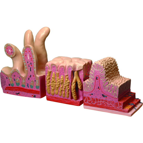
Digestive Canal Model
The Digestive Canal Model (Product Code: SLE/A/158) shows the structure of the digestive canal, the continuous tube through which food passes as it is broken down and absorbed. It is supplied in three parts, and teachers use it to take students through the canal region by region in nutrition and digestion lessons. Made by Jain Scientific Equipments Pvt Ltd (ScienceLab), Ambala, it can form the digestion component of a human body model kit for schools, colleges and nursing institutes in India.
Key Details
| Parameter | Detail |
|---|---|
| Type | Anatomical model of the human digestive canal |
| Shows | Structure of the digestive canal |
| Supplied as | Three parts |
| Suitable for | Digestion and nutrition lessons in schools, colleges and nursing institutes |
Typical Use
Start with the canal as a whole so students appreciate its length and continuity, then focus on individual regions to discuss what each contributes: mechanical and chemical breakdown of food, absorption of nutrients and the recovery of water before waste leaves the body. Asking learners to trace a meal’s journey along the model and name each region in order is a quick check of understanding at the end of the topic.
The model also anchors practical work. After studying it, students can test food samples for starch or protein, or run a starch-digestion test with an enzyme experiment kit, and then point to the region of the canal where that digestion takes place. Linking results to the model helps them remember where each process happens.
Manufacturer
ScienceLab is how Jain Scientific Equipments Private Limited trades; the company is located in Ambala, Haryana, and makes human anatomy models alongside a wider range of school and college laboratory equipment. You can learn more on our company profile page.
Related Laboratory Equipment
Models and slides that support the same digestion lessons:
- Human Digestive System Model — a broader digestive system model for comparison
- Human Digestive Tract Model — an alternative tract model for side-by-side teaching
- Human Stomach Model — closer study of the stomach
- Appendix and Caecum Model — detail of the region where the small and large intestines meet
- Prepared Microscope Slide Set – Digestion — a tissue-level view to accompany the model
Our Human Models range lists further anatomy models for biology departments.
Frequently Asked Questions
What does the model show?
It shows the structure of the human digestive canal, the tube that runs through the body from where food enters to where waste leaves.
How many parts is it supplied in?
Three parts. The arrangement of the parts is shown in the product photograph; contact us for a detailed breakdown.
Does it include the liver and pancreas?
The product documentation describes the digestive canal only. If you need the accessory organs as well, compare the Human Digestive System Model or ask us for advice.
Is a key or labelling supplied?
A key is not mentioned in our documentation, so please confirm with us before ordering if you need one.
Which classes can use it?
It fits the digestion and nutrition topics of middle and secondary school biology and is equally useful for introductory anatomy in nursing and paramedical courses.
Can a school buy several at once?
Yes. Send the quantity and delivery address and we will quote for supply within India or to international markets.
Request a Quote
Request a quotation for the Digestive Canal Model through our contact page, or collect it with other items in the enquiry cart and send one combined enquiry.
Last updated: 24 September 2026
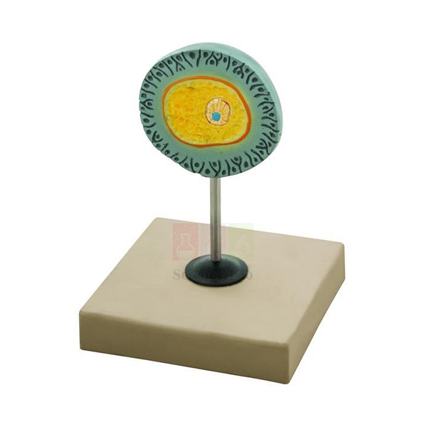
Human Egg Model
The Human Egg Model (Product Code: SLE/A/159) is a teaching model of the human egg, or ovum — the female gamete that joins with a sperm at fertilisation. Mounted on a base so it stands securely on a bench or display shelf, it gives teachers a clear visual starting point for lessons on human reproduction. It is manufactured by Jain Scientific Equipments Pvt Ltd (ScienceLab), Ambala, and is a straightforward item to include when planning a biology lab setup for a school in India.
Key Details
| Parameter | Detail |
|---|---|
| Type | Teaching model of the human egg (ovum) |
| Mounting | Mounted on base |
| Purpose | Visual aid for lessons on the female gamete, ovulation and fertilisation |
| Suitable for | School biology, college zoology, and nursing or health education classes |
Typical Use
A real human egg is barely visible to the naked eye, so a model is the easiest way to discuss it with a group. Teachers use it to explain that the egg is far larger than a sperm cell, that it carries stored nutrients and half of the genetic material a new individual needs, and that it is surrounded by protective layers a sperm must pass through before fertilisation.
Placed next to a sperm model, it lets students contrast the two gametes in size, shape and role; it is also a helpful prop when explaining the menstrual cycle, ovulation and the start of pregnancy in health education sessions. Within a human biology model set it pairs naturally with sperm, embryo and pelvis models.
Manufacturer
Jain Scientific Equipments Private Limited, which trades as ScienceLab, manufactures anatomical teaching models such as this one in Ambala, Haryana, for schools, colleges and distributors. Further information is available on the company profile.
Related Human Models
Models that complete a lesson on gametes and fertilisation:
- Human Sperm Model — the matching male gamete model for comparison lessons
- Embryo Model — shows what develops after fertilisation
- Human Female Genital Organs Model — the organs in which eggs mature and are released
- Female Pelvis Model — a median-section view of the female pelvic organs
Find more reproduction teaching models in our Human Models range.
Frequently Asked Questions
How is the model presented?
It is mounted on a base, so it can stand on a bench, shelf or teacher’s desk during lessons.
Is the egg shown enlarged, and by how much?
Because the human egg is so small, any teaching model of it is enlarged. The exact magnification is not stated in our documentation; contact us if you need it.
Does it come with a key or labelled structures?
This is not listed in our product documentation. Please ask us for details before ordering if labelling is essential.
Can it be quoted together with the Human Sperm Model?
Yes. They are separate items, but you can add both to the enquiry cart and we will quote them together.
How should students handle it?
Lift and carry it by the base rather than the model itself, and keep it away from heat and direct sunlight when stored.
Do you supply schools outside India?
Yes. Orders are shipped to schools, colleges and distributors in India and international markets.
Request a Quote
To get a price for the Human Egg Model, send us an enquiry or add it to the enquiry cart with any companion models.
Last updated: 24 September 2026
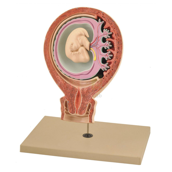
Human Foetus Model
The Human Foetus Model (Product Code: SLE/A/160) is a three-part model that shows the relationship between the foetus, the foetus membrane and the uterus. Rather than tracing growth over time, it concentrates on how the developing baby is enclosed and supported inside the mother, which makes it valuable for reproduction, pregnancy and nursing lessons. It is produced by Jain Scientific Equipments Pvt Ltd (ScienceLab), an anatomical models manufacturer in Ambala, India.
Specifications
| Parameter | Detail |
|---|---|
| Construction | Three-part model |
| Structures shown | Foetus, foetus membrane and uterus |
| Teaching focus | Relationship between the foetus, its membrane and the uterus |
Typical Use
Teachers use the model to explain, from the outside in, how the muscular uterus holds the pregnancy, how the membrane encloses the foetus and cushions it within a fluid-filled space, and how the foetus lies inside this protected environment. Students can then discuss why the membrane matters, what happens when it ruptures at the start of labour, and how the arrangement changes as the pregnancy advances.
In nursing, midwifery and health education courses, the model helps trainees picture what they will later palpate or see on an ultrasound image, and it works well with other biology teaching aids for pregnancy topics, such as placenta and developmental-stage models.
Manufacturer
ScienceLab is the trading name of Jain Scientific Equipments Private Limited (CIN U29309HR2020PTC087597), an Ambala, Haryana company that manufactures at its licensed factory at 449, HSIIDC Industrial Area, Saha, Ambala (Factory Licence AMB-ONLINE-CHD-J-414). See the company profile for details of the business.
Related Human Models
Models that build a fuller picture of pregnancy alongside this one:
- Human Placenta Model — explains the exchange between mother and foetus
- Foetus Model, Four Month — life-sized developmental stages shown within a womb model
- Period of Gestation Model — stand-mounted stages of embryonic and foetal development
- Human Birth Demonstration Model — continues the lesson to the process of birth
View the rest of our Human Models for anatomy and reproduction teaching.
Frequently Asked Questions
What does this model teach?
It shows how the foetus, the foetus membrane and the uterus relate to one another during pregnancy.
How many parts does it have?
It is a three-part model.
How is it different from the Foetus Model, Four Month?
This model focuses on the foetus within its membrane and the uterus, while the Foetus Model, Four Month presents a sequence of life-sized developmental stages beside a foetus in a womb model.
Which stage of pregnancy does the foetus represent, and is it life-size?
Neither is stated in our product documentation; please contact us for these details.
Is a key card included?
A key card is not mentioned in our documentation, so confirm with us if you need one for labelling.
What should a college include in its purchase enquiry?
The quantity, delivery location, billing details and any registration documents your procurement office needs; our company registration details are given above.
Is export supply possible?
Yes, we supply buyers in India and international markets.
Request a Quote
For a quotation on the Human Foetus Model, contact us or add it to your enquiry cart together with other pregnancy and development models.
Last updated: 24 September 2026
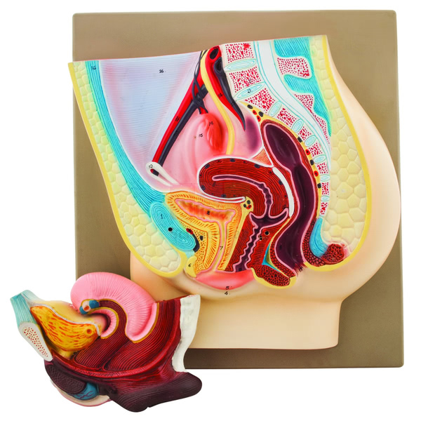
Female Pelvis Model
The Female Pelvis Model (Product Code: SLE/A/161) is a median section of the female pelvis. One half presents the genital organs together with the bladder and a removable rectum, while the other half is shown in its normal position in the pelvis, so students can compare a sectional view with the natural arrangement. A numbered key card is supplied. The model is manufactured by Jain Scientific Equipments Pvt Ltd (ScienceLab), Ambala, one of the human anatomical models manufacturers in India serving schools, colleges and medical training institutes.
Specifications
| Parameter | Detail |
|---|---|
| Section | Median section of a female pelvis |
| First half | Genital organs with bladder and removable rectum |
| Second half | Shown in the normal position in the pelvis |
| Removable part | Rectum |
| Identification | Numbered key card |
Typical Use
In reproductive anatomy classes, the median section lets students see how the internal genital organs sit between the bladder in front and the rectum behind — a relationship that is hard to grasp from surface drawings. Taking out the removable rectum gives a clearer view of the neighbouring structures, and comparing the sectioned half with the half in its normal position helps learners connect a cut-away view with the body as it really is.
Nursing, midwifery and paramedical students use the model to relate pelvic anatomy to clinical topics such as catheterisation, pregnancy and childbirth. The numbered key card makes it easy to run quick identification tests, which is why it is useful school biology lab equipment for senior classes as well as professional training.
Manufacturer
Jain Scientific Equipments Private Limited manufactures the Female Pelvis Model and sells it under the ScienceLab brand. The company is based in Ambala, Haryana, and supplies anatomy models and laboratory equipment to educational and training institutions. The company profile has more information.
Related Human Models
Models that support the same pelvic and reproductive anatomy lessons:
- Female Half Pelvis Model — an alternative half-pelvis presentation for comparison
- Human Adult Female Pelvis Structural Model — the bony pelvis for skeletal context
- Human Female Urogenital Model — brings the urinary and reproductive organs together
- Male Pelvis Section Model — the male counterpart for comparison lessons
See all models in our Human Models category.
Frequently Asked Questions
What does the median section show?
One half shows the genital organs with the bladder and a removable rectum; the other half is shown in its normal position in the pelvis.
Which part can be removed?
The rectum is removable.
Is a key supplied?
Yes, the model comes with a numbered key card for identifying the structures.
Is it natural size?
The scale is not stated in our product documentation; please contact us if you need dimensions before ordering.
Is it suitable for nursing and midwifery training?
Yes. The sectional view of the pelvic organs and the key card suit anatomy teaching in nursing, midwifery and paramedical courses as well as senior school biology.
How should it be cleaned and stored?
Dust it with a soft brush or wipe it with a lightly damp cloth, keep the removable rectum with the model, and store it covered away from heat.
Can institutions order in bulk?
Yes. We quote for single units and larger institutional orders in India and international markets.
Request a Quote
To receive a quotation for the Female Pelvis Model, write to us via the contact page or add it to the enquiry cart.
Last updated: 24 September 2026
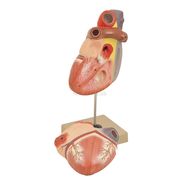
Heart 3 times Natural Size Model
The Heart 3 times Natural Size Model (Product Code: SLE/A/162) is a human heart enlarged 3 times its actual size and dissectible into 2 parts, showing the structure of the auricles and ventricles and, clearly, the chief blood vessels. Made up on a stand 30 cm high, it is large enough to demonstrate at the front of a class yet compact on the bench. It is manufactured by Jain Scientific Equipments Pvt Ltd (ScienceLab), Ambala, as a heart model for anatomy teaching in India’s schools, colleges and medical training institutes.
Specifications
| Parameter | Detail |
|---|---|
| Scale | Enlarged 3 times actual size |
| Construction | Dissectible into 2 parts |
| Internal structure | Auricles and ventricles shown |
| Vessels | Chief blood vessels shown clearly |
| Mounting | Made up on stand |
| Dimensions | Height 30 cm, width 12 cm |
Typical Use
With the model closed, students identify the major vessels entering and leaving the heart; opening it into its two parts reveals the auricles (atria) and ventricles, so the teacher can trace the double circulation — blood returning from the body to the right side, travelling to the lungs, coming back to the left side and leaving for the rest of the body. The threefold enlargement makes the chambers far easier to point out than on a natural-size specimen.
A good routine is to have students label a blank heart diagram first and then check their answers against the model, or to follow a drop of blood through the chambers and vessels aloud. Mounted on its stand, it serves both as demonstration apparatus and as a revision station among school biology lab equipment.
Manufacturer
Made by Jain Scientific Equipments Private Limited, which trades as ScienceLab from Ambala in Haryana, this heart model belongs to a wider range of anatomy models and laboratory apparatus the company produces for educational buyers. Company details are on our company profile.
Related Laboratory Equipment
Heart and circulation items that pair well with this model:
- Human Heart Model — an alternative heart model for comparison
- Human Heart, Pumping Model — demonstrates the pumping action of the heart
- Human Circulatory System Model — places the heart within the whole circulation
- Human Heart on Diaphragm Model — shows where the heart sits in relation to the diaphragm
- Prepared Microscope Slide Set – Circulatory System — slides for microscope work on heart and vessel tissue
Browse the Human Models range for more anatomy models.
Frequently Asked Questions
How much larger than a real heart is it?
It is enlarged 3 times natural size.
How many parts does it come apart into?
It is dissectible into 2 parts, which opens up the auricles and ventricles.
What are the dimensions on the stand?
It stands 30 cm high and is 12 cm wide.
Does it show the heart valves?
Our documentation lists the auricles, ventricles and chief blood vessels. For valve detail, please contact us or refer to the product photograph.
Is a key or label chart included?
A key is not mentioned in the product documentation; ask us if you need one.
Is there a warranty?
Warranty terms are not listed in our documentation and are confirmed with each quotation.
Can you supply several for a department or export them?
Yes. We supply single pieces and multiple units to buyers in India and international markets.
Request a Quote
For pricing on the Heart 3 times Natural Size Model, reach our team or add it to the enquiry cart with other circulation models.
Last updated: 24 September 2026
Human Sperm Model
The Human Sperm Model (Product Code: SLE/A/163) is a teaching model of the human sperm cell, the male gamete, mounted on a base. Because a real sperm can only be seen under a microscope, the model gives students a tangible reference for its overall form when fertilisation and human reproduction are taught. It is manufactured by Jain Scientific Equipments Pvt Ltd (ScienceLab), Ambala, and is a sensible inclusion when a school places a biology model kit bulk order for its reproduction units.
Key Details
| Parameter | Detail |
|---|---|
| Type | Teaching model of the human sperm cell (male gamete) |
| Mounting | Mounted on base |
| Purpose | Illustrates the male gamete in reproduction and fertilisation lessons |
| Suitable for | School biology, college zoology, nursing and health education |
Typical Use
Teachers use the model to show how a sperm cell’s form suits its job: a head that carries the father’s genetic material, a middle section that supplies energy, and a long tail that drives the cell towards the egg. Students can sketch and label the cell from the model before looking at real sperm on a slide or in micrographs, which helps them recognise the shape at a much smaller scale.
It is most effective alongside the spermatogenesis and egg models — the first explains how sperm are formed and the second shows the far larger cell they must reach — so students see the whole story from production to fertilisation. Among biology teaching aids for this topic, the base-mounted format also makes it easy to leave on a bench for self-study.
Manufacturer
The model is a product of Jain Scientific Equipments Private Limited, an Ambala, Haryana manufacturer whose trading name is ScienceLab and whose catalogue covers anatomy, biology and general science teaching equipment. Visit our company profile to find out more.
Related Laboratory Equipment
Items that complete a lesson on male gametes and fertilisation:
- Spermatogenesis Model — shows how sperm develop from spermatids
- Human Egg Model — the matching female gamete, also mounted on a base
- Human Male Genital Organs Model — the organs that produce and carry sperm
- Male Pelvis Section Model — a sectional view that includes the testicular ducts
- Ts Of Testis (Mammalian) — a prepared slide for seeing sperm-producing tissue under the microscope
More models are available in our Human Models category.
Frequently Asked Questions
What exactly is supplied?
A model of the human sperm cell mounted on a base.
What magnification does the model represent?
The magnification is not stated in our product documentation; please contact us if your syllabus or tender requires it.
How does it differ from the Spermatogenesis Model?
This model shows the sperm cell on its own, while the Spermatogenesis Model traces its development from spermatid to mature spermatozoon and comes with a key.
Which classes is it suited to?
Any class where human reproduction is taught, typically secondary school onward, as well as nursing and health education programmes.
What warranty applies?
Warranty terms are not given in our documentation; we will state them in your quotation on request.
Can you ship bulk orders?
Yes. We deliver single models and bulk orders to schools, colleges and distributors in India and international markets.
Request a Quote
Request a price for the Human Sperm Model through our contact page, or add it to the enquiry cart together with the egg and spermatogenesis models for a single quotation.
Last updated: 24 September 2026
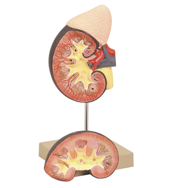
Kidney with Adrenal Gland Model
The Kidney with Adrenal Gland Model (Product Code: SLE/A/164) shows a kidney together with its adrenal gland, the renal and adrenal vessels and the upper portion of the ureter. The front half of the kidney is removable to expose the cortex, medulla, vessels and renal pelvis, so students can move from outer shape to internal structure with one model. Manufactured by Jain Scientific Equipments Pvt Ltd (ScienceLab), Ambala, it is built for excretion and endocrine lessons and reflects the company’s place among high school biology lab equipment manufacturers in India.
Specifications
| Parameter | Detail |
|---|---|
| Structures shown | Kidney with adrenal gland |
| Vessels | Renal and adrenal vessels |
| Outflow | Upper portion of the ureter |
| Removable part | Front half of the kidney |
| Internal view | Cortex, medulla, vessels and renal pelvis |
Typical Use
With the front half in place, students locate the adrenal gland resting on the kidney and follow the renal vessels in and out. Lifting the front half away then shows the cortex and medulla, where urine is formed, and the renal pelvis that collects it before it drains into the ureter. This sequence works well as a guided dissection substitute when real specimens are not available.
Because the adrenal gland is included, the model bridges two syllabus topics — excretion and the endocrine system — and lets teachers explain why two organs with very different jobs sit side by side. Pairing it with a nephron model or prepared kidney slides gives a complete run from organ to microscopic structure, making it versatile biology lab equipment for schools and colleges in India.
Manufacturer
Jain Scientific Equipments Private Limited manufactures this model under its ScienceLab trading name in Ambala, Haryana. The company is a registered MSME (Udyam UDYAM-HR-01-0003714), and its export consignments are handled under Importer-Exporter Code AAECJ8618A issued by the DGFT. More background is available on the company profile.
Related Laboratory Equipment
Models and slides that extend kidney and adrenal lessons:
- Human Kidney, Nephron, and Glomerulus Model Set — takes the lesson down to the nephron and glomerulus
- Denoyer-Geppert Free-Standing Human Urinary System Model — shows the kidney within the complete urinary system
- Human Kidney with Adrenal Gland Model — an alternative kidney and adrenal model from another of our ranges
- Human Endocrine Organs Model — shows the adrenal glands alongside the other endocrine organs
- Prepared Microscope Slide Set – Skin and Excretion — tissue slides for excretion practicals
For more anatomy models, visit our Human Models category.
Frequently Asked Questions
Which part of the model can be removed?
The front half of the kidney is removable.
What can be seen inside the kidney?
Removing the front half exposes the cortex, medulla, vessels and renal pelvis.
Which blood vessels are included?
Both the renal and adrenal vessels are shown, together with the upper portion of the ureter.
What size is the model?
Dimensions and scale are not given in our product documentation; please contact us for them.
Is a numbered key provided?
A key is not listed in our documentation, so please check with us before ordering if you need one.
What do you need from us to prepare a tender quotation?
Send the quantity, delivery address and the vendor documents your tender asks for; our registration details above can be quoted in your vendor forms.
Do you ship outside India?
Yes. We supply buyers in India and international markets.
Request a Quote
Ask for a quotation on the Kidney with Adrenal Gland Model via our contact page, or add it to the enquiry cart with related excretion and endocrine models.
Last updated: 24 September 2026
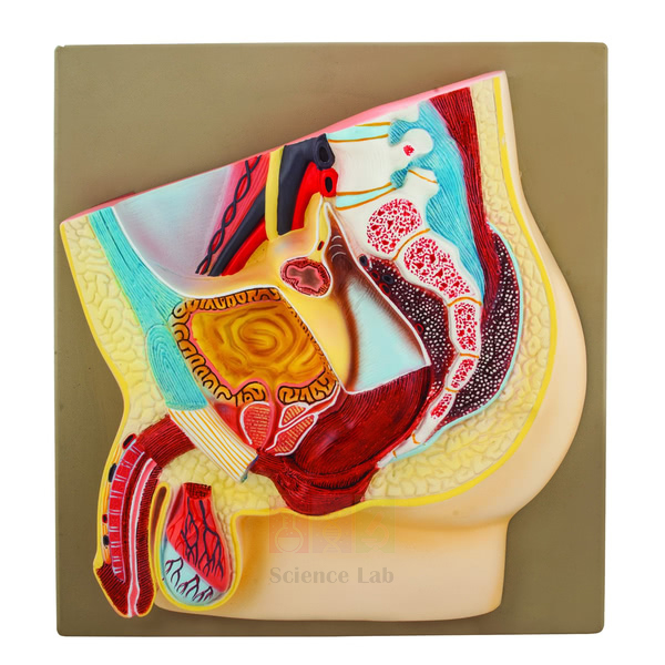
Male Pelvis Section Model
The Male Pelvis Section Model (Product Code: SLE/A/165) is a natural size sectional model on a base that shows the rectum, the bladder with the prostate, the testicular ducts and the external genital organs. A numbered key card is supplied so each structure can be identified quickly. Manufactured by Jain Scientific Equipments Pvt Ltd (ScienceLab), Ambala, it serves colleges, nursing schools and senior biology classes that source anatomy models from a biology lab equipment supplier in India.
Specifications
| Parameter | Detail |
|---|---|
| Scale | Natural size |
| Mounting | On base |
| Structures shown | Rectum; bladder with prostate; testicular ducts; external genital organs |
| Identification | Numbered key card |
Typical Use
Because the model is natural size, students gain a realistic sense of how closely the bladder, prostate and rectum lie to one another — knowledge that matters in nursing and paramedical training for procedures such as urinary catheterisation. Teachers can trace the route sperm take from the testicular ducts past the prostate to the exterior, and explain why enlargement of the prostate can affect the flow of urine from the bladder.
Set it beside a female pelvic section to compare the two pelvic layouts in one lesson, and use the numbered key card for a timed identification exercise at the end. Used this way, it is a practical piece of school biology lab equipment for senior classes as well as a reference model for professional courses.
Manufacturer
ScienceLab is the brand of Jain Scientific Equipments Private Limited, a manufacturer of anatomy models and science laboratory equipment located in Ambala, Haryana, India. Our company profile explains more about the company and its range.
Related Human Models
Models that round out male pelvic and reproductive anatomy lessons:
- Human Male Reproductive System Model Set — a broader set covering the male reproductive organs
- Male Skeletal Pelvis — the bony framework that surrounds these organs
- Human Male and Female Urogenital Systems Model — compares male and female urogenital anatomy
- Female Pelvis Model — the female median-section counterpart
Explore further models in the Human Models category.
Frequently Asked Questions
Is the model life-size?
Yes, it is a natural size model mounted on a base.
Which structures does it show?
The rectum, the bladder with the prostate, the testicular ducts and the external genital organs.
How are the structures identified?
A numbered key card is supplied, matching numbers on the model to structure names.
Does it show the testes in section?
Our documentation lists the testicular ducts and external genital organs; for a detailed list of what is sectioned or numbered, please contact us.
Is it appropriate for nursing schools?
Yes. The natural size and clear organ relationships support nursing, paramedical and introductory medical anatomy teaching.
How do I order several pieces or arrange export?
Send us the quantity and destination; we supply institutions and distributors in India and international markets.
Request a Quote
For a price on the Male Pelvis Section Model, use our contact page or add it to the enquiry cart alongside the Female Pelvis Model for a comparison pair.
Last updated: 24 September 2026
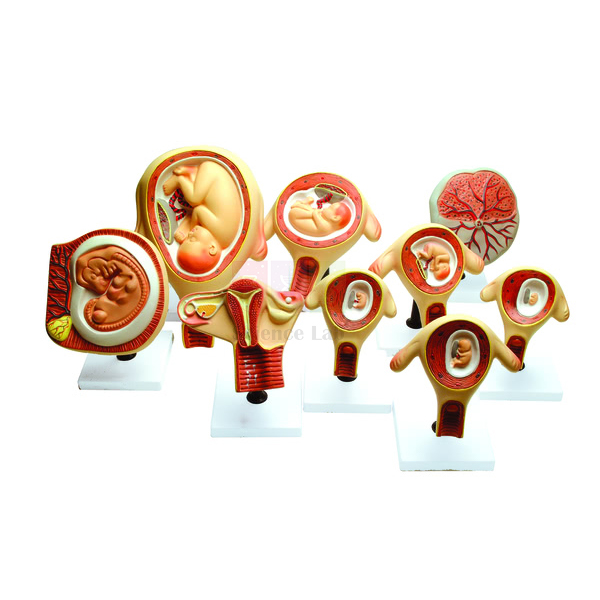
Period of Gestation Model
The Period of Gestation Model (Product Code: SLE/A/166) is a set of nine models, each mounted on its own stand, showing various stages of embryonic and foetal development. Arranged in order, the nine stands turn prenatal development into a timeline that students can walk along and compare stage by stage. The set is manufactured by Jain Scientific Equipments Pvt Ltd (ScienceLab), Ambala, and is used as biology lab equipment in India for reproduction and human development topics.
Specifications
| Parameter | Detail |
|---|---|
| Number of models | Nine |
| Mounting | Each model mounted on its own stand |
| Coverage | Various stages of embryonic and foetal development |
Typical Use
Lined up along a bench, the nine models let a class follow development from the embryonic period into the foetal period, noting how the body takes shape, how limbs and features form and how overall size increases. Because every stage has its own stand, individual models can be handed to small groups for close study and then returned to their place in the sequence.
A simple assessment is to mix up the stands and ask students to put them back in the correct order, explaining the clues they used. The set also supports health education sessions on prenatal care, where seeing each stage helps learners understand why the early weeks of pregnancy are so important. It sits well with other biology teaching aids on reproduction, such as placenta and pelvis models.
Manufacturer
This gestation set is manufactured in Ambala, Haryana, by Jain Scientific Equipments Private Limited, which trades under the ScienceLab name and supplies teaching models and laboratory equipment to institutions. The company profile provides more background.
Related Human Models
Models that fill in the rest of the story around the gestation sequence:
- Human Embryo Development Set — additional models focused on early development
- Human Embryo Set, PVC — an alternative embryo set in PVC
- Foetus Model, Four Month — shows a foetus positioned within a womb model
- Human Birth Demonstration Model — the final stage, birth
Our complete Human Models range includes more reproduction and anatomy models.
Frequently Asked Questions
How many models are in the set?
Nine, each showing a different stage of embryonic or foetal development.
Are the models free-standing?
Yes. Each one is mounted on its own stand.
Which weeks or months do the stages represent?
The specific stages are not listed in our product documentation; please contact us for the stage list before ordering.
Are the models life-size?
Scale is not stated in our documentation, so confirm with us if this matters for your syllabus.
Can individual stages be purchased separately?
The item is described as a set of nine; ask us if you need a single stage or a replacement model.
What do you need to prepare a quotation?
The number of sets, the delivery location and the billing details; we quote for buyers in India and international markets.
Request a Quote
To price the Period of Gestation Model, contact us or place it in the enquiry cart together with any related embryology models.
Last updated: 24 September 2026
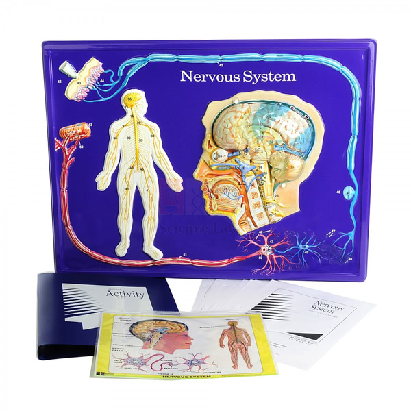
Nervous System Model
The Nervous System Model (Product Code: SLE/A/167) is a full-colour, 600 x 450 mm markable model made of durable vinyl that illustrates the structure of the central and peripheral nervous systems. It includes a close examination of the human brain, with a removable transparent overlay defining the brain lobes, and comes with an activity guide for teachers. Manufactured by Jain Scientific Equipments Pvt Ltd (ScienceLab), Ambala, it fits into a human biology model set for coordination and nervous system topics.
Specifications
| Parameter | Detail |
|---|---|
| Format | Full-colour markable model of durable vinyl |
| Size | 600 x 450 mm |
| Systems illustrated | Central and peripheral nervous systems |
| Brain detail | Close examination of the human brain with a removable transparent overlay defining the brain lobes |
| Locations shown in diagrams | Brain, spinal cord and spinal nerves, dendrites, axons and synapses |
| Included | Activity guide (contents listed below) |
The activity guide contains:
- Background information
- Basic understandings
- Teacher-developed activities
- A glossary
- A key to model structures
- Colour transparencies
- A blackline master
Typical Use
Because the surface is markable, students can trace pathways directly on the model — for example, following a reflex arc from a sensory nerve into the spinal cord and back out to a muscle, or shading where the central system ends and the peripheral system begins. The transparent overlay lets the teacher present the brain first as a whole and then with its lobes defined, before linking each lobe to functions such as movement, sensation, hearing and vision.
The guide’s teacher-developed activities and glossary make it straightforward to plan structured lessons, the blackline master can be copied for student worksheets, and the colour transparencies suit projection for a large class. Test any marker on a small corner first and clean the surface after each session. These features make it one of the more interactive biology teaching aids for neuroscience basics.
Manufacturer
This model is manufactured by Jain Scientific Equipments Private Limited, known commercially as ScienceLab and based in Ambala, Haryana, as part of its range of biology teaching resources for schools and colleges. Read more on our company profile.
Related Human Models
Models that deepen the study of the brain and spinal cord:
- Human Nervous System Model — an alternative nervous system model for comparison
- Human Regional Brain Model — extends the lobe overlay to detailed brain regions
- Human Spinal Cord Section Model — a closer look at spinal cord structure
- Human Vertebral And Partial Spinal Cord Model — shows how the cord sits within the vertebrae
- Endocrine System Model Activity Set — a companion set on the body’s hormonal control system
Discover more in our Human Models category.
Frequently Asked Questions
How large is the model?
It measures 600 x 450 mm and is made of durable vinyl in full colour.
What is in the activity guide?
Background information, basic understandings, teacher-developed activities, a glossary, a key to model structures, colour transparencies and a blackline master.
Which markers can be used on it?
The model is markable, but the recommended marker type is not stated in our documentation; please ask us, and test any marker on a corner first.
Does it show the lobes of the brain?
Yes. A removable transparent overlay defines the brain lobes over the close-up view of the brain.
Does it cover nerve cells as well as organs?
Yes. Its diagrams illustrate the locations of the brain, spinal cord and spinal nerves, along with dendrites, axons and synapses.
Can we order class sets or ship overseas?
Yes. We quote for multiple units and supply buyers in India and international markets.
Request a Quote
For a quotation on the Nervous System Model, send us your requirement or add it to the enquiry cart with other nervous system models.
Last updated: 24 September 2026