- Physics Lab Equipment
- Chemistry Lab Equipment
- Biology Lab Equipment
- Anatomical Lab Equipment
- Learning
- Educational Lab Equipment
- Magnets
- Agriculture
- Astronomy
- Earth Science
- Geography Lab Equipment
- Safety Lab Coat
- Laboratory
- Meteorological
- Electricity & Magnetism
- Heat
- School Lab Equipment
- Lab Glassware
- Science Lab Equipment
- Tvet Lab Equipment
- Robotics Kits Supplier and Manufacturer for Schools India
- Math Lab Equipment Manufacturers and Exporters in Ambala
- STEM Kits Manufacturers and Supplier India
- Engineering Lab Equipment
- Hospital Lab Equipment
- Water, Sanitation & Hygiene Supplies
- Cold Chain, Solar & Communication Equipment
- Nutrition
Last updated: 22 October 2026
Quick Answer
Jain Scientific Equipments Pvt Ltd (ScienceLab), Ambala, Haryana, an anatomical models manufacturer, lists 12 digestive system models for school, college, university and health-science teaching, arranged in 3 groups from stomach and whole digestive system models to liver, gallbladder, pancreas and upper-abdomen models and clinical examples such as gallstones, a gastric band and liver pathology. All 12 items are manufactured by ScienceLab at its factory at Saha, Ambala. Each item has its own product page, linked from the product listing under this description. The company supplies buyers in India and in 110 countries.
Quick links: Request a quotation · Jump to specifications by group · Open your enquiry cart
Explore the Digestive System Models Range
| Range | Items | What it covers |
|---|---|---|
| Stomach and whole digestive system | 4 | Two-part stomach models, including one on a stand showing the muscle layers, and a life-size digestive system model in graphic relief on a baseboard. |
| Liver, gallbladder, pancreas and upper abdomen | 5 | Liver models with the gall bladder, bile and pancreatic ducts, an enlarged dissected liver and gallbladder, a pancreas, spleen and duodenum model and the rear organs of the upper abdomen. |
| Clinical and patient-education models | 3 | A gallstone model of the biliary system, a two-part stomach model with a gastric band and a life-size liver pathology model. |
| Total | 12 | All items listed in the Digestive System range |
Digestive System Models Specifications
Key specifications below are taken from each product's own page; open a product for its full specifications and product code.
Stomach and whole digestive system (4)
| Product | Key specification |
|---|---|
| Digestive System Model 2 part | Weight: 4.4 kg; Scale: Life size; Format: Graphic relief, mounted on baseboard |
| Human Stomach Model | Size: 19 x 18 x 34 cm; Construction: Dissectible into 2 parts; Scale: Life size |
| Human Stomach, on Stand | Construction: Separates into two parts; Muscle layers: Longitudinal and circular muscle layers |
| Stomach Model 2 Part | Construction: 2 parts; front half removable; Weight: 0.6 kg; Size: 25 x 22 x 12 cm |
Liver, gallbladder, pancreas and upper abdomen (5)
| Product | Key specification |
|---|---|
| Human Liver and Gallbladder Model | Scale: 1-1/2x life size, enlarged; Size: 19 x 26 x 29 cm; Construction: One-part, dissected model |
| Human Liver Model | Structures shown: Liver, gall bladder, right hepatic duct, left hepatic duct, cystic duct, duodenum, pancreatic duct, pancreas |
| Human Liver With Gall Bladder Model | Lobes shown: 4 lobes; Other structures: Gall bladder, extra hepatic ducts, hilus vessels; Finish: Appropriately coloured |
| Human Pancreas, Spleen and Duodenum Model | Organs shown: Spleen, pancreas and duodenum (3 abdominal organs); Finish: Appropriately coloured and numbered |
| Rear Organs of the Upper Abdomen | Scale: Natural size; Region: Rear organs of the upper abdomen; Duodenum: Partially opened |
Clinical and patient-education models (3)
| Product | Key specification |
|---|---|
| Gallstone Model | Scale: Half natural size; Mounting: Mounted on base; Anatomy shown: Biliary system and its surroundings |
| Gastric Band Model | Weight: 0.51 kg; Dimensions: 18 x 16 x 25 cm; Base model: Two-part stomach model |
| Human Liver Pathology Model | Size: 8 x 4-1/2 x 5-1/2 in; Scale: Life size; Organs: Human liver and gallbladder |
Related Equipment in Other ScienceLab Ranges
Digestion lessons often also use these models and slides from other ScienceLab ranges:
Who Buys This Range
Schools use the Human Stomach Model and the Digestive System Model 2 part to follow food from the mouth cavity and pharynx through the GI tract. Colleges, universities and health-science institutes add the dissected Human Liver and Gallbladder Model, the Human Pancreas, Spleen and Duodenum Model and the Rear Organs of the Upper Abdomen for detailed anatomy teaching. Clinics and patient-education programmes use the Gallstone Model, the Gastric Band Model and the Human Liver Pathology Model to explain conditions and treatments. Dealers and tender buyers order single organ models or complete digestive sets in quantity.
Other ScienceLab Equipment Ranges
| Range | What it covers |
|---|---|
| Human Models | Human anatomy models, including torsos, organs, sense organs, teeth, pathology and CPR trainers |
| Brain Models | Neuroanatomy models, including multi-part brains, brains with arteries and head sections |
| Bone Model | Articulated and disarticulated skeletons, spines, pelves and joints |
| Skulls | Adult, fetal and child skulls, skulls with spine or muscles and hominid crania |
| Animal Model | Zoology models, including frog, fish and rat dissection models and heart and brain sets |
| Anatomical Lab Equipment | Anatomical models hub, from organs, skulls and skeletons to brains and animals |
| Prepared Slides | Histology and digestion slide sets, Microslide image sets and slide storage |
Frequently Asked Questions
What does the Digestive System range include?
12 models in 3 groups: three stomach models and the life-size Digestive System Model 2 part, five liver, gallbladder, pancreas and upper-abdomen models, and three clinical models showing gallstones, a gastric band and liver pathology.
Are the digestive system models manufactured by ScienceLab?
Yes. All 12 models are manufactured by ScienceLab, from the Stomach Model 2 Part (SLE/DS/01) and the Human Liver Model (SLE/BL/65) to the Human Liver Pathology Model (SLE/A/110).
How do the stomach models differ?
The Human Stomach Model is life size (19 x 18 x 34 cm) and dissectible into 2 parts. The Human Stomach, on Stand separates into two parts to show the longitudinal and circular muscle layers and the cardiac and pyloric parts, while the Stomach Model 2 Part has a removable front half showing the layers of the stomach wall, vessels, nerves and lower esophagus.
Which liver models are available?
The Human Liver Model shows the liver with the gall bladder, hepatic and cystic ducts, duodenum, pancreatic duct and pancreas. The Human Liver With Gall Bladder Model shows 4 lobes, the extra hepatic ducts and hilus vessels, and the Human Liver and Gallbladder Model is a dissected model enlarged to 1-1/2x life size.
Which models show disease or treatment?
The Gallstone Model shows the biliary system at half natural size with typical gallstone locations, the Gastric Band Model adds a gastric band to a two-part stomach model, and the Human Liver Pathology Model depicts common pathologies of the liver and gallbladder.
Which models are life size?
The Digestive System Model 2 part, the Human Stomach Model and the Human Liver Pathology Model are life size, the Rear Organs of the Upper Abdomen is natural size and the Gallstone Model is half natural size.
Do the models come with key cards or stands?
The Human Liver Model and the Human Pancreas, Spleen and Duodenum Model have numbered structures with a key card, and the Human Liver Pathology Model has a full-colour, double-sided illustrated key card. The Human Stomach, on Stand and the Gallstone Model are mounted on a base, and the Stomach Model 2 Part is delivered on a stand.
What should I include in a quotation request?
Model names or codes (for example SLE/DS/03 for the Digestive System Model 2 part), quantities, the delivery location and any tender specification. Single models, complete digestive sets, bulk quantities, tender enquiries and export orders are accepted; product specifications are provided on request.
Request a Quote
Send product names or codes and quantities through the contact page, or add items to your enquiry cart and submit it.
ScienceLab (Jain Scientific Equipments Pvt Ltd)
1226/1-5, Bengali Mohalla, Science Market, Ambala Cantt, Haryana 133001, India
Phone: +91-7082934803
Email: sales@scilabexport.com
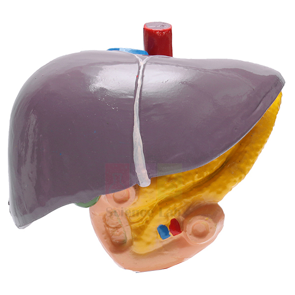
Human Liver Model
Human Liver Model (Product Code: SLE/BL/65) shows the liver together with the ducts and neighbouring organs that carry bile and pancreatic juice into the gut: the gall bladder, right and left hepatic ducts, cystic duct, duodenum, pancreatic duct and pancreas. Every structure is coloured to aid recognition and numbered against a key card, and the model is mounted on a base for display on a bench or in a cabinet. It is one of the practical biology teaching aids India's schools, nursing colleges and medical departments use to explain how the liver connects to the rest of the digestive system. It is manufactured by Jain Scientific Equipments Pvt Ltd (ScienceLab), Ambala.
Key Details
| Parameter | Detail |
|---|---|
| Type | Anatomical model of the liver with biliary and pancreatic structures |
| Structures shown | Liver, gall bladder, right hepatic duct, left hepatic duct, cystic duct, duodenum, pancreatic duct, pancreas |
| Finish | Appropriately coloured |
| Identification | Numbered structures with key card |
| Mounting | Mounted on a base |
| Suitable for | School biology, nursing, pharmacy and medical anatomy teaching |
Typical Use
The model is built around the route bile takes. Teachers can start at the liver, follow the right and left hepatic ducts, show how bile is diverted along the cystic duct for storage in the gall bladder, and then trace it onward into the duodenum, where the pancreatic duct from the pancreas also delivers its enzymes. This single view helps students understand fat digestion, why a blocked duct causes jaundice, and how gallstones or pancreatic disease produce the symptoms they read about in physiology and pathology. Placed alongside heart, lung and kidney pieces, it rounds out a human biology model set in India's secondary and higher-secondary labs.
For revision, cover the key card and ask students to name each numbered structure, or have them point out the path of bile in sequence. Keep the model on its base in a closed cabinet between lessons, dust it with a soft dry cloth and avoid solvents or strong cleaners that could affect the painted surface.
Manufacturer
Jain Scientific Equipments Private Limited produces anatomical and biology teaching models under its ScienceLab trading name in Ambala, Haryana, and supplies them to schools, colleges and distributors. To know more about the company and its manufacturing base, see our company profile.
Related Digestive System Models
Teachers building a lesson on the liver and biliary system often add these models:
- Human Liver With Gall Bladder Model — focuses on the lobes of the liver and the hilus vessels.
- Human Liver Pathology Model — compares healthy tissue with disease changes.
- Gallstone Model — shows where stones form and block the biliary tract.
- Rear Organs of the Upper Abdomen — places the pancreas and duodenum in their wider setting.
- Digestive System Model 2 part — the full digestive tract for overview lessons.
The complete range is in our Digestive System category.
Frequently Asked Questions
Which structures does the model show?
The liver, gall bladder, right and left hepatic ducts, cystic duct, duodenum, pancreatic duct and pancreas, each coloured and numbered.
Does it come with a key?
Yes. The numbered structures are identified on a key card supplied with the model.
What size and material is the model?
The listing does not state dimensions or material. Please ask us through the contact page and we will confirm them before you order.
Can parts be removed?
The description covers a mounted display model and does not mention removable parts. Check with us if your syllabus needs a dissectible version.
Is it suitable for school-level biology?
Yes. The coloured, numbered layout suits secondary students, and the level of detail on the duct system is also useful for nursing and pharmacy courses.
Can you supply several models for a college or export order?
Yes, we supply institutions, tenders and distributors in India and international markets. Include product code SLE/BL/65 and the quantity in your enquiry.
Request a Quote
For a quotation on the Human Liver Model, reach us via the contact page or add it to the enquiry cart with other anatomy models.
Last updated: 24 September 2026
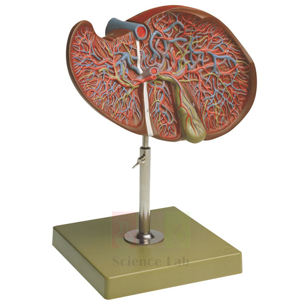
Human Liver With Gall Bladder Model
Human Liver With Gall Bladder Model (Product Code: SLE/BL/66) concentrates on the liver itself, showing its 4 lobes together with the gall bladder, the extra hepatic ducts and the vessels at the hilus. Coloured to separate tissue types and numbered against a key card, the model sits on a base so that students can view the organ from different sides during a lesson. It is a useful piece of school biology lab equipment in India and is equally at home in nursing, paramedical and medical anatomy rooms where the external surfaces of the liver are studied. It is manufactured by Jain Scientific Equipments Pvt Ltd (ScienceLab), Ambala.
Key Details
| Parameter | Detail |
|---|---|
| Type | Anatomical liver model with gall bladder |
| Lobes shown | 4 lobes |
| Other structures | Gall bladder, extra hepatic ducts, hilus vessels |
| Finish | Appropriately coloured |
| Identification | Numbered with key card |
| Mounting | Mounted on a base |
Typical Use
Anatomy classes use this model to identify the lobes of the liver and to show how the gall bladder sits on its under-surface. The hilus, or porta hepatis, is where vessels enter and bile ducts leave the organ, and seeing these together on one model helps students link the liver's double blood supply to its role in processing nutrients absorbed from the gut. Following the extra hepatic ducts from the hilus to the gall bladder also prepares learners for topics such as bile storage, obstruction and surgical removal of the gall bladder.
A good classroom routine is to have students sketch the organ from the model, label the four lobes and the hilus structures using the numbered key, and then compare their drawings with textbook diagrams. Schools placing a biology model kit bulk order often combine this piece with a whole-tract digestive model so that the liver can be discussed both in detail and in position. Store it covered to keep dust off, and clean it only with a soft dry cloth.
Manufacturer
The model is made by Jain Scientific Equipments Private Limited, which trades as ScienceLab and is based in Ambala, Haryana. The company is registered as an MSME enterprise with manufacturing as its major activity (Udyam UDYAM-HR-01-0003714) and holds Importer-Exporter Code AAECJ8618A from the DGFT for overseas orders. Read more on the ScienceLab profile.
Related Digestive System Models
These models extend a lesson on the liver and gall bladder:
- Human Liver Model — adds the duodenum, pancreas and full duct system.
- Human Liver and Gallbladder Model — an alternative liver and gall bladder design.
- Human Liver Pathology Model — for comparing normal anatomy with diseased tissue.
- Gallstone Model — illustrates stones in the gall bladder and ducts.
- Human Digestive System Model — shows where the liver lies within the whole tract.
See every model in the Digestive System category.
Frequently Asked Questions
How many lobes does the model show?
It shows the 4 lobes of the liver, together with the gall bladder, extra hepatic ducts and hilus vessels.
How does it differ from the Human Liver Model?
This model concentrates on the liver's lobes and hilus. The Human Liver Model also includes the duodenum, pancreas and pancreatic duct, so it suits lessons on the whole biliary and pancreatic drainage route.
Are the parts labelled?
The structures are numbered and identified on the key card that comes with the model.
What are its dimensions?
Size and material are not given in the product data. Contact us via the contact page and we will confirm them with your quotation.
What should a school include in a quotation request?
Give product code SLE/BL/66, the quantity, the delivery address and any other models needed so we can quote everything together.
Do you ship outside India?
Yes, we supply institutions and distributors in India and international markets.
Request a Quote
To price the Human Liver With Gall Bladder Model, write to us through the contact page or add it to the enquiry cart.
Last updated: 24 September 2026
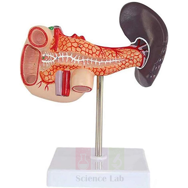
Human Pancreas, Spleen and Duodenum Model
Human Pancreas, Spleen and Duodenum Model (Product Code: SLE/BL/78) is an anatomical model of three neighbouring abdominal organs — the spleen, pancreas and duodenum — used to teach how the pancreas delivers its secretions into the small intestine during digestion, in school, nursing and medical classes. The duodenum shows its internal structures, the position of the pancreatic ducts and the vessels. It is made by Jain Scientific Equipments Pvt Ltd (ScienceLab), an OEM anatomical models manufacturer, at its own factory in Ambala, India.
Specifications
| Parameter | Detail |
|---|---|
| Organs shown | Spleen, pancreas and duodenum (3 abdominal organs) |
| Duodenum detail | Internal structures, position of the pancreatic ducts, and vessels |
| Finish | Appropriately coloured and numbered |
| Identification | Key card for the numbered structures |
| Mounting | Mounted on a base |
| Teaching purpose | Understanding the process of digestion |
| Product code | SLE/BL/78 |
| Manufacturer | Jain Scientific Equipments Pvt Ltd (ScienceLab) — OEM |
| Country of origin | India (made in Ambala, Haryana) |
What Is the Pancreas, Spleen and Duodenum Model Used For?
This model fits the part of a digestion lesson that follows the stomach: food enters the duodenum, where pancreatic juice arrives through the pancreatic ducts to continue breaking it down. With the duodenum displayed to show its interior and duct openings, a teacher can trace that route on the model while students follow the numbers on the key card. The spleen alongside lets the class discuss why an organ next to the digestive tract belongs to the lymphatic and circulatory story rather than to digestion itself.
Biology teachers use it for senior secondary human physiology, while nursing, pharmacy and paramedical colleges use it for abdominal anatomy revision. Among biology teaching aids for the digestive system, it complements a stomach or full digestive-system model rather than replacing it.
OEM Manufacturer
Jain Scientific Equipments Private Limited, marketed under the ScienceLab name, is the OEM (original equipment manufacturer) of this model and produces it at its own licensed factory at Saha, Ambala, Haryana, India, so schools, colleges, dealers and importers receive it factory-direct. ScienceLab is a registered MSME manufacturer (Udyam UDYAM-HR-01-0003714), and its export consignments move under Importer-Exporter Code AAECJ8618A issued by the DGFT.
A replacement key card or a repeat order for another laboratory is handled by the maker itself — just ask. More detail is on our company profile page.
Related Digestive System Models
Other models used when teaching the digestive system and its accessory organs:
- Human Stomach, on Stand — the organ that empties into the duodenum, for the preceding stage of digestion
- Human Liver With Gall Bladder Model — shows the bile-producing organs that also drain into the duodenum
- Digestive System Model 2 part — places these organs within the complete digestive tract
- Pathology of the Human Pancreas, Duodenum, and Gallbladder Model — moves the same region on to disease-related teaching
- Prepared Microscope Slide Set – Digestion (Liver, Pancreas And Tongue) — pancreatic tissue at microscope level
See the full Digestive System range of anatomy models.
Frequently Asked Questions
Which organs does the Human Pancreas, Spleen and Duodenum Model include?
The Human Pancreas, Spleen and Duodenum Model includes three abdominal organs — the spleen, the pancreas and the duodenum — mounted together on one base, so students can see how they sit in relation to one another during a digestion lesson.
How are the parts of the Human Pancreas, Spleen and Duodenum Model identified?
The structures on the Human Pancreas, Spleen and Duodenum Model are coloured and numbered, and a key card lists what each number refers to, so students can test themselves without a separate chart or teacher prompt.
Can the pancreatic ducts be seen on the Human Pancreas, Spleen and Duodenum Model?
Yes. On the Human Pancreas, Spleen and Duodenum Model, the duodenum is presented to show its internal structures together with the position of the pancreatic ducts and the vessels, which lets a teacher trace the route of pancreatic juice.
What size is the Human Pancreas, Spleen and Duodenum Model?
Dimensions of the Human Pancreas, Spleen and Duodenum Model are not stated in our current listing for SLE/BL/78. Contact us and the manufacturer will confirm the size before you place an order.
How should the Human Pancreas, Spleen and Duodenum Model be cared for?
Dust the Human Pancreas, Spleen and Duodenum Model regularly with a soft brush or dry cloth and keep it out of strong direct sunlight. Avoid solvent-based cleaners on the coloured surfaces, and keep the key card with the model.
Who manufactures the Human Pancreas, Spleen and Duodenum Model?
The Human Pancreas, Spleen and Duodenum Model is manufactured by Jain Scientific Equipments Pvt Ltd (ScienceLab), the OEM, at its own factory at Saha, Ambala, Haryana, India. Quotations and repeat orders come straight from the manufacturer.
Can schools and colleges order the Human Pancreas, Spleen and Duodenum Model in bulk?
Yes. ScienceLab accepts orders for the Human Pancreas, Spleen and Duodenum Model from institutions, dealers and importers in India and international markets. Tell us the quantity and destination so the manufacturer can quote accordingly.
Request a Quote
For a quotation direct from the manufacturer on the pancreas, spleen and duodenum model, send us your requirement or place it in the enquiry cart.
Last updated: 24 September 2026
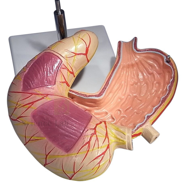
Human Stomach, on Stand
Human Stomach, on Stand (Product Code: SLE/BL/86) is a two-part stomach model that opens to reveal the inside of the organ as well as the layers of its wall. It shows both the longitudinal and circular muscle layers, the cardiac and pyloric parts, the mucous membrane and gastric canal, the supplying arteries and nerves, and the position of the peritoneum. The model is coloured, numbered with a key card and mounted on a base for bench or cabinet display. As an anatomical models manufacturer, ScienceLab designs pieces like this for biology, nursing and medical teaching; it is manufactured by Jain Scientific Equipments Pvt Ltd (ScienceLab), Ambala.
Key Details
| Parameter | Detail |
|---|---|
| Type | Human stomach model, mounted on a base |
| Construction | Separates into two parts |
| Muscle layers | Longitudinal and circular muscle layers |
| Regions shown | Cardiac and pyloric parts |
| Internal features | Mucous membrane, gastric canal |
| Vessels and nerves | Arteries and nerves |
| Other | Position of the peritoneum |
| Finish and key | Appropriately coloured, numbered with key card |
Typical Use
Teachers use the outer surface to name the regions of the stomach and trace its blood and nerve supply, then open the two halves to show the mucous membrane lining and the gastric canal along which liquids pass. Because the longitudinal and circular muscle layers are shown separately, the model makes it easy to explain how coordinated contractions churn food and push it towards the pylorus, and how the cardiac and pyloric regions control entry and exit. The peritoneal covering helps students understand how the stomach is suspended within the abdominal cavity.
It fits naturally into a biology lab setup for schools in India as well as nursing and pharmacy teaching rooms, where it supports topics such as digestion, gastric secretion, ulcers and pyloric narrowing. Encourage students to handle the halves over a table, match each numbered feature to the key card, and close the model after use. Dust it with a soft cloth and keep it away from direct sunlight to preserve the colours.
Manufacturer
Jain Scientific Equipments Private Limited, incorporated under CIN U29309HR2020PTC087597, trades as ScienceLab and is based in Ambala, Haryana. Its licensed factory at 449, HSIIDC Industrial Area, Saha, Ambala operates under Factory Licence AMB-ONLINE-CHD-J-414. Our company profile has further details.
Related Digestive System Models
These items build a fuller lesson around the stomach:
- Human Stomach Model — another stomach design for comparison or group work.
- Stomach Model 2 Part — an alternative opening model of the stomach.
- Gastric Band Model — links stomach anatomy to a common surgical procedure.
- Human Digestive Tract Model — shows the stomach's place in the whole tract.
- Microslide The Digestive System — lets students see the stomach lining under the microscope.
Browse all models in the Digestive System category.
Frequently Asked Questions
What can be seen when the model is opened?
Opening the two parts shows the mucous membrane, the gastric canal and the cardiac and pyloric regions from the inside.
Which layers of the stomach wall are shown?
Both the longitudinal and the circular muscle layers are displayed, which helps explain how the stomach mixes and moves food.
Does it show blood supply and nerves?
Yes. Arteries and nerves are shown on the model, along with the position of the peritoneum.
How are the features identified?
They are numbered and listed on a key card supplied with the model.
What are the dimensions and material?
These are not stated in the listing; ask us through the contact page for confirmation before ordering.
What if the model arrives damaged?
Report any transit damage to our team promptly through the contact page with photographs, and we will advise on the next steps.
Do you take bulk and export orders?
Yes, for institutions and distributors in India and international markets. Include product code SLE/BL/86 and your quantity.
Request a Quote
Request a price for the Human Stomach, on Stand via our contact page, or add it to the enquiry cart with other digestive system models.
Last updated: 24 September 2026
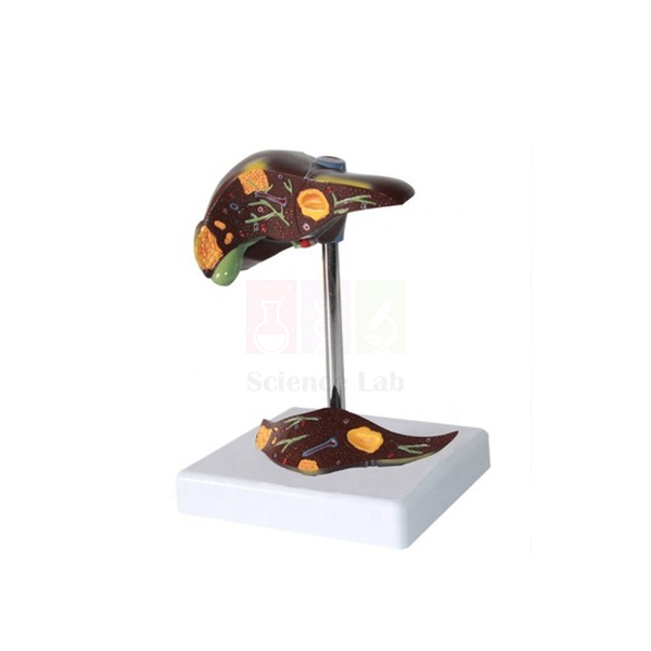
Human Liver Pathology Model
Human Liver Pathology Model (Product Code: SLE/A/110) is a life-size model of the human liver and gallbladder that depicts common pathologies of these organs, supplied with a full-colour, double-sided, illustrated key card that explains what each area shows. It is manufactured by Jain Scientific Equipments Pvt Ltd (ScienceLab), Ambala, for medical and nursing education, hepatology and gastroenterology patient counselling, and advanced biology teaching where disease is taught alongside normal anatomy.
Specifications
| Parameter | Detail |
|---|---|
| Scale | Life size |
| Organs | Human liver and gallbladder |
| Content | Common pathologies of the liver and gallbladder |
| Key | Full-colour, double-sided, illustrated key card included |
| Size | 8 x 4-1/2 x 5-1/2 in |
Typical Use
Pathology is easier to remember when it can be seen on a real-sized organ. Tutors hand out the illustrated key card, and students locate each depicted condition on the model before discussing its causes and consequences. Used next to a healthy liver model, it shows clearly how disease changes the appearance of liver and gallbladder tissue, which supports lectures on alcohol, infection, diet and other risk factors for liver disease.
In clinics, a doctor or dietitian can use the model and card to explain a diagnosis to a patient in plain terms. For colleges assembling a human biology model set with a disease component, it covers the digestive organs that are most often asked about. Store the key card with the model so the two are never separated, and clean the surface with a dry cloth only.
Manufacturer
Jain Scientific Equipments Private Limited manufactures this model under its ScienceLab brand in Ambala, Haryana, where the company develops teaching models and laboratory apparatus. Procurement teams completing vendor registration can use the company’s GSTIN 06AAECJ8618A1ZT and LEI 3358004HRD1JIVXKYX49. Further information is on our company profile.
Related Digestive System Models
Models that help teach liver and biliary disease against normal anatomy:
- Human Liver and Gallbladder Model — enlarged healthy internal anatomy for comparison
- Human Liver Model — a normal liver reference for introductory lessons
- Gallstone Model — focuses on gallstones and gallbladder inflammation in the biliary system
- Pathology of the Human Pancreas, Duodenum, and Gallbladder Model — extends pathology teaching to neighbouring digestive organs
See the complete Digestive System category for more organ models.
Frequently Asked Questions
Which pathologies are shown?
The model depicts common pathologies of the liver and gallbladder, identified on the included key card. For the full list before ordering, contact us and we will share it.
What is on the key card?
It is a full-colour, double-sided, illustrated card that explains the features shown on the model, so the model can be used without a separate textbook.
What are the dimensions?
The model is life size and measures 8 x 4-1/2 x 5-1/2 inches.
Is the model a single piece?
Our description does not state that it separates into parts. Please check with us if you need a take-apart version.
Can you provide vendor registration details for a tender?
Yes. Our GSTIN and LEI appear in the Manufacturer section above, and further documents required by your tender can be discussed with our team.
Is bulk supply to several campuses possible?
Yes. Give us the quantity and delivery points, in India or abroad, and we will quote accordingly.
Request a Quote
Request pricing for the Human Liver Pathology Model through our contact page, or add it to your enquiry cart to combine it with healthy liver models in one quotation.
Last updated: 24 September 2026
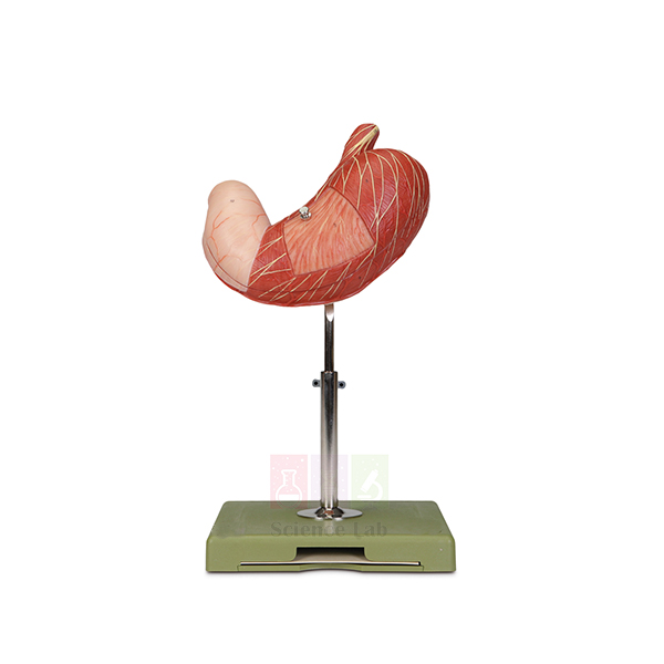
Human Stomach Model
Human Stomach Model (Product Code: SLE/A/149) is a life-size stomach that is dissectible into 2 parts, letting students examine both the external shape of the organ and its internal anatomy on the same model. A numbered key is included for identifying the structures. It is manufactured by Jain Scientific Equipments Pvt Ltd (ScienceLab), Ambala, an anatomical models manufacturer, for school biology classrooms, nursing and medical colleges, and dietetics training, and fits easily into a human biology model set on the digestive system.
Specifications
| Parameter | Detail |
|---|---|
| Scale | Life size |
| Size | 19 x 18 x 34 cm |
| Construction | Dissectible into 2 parts |
| Anatomy shown | Internal and external anatomy of the human stomach |
| Key | Numbered key included |
Typical Use
A typical lesson starts with the closed model: students trace where food enters from the esophagus and where it leaves towards the duodenum, and note the curved shape that lets the stomach expand after a meal. Opening the 2 parts then reveals the internal anatomy, which the class identifies using the numbered key. This sequence supports explanations of how the stomach stores food, mixes it with gastric juice and releases it gradually into the small intestine.
Being life size, the model gives a realistic sense of how large the stomach actually is, which is useful in nutrition and dietetics classes as well as in anatomy. Reassemble the two halves after each session so the joining edges are not chipped, and keep the key with the model.
Manufacturer
Our stomach model is produced by Jain Scientific Equipments Private Limited, the Ambala-based company in Haryana, India, that markets its teaching models and lab apparatus as ScienceLab. Find out more about the company on our profile page.
Related Digestive System Models
Other models used in the same digestion lessons:
- Stomach Model 2 Part — shows the individual layers of the stomach wall, with a removable front half
- Human Stomach, on Stand — a stand-mounted stomach model for display
- Gastric Band Model — adds a surgical treatment topic to stomach anatomy
- Digestive System Model 2 part — places the stomach within the whole digestive tract
Browse all organ models in the Digestive System category.
Frequently Asked Questions
How many parts does the model have?
It is dissectible into 2 parts, revealing the internal anatomy when opened.
What are its dimensions?
The life-size model measures 19 x 18 x 34 cm.
Is a key included?
Yes, a numbered key is supplied so each marked structure can be named.
Does it come on a stand?
A stand is not mentioned in the description for this code. If you need a stand-mounted version, see Human Stomach, on Stand, or ask us to confirm.
How is it different from the Stomach Model 2 Part?
That model concentrates on the layers of the stomach wall, vessels and nerves. This one is a straightforward life-size stomach opening into 2 parts, with a numbered key, suited to general anatomy teaching.
How should it be cleaned?
Use a soft cloth, lightly dampened if needed, and dry the surface afterwards. Avoid solvents that could lift the paint.
Can schools order it in quantity?
Yes. We supply multiple units for schools, colleges and distributors; include the quantity and delivery address in your enquiry.
Request a Quote
For a quotation on the Human Stomach Model, send your details via the contact page or add it to the enquiry cart along with other digestive system models.
Last updated: 24 September 2026
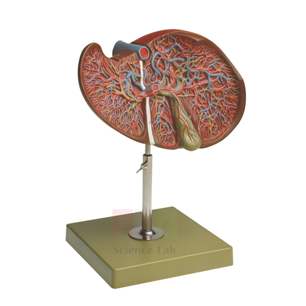
Human Liver and Gallbladder Model
Human Liver and Gallbladder Model (Product Code: SLE/A/153) is a one-part, dissected model enlarged to 1-1/2x life size, showing the internal anatomy of the human liver together with its gallbladder. The enlargement makes internal detail easier to point out to a group, and a numbered key is included. It is manufactured by Jain Scientific Equipments Pvt Ltd (ScienceLab), Ambala, for anatomy and physiology teaching in schools, nursing colleges and medical institutions, and is a useful piece among biology teaching aids in India for the digestive system.
Specifications
| Parameter | Detail |
|---|---|
| Scale | 1-1/2x life size, enlarged |
| Size | 19 x 26 x 29 cm |
| Construction | One-part, dissected model |
| Anatomy shown | Internal anatomy of the human liver with gallbladder |
| Key | Numbered key included |
Typical Use
The liver is large and solid, so its interior is hard to picture from outside views alone. Because this model is already dissected, the internal anatomy is visible the moment it is placed on the bench, with no parts to remove. Teachers use it to explain the liver’s role in producing bile, the gallbladder’s role in storing it, and how both connect to digestion of fats. Students then work through the numbered key to name each marked structure.
The one-part build suits frequent handling by younger classes, and the enlarged scale helps at the back of a lecture room. Pairing it with a liver pathology model turns a single lesson into a normal-versus-disease comparison. Clean it with a soft dry cloth and keep it away from heat sources.
Manufacturer
Manufactured by Jain Scientific Equipments Private Limited, which trades as ScienceLab from Ambala, Haryana, and produces anatomy models and teaching apparatus for institutions in India and international markets. Details about the company are on our company profile.
Related Digestive System Models
Models that fit into the same liver and biliary lessons:
- Human Liver Pathology Model — life-size liver and gallbladder showing common pathologies, for comparison
- Human Liver With Gall Bladder Model — an alternative liver and gall bladder model
- Rear Organs of the Upper Abdomen — shows the opened bile ducts, duodenum and pancreas that connect to the gallbladder
- Gallstone Model — covers gallstones and gallbladder inflammation in the biliary system
More models are listed in our Digestive System category.
Frequently Asked Questions
Why is the model enlarged?
At 1-1/2x life size, the internal features of the liver and gallbladder are larger and easier to identify during group teaching.
Does it separate into parts?
No. It is a one-part model that is already dissected to show the internal anatomy.
What size is it?
It measures 19 x 26 x 29 cm.
Is there a key?
Yes, a numbered key comes with the model.
Does it show liver disease?
No, it shows normal internal anatomy. For disease teaching, see the Human Liver Pathology Model listed above.
What about warranty and delivery time?
These are confirmed in each quotation rather than fixed on this page. Contact us with your requirement.
Request a Quote
Ask for a price on the Human Liver and Gallbladder Model through our contact page, or add it to the enquiry cart if you are ordering several anatomy models together.
Last updated: 24 September 2026
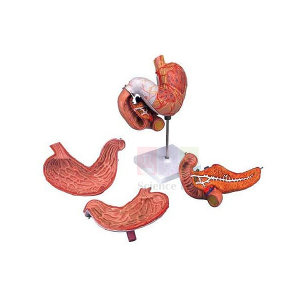
Stomach Model 2 Part
Stomach Model 2 Part (Product Code: SLE/DS/01) is an anatomical replica of the human stomach that shows the different, individual layers of the stomach wall. Its front half is removable for detailed study of the interior, and the model also depicts vessels, nerves and the lower esophagus. Delivered on a stand and weighing 0.6 kg, it is light enough to move between a classroom and a doctor’s consulting room. It is manufactured by Jain Scientific Equipments Pvt Ltd (ScienceLab), Ambala, as part of its human biology model set range for the digestive system.
Specifications
| Parameter | Detail |
|---|---|
| Construction | 2 parts; front half removable |
| Stomach wall | Different and individual layers shown |
| Structures depicted | Vessels, nerves, lower esophagus |
| Mounting | Delivered on stand |
| Weight | 0.6 kg |
| Size | 25 x 22 x 12 cm |
| Suitable for | Doctor’s office or classroom |
Typical Use
The layered wall is the feature that sets this model apart. With the front half lifted away, tutors can show how the stomach wall is built up from its inner lining to its outer covering, which is the basis for explaining how ulcers and inflammation affect different depths of tissue. The vessels and nerves show how the organ is supplied and controlled, and the lower esophagus marks where swallowed food enters.
In a clinic, a doctor can hold the model up during a consultation to explain a gastroscopy finding or a diagnosis in plain language; in the classroom it supports digestion lessons for biology and nursing students. Refit the front half carefully after use and keep the model on its stand to avoid knocks.
Manufacturer
This two-part stomach is made by Jain Scientific Equipments Private Limited of Ambala, Haryana, India, whose anatomy models and laboratory products carry the ScienceLab name. Our company profile has more about who we are.
Related Digestive System Models
Items often chosen together with this stomach model:
- Gastric Band Model — a two-part stomach with an added gastric band, for bariatric counselling
- Human Stomach Model — a life-size 2-part stomach with numbered key for general anatomy
- Human Stomach, on Stand — another stand-mounted stomach option
- Digestive System Model 2 part — shows where the stomach sits in the complete digestive tract
Explore the full Digestive System category.
Frequently Asked Questions
What does the removable front half reveal?
Removing it opens the stomach for detailed study of its interior and of the individual layers of the stomach wall.
Which other structures are shown?
The model depicts vessels, nerves and the lower esophagus.
Is it heavy to carry around?
No. It weighs 0.6 kg and measures 25 x 22 x 12 cm, so it moves easily between rooms.
Does it include a stand?
Yes, the stomach is delivered on a stand.
Is a numbered key provided?
A key is not listed in our description of this model. Please contact us if you need one.
Can hospitals and training centres order several units?
Yes. We quote for single pieces and multiple units for clinics, colleges and distributors in India and international markets.
Request a Quote
To order the Stomach Model 2 Part, send your quantity and delivery details through our contact page, or add it to your enquiry cart for a combined quotation.
Last updated: 24 September 2026
Rear Organs of the Upper Abdomen
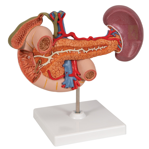
Rear Organs of the Upper Abdomen (Product Code: SLE/DS/02) is a natural-size anatomical model of the organs that lie at the back of the upper abdomen: the duodenum (partially opened), the gall bladder and bile ducts (both opened), the pancreas with its large ducts revealed, the spleen and the surrounding vessels. It is manufactured by Jain Scientific Equipments Pvt Ltd (ScienceLab), Ambala, for anatomy, surgery and gastroenterology teaching in medical and nursing colleges and for advanced biology lab equipment in India where the biliary and pancreatic systems are studied.
Specifications
| Parameter | Detail |
|---|---|
| Scale | Natural size |
| Region | Rear organs of the upper abdomen |
| Duodenum | Partially opened |
| Gall bladder and bile ducts | Opened |
| Pancreas | Shown revealing its large ducts |
| Also shown | Spleen and surrounding vessels |
Typical Use
These organs sit deep in the abdomen and are hidden behind the stomach and intestines on most torso models, so a dedicated model makes them far easier to teach. With the bile ducts, gall bladder and duodenum opened and the pancreatic ducts exposed, students can trace how bile and pancreatic juice travel into the duodenum, which is central to understanding fat digestion and conditions such as gallstones, bile duct obstruction and pancreatitis.
Surgical tutors use the model to explain the close relationships between the duodenum, pancreas, bile ducts and major vessels before students see them in theatre or on imaging. Place it where it can be viewed from the front at eye level, and wipe it with a dry cloth after handling.
Manufacturer
Behind the ScienceLab brand is Jain Scientific Equipments Private Limited, a model and laboratory equipment maker located in Ambala, Haryana. The company is enrolled as an MSME (Udyam UDYAM-HR-01-0003714) and ships international orders under its DGFT Importer-Exporter Code AAECJ8618A. More about us on the company profile.
Related Digestive System Models
Models that cover the same hepatobiliary and pancreatic topics:
- Gallstone Model — shows typical gallstone sites along the biliary system
- Pathology of the Human Pancreas, Duodenum, and Gallbladder Model — disease changes in the same organ group
- Human Liver and Gallbladder Model — the liver side of the biliary system in enlarged form
- Digestive System Model 2 part — the complete digestive tract for orientation
View all models in our Digestive System category.
Frequently Asked Questions
Which organs are included?
The duodenum, gall bladder, bile ducts, pancreas and spleen, together with the surrounding vessels.
What does “natural size” mean here?
The organs are shown at the size they are in the body, so students get a realistic sense of their proportions.
Which parts are opened?
The duodenum is partially opened, the gall bladder and bile ducts are opened, and the pancreas reveals its large ducts.
What are the model’s dimensions and mounting?
Overall dimensions and base details are not given in our current description. Contact us and we will confirm them before you order.
Is it suitable for school use?
It is aimed mainly at medical, nursing and advanced biology courses, though senior school teachers can use it to extend a digestion lesson beyond the stomach.
Which registration details can you share for vendor onboarding?
Our Udyam and IEC numbers are given above; other documents your institution requires can be requested through the contact page.
Request a Quote
To receive pricing for the Rear Organs of the Upper Abdomen model, write to us via the contact page or add it to your enquiry cart with any related biliary models.
Last updated: 24 September 2026
Digestive System Model 2 part
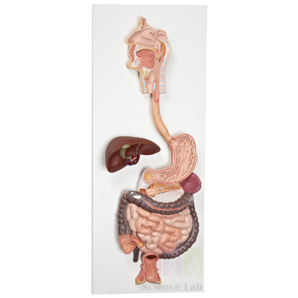
Digestive System Model 2 part (Product Code: SLE/DS/03) is a life-size model that presents the entire human digestive system in graphic relief on a baseboard, from the nose, mouth cavity and pharynx through the esophagus and GI tract, with the liver and gall bladder, spleen and pancreas in place. The duodenum, caecum and rectum are opened to show their interior, and the transverse colon is removable. Manufactured by Jain Scientific Equipments Pvt Ltd (ScienceLab), Ambala, an anatomical models manufacturer, it is a core wall or bench model for schools and colleges choosing biology teaching aids in India for the digestion syllabus.
Specifications
| Parameter | Detail |
|---|---|
| Scale | Life size |
| Format | Graphic relief, mounted on baseboard |
| Features | Nose, mouth cavity and pharynx, esophagus, GI tract, liver with gall bladder, spleen, pancreas |
| Opened sections | Duodenum, caecum and rectum |
| Removable part | Transverse colon |
| Weight | 4.4 kg |
| Dimensions | 81 x 33 x 10 cm |
Typical Use
Because the whole tract is laid out in one relief, teachers can follow a meal step by step: chewing in the mouth cavity, swallowing through the pharynx and esophagus, storage and mixing in the stomach, digestion and absorption in the small intestine, and water recovery in the large intestine down to the rectum. The opened duodenum, caecum and rectum let students see inside key sections, while lifting off the transverse colon gives a clearer view of the organs it normally covers.
The flat baseboard format suits permanent display in a biology room or corridor, where it acts as a reference for the whole digestion unit, and it works equally well laid on a bench for small-group revision. At 4.4 kg it is best lifted with both hands by the baseboard, not by the organs.
Manufacturer
The model is a product of Jain Scientific Equipments Private Limited, an Ambala, Haryana company that builds educational models and laboratory equipment under the ScienceLab name. Read about the company on our profile page.
Related Digestive System Models
Organ-level models that zoom in on parts of this system:
- Stomach Model 2 Part — detailed wall layers of the stomach
- Rear Organs of the Upper Abdomen — duodenum, pancreas, bile ducts and spleen at natural size
- Human Liver and Gallbladder Model — enlarged internal anatomy of the liver
- Human Stomach Model — a life-size stomach that opens into 2 parts
- Gallstone Model — adds a common digestive disorder to the lesson
See every model in the Digestive System category.
Frequently Asked Questions
What does the model cover?
The entire digestive system in relief: nose, mouth cavity and pharynx, esophagus, GI tract, liver with gall bladder, spleen and pancreas.
Which part is removable?
The transverse colon can be removed. The duodenum, caecum and rectum are shown opened.
How big and heavy is it?
It measures 81 x 33 x 10 cm and weighs 4.4 kg.
Can it be hung on a wall?
It is mounted on a baseboard. Wall-fixing hardware is not listed in our description, so contact us to confirm how it is supplied.
Is a key or teaching guide included?
Our description does not mention one; please ask at enquiry stage if you need labelling support.
Do you pack it safely for long-distance delivery?
Packing is arranged for each order, including export consignments to international markets, and is described in the quotation.
Request a Quote
For a quotation on the Digestive System Model 2 part, message us through the contact page, or add it to the enquiry cart with any organ-level models you need.
Last updated: 24 September 2026
Gastric Band Model
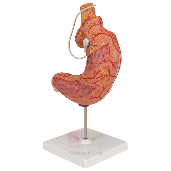
Gastric Band Model (Product Code: SLE/DS/04) is a two-part stomach model to which a gastric band has been added, created to explain this surgical device to patients. A gastric band is inserted surgically to treat morbid obesity when conservative treatments have not been successful, and the model lets a surgeon, dietitian or nurse show exactly where the band sits on the stomach. It is manufactured by Jain Scientific Equipments Pvt Ltd (ScienceLab), Ambala, for bariatric clinics, hospitals and medical and nursing colleges, and is one of the more specialised items among biology teaching aids in India.
Specifications
| Parameter | Detail |
|---|---|
| Base model | Two-part stomach model |
| Added feature | Gastric band |
| Clinical context | Gastric band inserted surgically to treat morbid obesity if conservative treatments are unsuccessful |
| Main purpose | Explaining the gastric band device to patients |
| Weight | 0.51 kg |
| Dimensions | 18 x 16 x 25 cm |
Typical Use
Patients considering weight-loss surgery often find it hard to picture what a band does. In a pre-operative consultation the clinician can point to the band on the stomach, explain how it restricts the upper part of the organ so that smaller meals bring a feeling of fullness, and answer questions about eating after surgery. Because the base is a two-part stomach model, the stomach can also be opened to discuss its normal anatomy in the same conversation.
In nursing and medical education the model supports lectures on obesity management and on the care of patients after bariatric procedures. At 0.51 kg it is easy to hold up during a consultation or pass round a seminar table. Wipe it with a soft cloth between patients and store it away from direct sunlight.
Manufacturer
Jain Scientific Equipments Private Limited, trading as ScienceLab, manufactures this model in Ambala, Haryana, alongside its wider line of anatomical models and laboratory apparatus for customers in India and international markets. Our company profile tells you more.
Related Digestive System Models
Useful companions for patient education and stomach anatomy:
- Stomach Model 2 Part — a two-part stomach without a band, for explaining normal anatomy and wall layers
- Human Stomach Model — a life-size 2-part stomach with numbered key
- Human Stomach, on Stand — a stand-mounted stomach for clinic or classroom display
- Digestive System Model 2 part — shows how the stomach connects to the rest of the digestive tract
Find more in our Digestive System category.
Frequently Asked Questions
Is this a medical device?
No. It is an educational model for explaining a gastric band; it is not a band for surgical use.
What is the model based on?
It was developed from a two-part stomach model, with a gastric band added.
How large and heavy is it?
It measures 18 x 16 x 25 cm and weighs 0.51 kg.
Can the band be removed from the stomach?
Our description does not say whether the band detaches. Contact us if you need a removable band for demonstrations.
Is it appropriate for school biology classes?
It is designed mainly for patient explanation and clinical education; schools teaching basic stomach anatomy usually choose a standard stomach model instead.
Can clinics order more than one?
Yes. Hospital groups and distributors can request multiple units; share the quantity and delivery locations in your enquiry.
Request a Quote
For pricing on the Gastric Band Model, contact our team through the contact page or add it to the enquiry cart to request it with other patient-education models.
Last updated: 24 September 2026
Gallstone Model
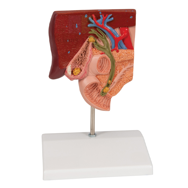
Gallstone Model (Product Code: SLE/DS/05) is a patient-education model, mounted on a base, that shows the anatomy of the biliary system and its surroundings at half natural size. It explains where gallstones are typically found — in the fundus of the gall bladder, around the spiral valve, in the common bile duct and at the papillary opening into the small intestine — and lets viewers identify both acute inflammation (cholecystitis) and the tissue changes of chronic inflammation in the gallbladder wall. It is manufactured by Jain Scientific Equipments Pvt Ltd (ScienceLab), Ambala, for surgical and gastroenterology clinics and for medical and nursing teaching, extending a human biology model set into common digestive disease.
Specifications
| Parameter | Detail |
|---|---|
| Scale | Half natural size |
| Mounting | Mounted on base |
| Anatomy shown | Biliary system and its surroundings |
| Typical gallstone locations | Fundus area of the gall bladder; area of the spiral valve; common bile duct; papillary opening to the small intestine |
| Gallbladder wall | Acute inflammation (cholecystitis) and tissue changes caused by chronic inflammation |
| Intended use | Patient education and clinical teaching |
Typical Use
The model is built around the questions patients ask after a gallstone diagnosis: where the stones are, why they cause pain and what surgery would involve. A clinician can move along the biliary system from the fundus of the gall bladder, past the spiral valve and down the common bile duct to the papillary opening, explaining why a stone in each position produces different symptoms. The inflamed gallbladder wall helps explain the difference between a sudden attack of cholecystitis and long-standing chronic inflammation.
For students, the model links biliary anatomy to one of the most frequently encountered digestive conditions. Keep it on its base on a consulting-room desk or in a teaching cabinet, and wipe it clean with a dry cloth.
Manufacturer
ScienceLab gallstone models are made by Jain Scientific Equipments Private Limited, whose production and offices are in Ambala, Haryana, India, and which supplies clinics, colleges and distributors under the ScienceLab brand. Visit the company profile for more.
Related Digestive System Models
Models used alongside this one in biliary and hepatobiliary teaching:
- Rear Organs of the Upper Abdomen — the opened gall bladder, bile ducts, duodenum and pancreas at natural size
- Human Liver and Gallbladder Model — enlarged normal anatomy of the liver with gallbladder
- Human Liver Pathology Model — common pathologies of the liver and gallbladder with a key card
- Pathology of the Human Pancreas, Duodenum, and Gallbladder Model — related disease changes in neighbouring organs
Browse our Digestive System category for further models.
Frequently Asked Questions
Where are the typical gallstone locations on this model?
In the fundus area of the gall bladder, the area of the spiral valve, the common bile duct and the papillary opening to the small intestine.
What scale is the model?
It shows the biliary system and its surroundings at half natural size.
Does it show inflammation?
Yes. Acute inflammation (cholecystitis) and the tissue changes caused by chronic inflammation can be identified in the gallbladder wall.
Is it mounted?
Yes, the model is mounted on a base.
What are the overall dimensions?
These are not listed in our current description. Please contact us for the measurements.
Can distributors stock this model?
Yes. We work with distributors and institutional buyers in India and international markets; ask for a quotation based on your volumes.
Request a Quote
To price the Gallstone Model for your clinic or college, get in touch via the contact page, or add it to your enquiry cart with other biliary models.
Last updated: 24 September 2026