- Physics Lab Equipment
- Chemistry Lab Equipment
- Biology Lab Equipment
- Anatomical Lab Equipment
- Learning
- Educational Lab Equipment
- Magnets
- Agriculture
- Astronomy
- Earth Science
- Geography Lab Equipment
- Safety Lab Coat
- Laboratory
- Meteorological
- Electricity & Magnetism
- Heat
- School Lab Equipment
- Lab Glassware
- Science Lab Equipment
- Tvet Lab Equipment
- Robotics Kits Supplier and Manufacturer for Schools India
- Math Lab Equipment Manufacturers and Exporters in Ambala
- STEM Kits Manufacturers and Supplier India
- Engineering Lab Equipment
- Hospital Lab Equipment
- Water, Sanitation & Hygiene Supplies
- Cold Chain, Solar & Communication Equipment
- Nutrition
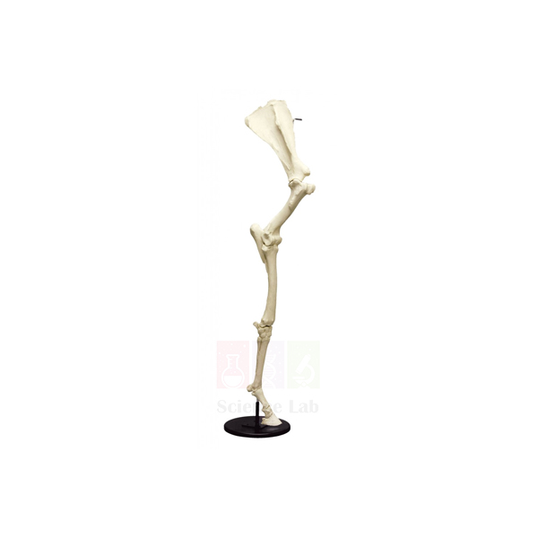
Horse Forelimb Skeleton
An articulated forelimb of an adult horse in natural bone, held on a steel support rod above a square wooden base. The hoof capsule is mounted separately on the base and lifts off, so students can set the outer hoof beside the bones it once enclosed. The mount measures around 13 x 51-1/2 in.
Horse forelimb mount specifications
| Parameter | Detail |
|---|---|
| Material | Natural bone |
| Specimen | Adult horse forelimb, articulated |
| Size | Around 13 x 51-1/2 in. (about 33 x 131 cm) |
| Base | Square wooden base, 11-1/4 x 11-1/4 in. (about 28.6 x 28.6 cm) |
| Support | Steel support rod |
| Hoof capsule | Mounted separately on the base, removable for close examination |
| Product code | SLE/A/120 |
| Minimum order | 10 units |
| Dispatch | Usually within 7 to 10 working days |
| Supplied by | ScienceLab (Jain Scientific Equipments Pvt Ltd), Ambala, India |
Osteology exercises with the horse forelimb
Naming the bones. Working down the mount, students label the humerus, the radius with its reduced ulna, the two rows of carpal bones, the cannon bone with a splint bone on each side, and the three phalanges. They then find the paired sesamoid bones behind the fetlock.
Homologous limbs. Stand the horse limb beside a bat skeleton, a pigeon skeleton and the arm of a rod-supported human skeleton. Students colour-code matching bones on a worksheet and explain how one basic limb plan has been changed for running, flying and grasping.
Joints and stance. Students locate the elbow, the carpus that horse owners call the knee, the fetlock, the pastern joint and the coffin joint. For each one they note the direction it bends and relate the joint angles to how a horse stands and strides.
Hoof and coffin bone. Lift the hoof capsule off the base and hold it next to the end of the limb. Farriery and veterinary students compare the curve of the hoof wall with the shape of the coffin bone, then sketch both in side view. A comparison model of vertebrate limbs can follow for a class summary.
Frequently Asked Questions About the Horse Forelimb Skeleton
Why does a horse stand on a single toe?
The horse is an odd-toed ungulate that carries its weight on the third digit alone. The side digits survive only as the slim splint bones. A long, single-toed limb is light at the lower end and suits fast running, whereas cattle and other even-toed ungulates stand on two toes.
What are the splint bones beside the cannon bone?
They are the second and fourth metacarpals. Each is a thin bone lying along the back of the large third metacarpal, or cannon bone, and it tapers to a small knob partway down. In ruminants the cannon bone forms instead from two fused metacarpals.
Are the radius and ulna separate in the horse forelimb?
No. The ulna is reduced and fused to the back of the radius, and only its upper end, the point of the elbow, stands clear. Because of this fusion the forearm cannot rotate, so the limb swings in one plane, unlike the forearm of a dog or a person.
Which bones lie inside the hoof capsule?
In the living foot the hoof encloses the distal phalanx, also called the coffin or pedal bone, and the small navicular bone, the distal sesamoid. The lower end of the middle phalanx, or short pastern, also sits inside. On this mount the capsule sits apart, so students can see both the inner surface of the hoof and the bones.
How do I protect a natural-bone specimen from damp and mould?
Keep it in a dry, airy room, out of direct sun and away from sinks. In humid weather, check it every week. If it gets damp, let it dry slowly at room temperature rather than with a heater, and handle the bones with clean, dry hands so skin oil does not stain them.
Skeletons and a model for comparing vertebrate limbs
- Bat Skeleton
- Pigeon Skeleton
- Human Skeleton, Rod-Supported
- Frog Skeleton
- Comparison Model of Vertebral Limbs
To request a quotation, send the product name, code and quantity through our contact page or add the item to your enquiry cart.
Last updated: 14 November 2026