- Physics Lab Equipment
- Chemistry Lab Equipment
- Biology Lab Equipment
- Anatomical Lab Equipment
- Learning
- Educational Lab Equipment
- Magnets
- Agriculture
- Astronomy
- Earth Science
- Geography Lab Equipment
- Safety Lab Coat
- Laboratory
- Meteorological
- Electricity & Magnetism
- Heat
- School Lab Equipment
- Lab Glassware
- Science Lab Equipment
- Tvet Lab Equipment
- Robotics Kits Supplier and Manufacturer for Schools India
- Math Lab Equipment Manufacturers and Exporters in Ambala
- STEM Kits Manufacturers and Supplier India
- Engineering Lab Equipment
- Hospital Lab Equipment
- Water, Sanitation & Hygiene Supplies
- Cold Chain, Solar & Communication Equipment
- Nutrition
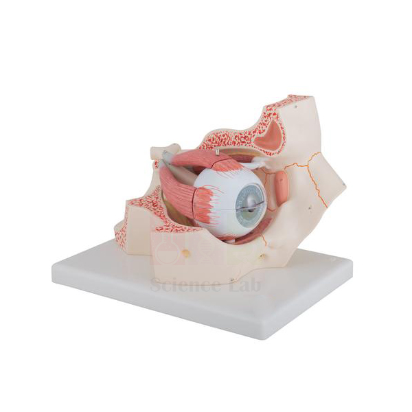
Human Eye in Orbit Model
Human Eye in Orbit Model (Product Code: SLE/A/81) is a large, 3x life size eye that sits in a section of the bony orbit, with the optic nerve shown in its natural position against the orbital floor and medial wall. The eyeball dissects into 7 pieces, so students can open it layer by layer from the sclera inwards to the lens and vitreous humor. It is a dependable biology teaching aid for schools, optometry courses and nursing colleges, made by Jain Scientific Equipments Pvt Ltd (ScienceLab), Ambala.
Specifications
| Parameter | Detail |
|---|---|
| Scale | 3x life size |
| Dissectible into | 7 pieces |
| Sclera | 2-part, with cornea and eye muscle attachments |
| Choroid | 2-part, with iris and retina |
| Other removable parts | Lens; vitreous humor |
| Orbit | Bony orbit (floor and medial wall) with the optic nerve in its natural position |
Typical Use
This model lets a teacher explain the eye both as an organ and as a structure housed in the skull. Lifting away the two halves of the sclera reveals the choroid, iris and retina; removing those exposes the lens and the vitreous humor, which is the natural order for a lesson on the three coats of the eyeball and the path of light to the retina. The muscle attachments on the sclera help when discussing how the eye is moved, and the orbit shows how the optic nerve leaves the eye towards the brain.
In practical exams it works well as a spotting station: students name each removed part and state its function. For school biology lab equipment in India where one eye model must serve many batches, keep the small parts in a labelled box beside the model so the lens and vitreous body are not misplaced between classes.
Manufacturer
The Human Eye in Orbit Model comes from Jain Scientific Equipments Private Limited, better known by its trading name ScienceLab, whose works are located in Ambala, Haryana. An overview of what the company makes and how it operates is available on the company profile page.
Related Human Models
Useful companions for a complete unit on the eye and vision:
- Eye Microanatomy Model — magnified retina views to follow up the retina shown inside this eye
- Human Eye Model with Eyelids — adds the eyelids and external features of the eye
- Human Eye Model — a separate eyeball model for group work at student benches
- Cataract Eye Model with 4 Forms of Cataract — relates the lens to cataract formation
- Human Brain Model in Head — continues the optic nerve story into the brain
Our anatomy models range lists further sense organ and system models.
Frequently Asked Questions
Which parts can be removed?
The 2-part sclera with cornea and muscle attachments, the 2-part choroid with iris and retina, the lens and the vitreous humor — 7 pieces in all.
How large is the model?
The eye is 3x life size, which keeps the lens and inner layers easy to see from the back of a classroom. Ask us for packed dimensions if you need them for storage planning.
Is the optic nerve shown?
Yes. It is shown in its natural position within the bony orbit, formed here by the orbital floor and medial wall.
Is it suitable for junior classes?
Yes, with supervision. The small removable parts need careful handling, so younger students are best shown the dissection by the teacher.
How should it be cleaned?
Wipe the outer surfaces with a soft, barely damp cloth and let each part dry fully before reassembly. Do not soak the pieces.
What warranty is offered?
Warranty terms are given with each quotation; please ask via the contact page for the current conditions.
Can you export it?
Yes. We supply institutions and distributors in India and international markets; send your destination and quantity for a shipping-inclusive quote.
Request a Quote
To order or get a price, message us on the contact page or add the model to the enquiry cart and submit your list in one go.
Last updated: 24 September 2026