- Physics Lab Equipment
- Chemistry Lab Equipment
- Biology Lab Equipment
- Anatomical Lab Equipment
- Learning
- Educational Lab Equipment
- Magnets
- Agriculture
- Astronomy
- Earth Science
- Geography Lab Equipment
- Safety Lab Coat
- Laboratory
- Meteorological
- Electricity & Magnetism
- Heat
- School Lab Equipment
- Lab Glassware
- Science Lab Equipment
- Tvet Lab Equipment
- Robotics Kits Supplier and Manufacturer for Schools India
- Math Lab Equipment Manufacturers and Exporters in Ambala
- STEM Kits Manufacturers and Supplier India
- Engineering Lab Equipment
- Hospital Lab Equipment
- Water, Sanitation & Hygiene Supplies
- Cold Chain, Solar & Communication Equipment
- Nutrition
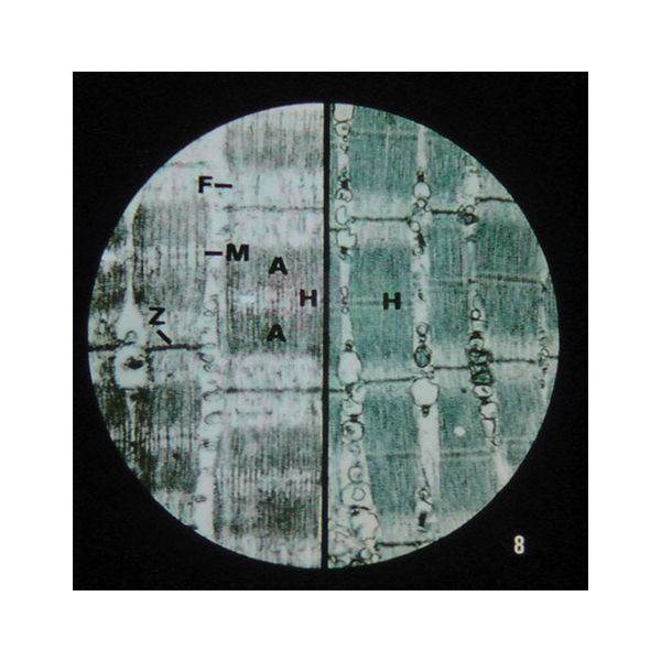
Microslide Nerve & Muscle Action
Microslide Nerve & Muscle Action (Product Code: SLE/PS/97) is a set of 8 related 35 mm images taken through a microscope, used to follow a nerve impulse from a single nerve cell to a contracting muscle in biology and physiology lessons. Its distinguishing frames show sarcomeres in both the relaxed and contracted state, with synapses, synaptic knobs and the nerve-muscle junction along the way. The set is supplied by Jain Scientific Equipments Pvt Ltd (ScienceLab), Ambala, India.
Specifications
| Parameter | Detail |
|---|---|
| What you get | 8 related 35 mm images photographed through a microscope |
| Nerve images | Single Cell; Synapse; Axons; Myelinated Axons; Synaptic Knobs |
| Junction image | Nerve-Muscle Junction |
| Muscle images | Striated Muscle Cell; Sarcomeres – relaxed and contracted |
| On-image help | Arrows and callouts locate the important structures |
| Text folder | Detailed folder that informs and questions students, with a handy pocket for the Microslides |
| Viewed with | Microslide Viewer |
| Product code | SLE/PS/97 |
| Supplied by | Jain Scientific Equipments Pvt Ltd (ScienceLab), Ambala, India |
What Is the Microslide Nerve & Muscle Action Used For?
Teachers use the set to tell one continuous story. Students start with a whole nerve cell, compare plain axons with myelinated axons, and then look closely at a synapse and the synaptic knobs where the signal passes from one cell to the next. The nerve-muscle junction image shows where that signal reaches a muscle fibre.
The last frames switch to the muscle itself: a striated muscle cell, followed by sarcomeres shown relaxed and contracted, which makes the shortening of the sarcomere during contraction easy to explain. With the callouts as a guide and the text folder questions to answer, this set suits both class teaching and self-study at senior secondary and early college level, and pairs well with a synapse model on the demonstration bench.
Supplier
ScienceLab is the trading name of Jain Scientific Equipments Private Limited, Ambala, Haryana, which supplies Microslide Nerve & Muscle Action to schools, colleges and distributors. Shipments outside India are handled under Importer-Exporter Code AAECJ8618A (DGFT), and the company is GST-registered under GSTIN 06AAECJ8618A1ZT. Background on the business is on our company profile.
Related Prepared Slides
Items that help students connect these images to three-dimensional structures:
- Microslide Viewer — the viewer these 35 mm images are made for
- Human Nerve Ending and Synapse Model — a model version of the synapse frames
- Skeletal Muscle Fiber Model — enlarges the striated muscle structure seen in the set
- Microslide Animal Tissue Set 2 Muscle, Bone, Connective — more muscle and tissue images for the same unit
More topic sets are listed under Prepared Slides.
Frequently Asked Questions
What does Microslide Nerve & Muscle Action show?
Microslide Nerve & Muscle Action shows 8 views: a single nerve cell, a synapse, axons, myelinated axons, the nerve-muscle junction, synaptic knobs, a striated muscle cell, and sarcomeres in relaxed and contracted states.
Are magnifications stated for Microslide Nerve & Muscle Action?
No magnification values are given in our listing for Microslide Nerve & Muscle Action. If your syllabus or worksheet needs them, ask us through the contact page and we will check the details for the sets we quote.
Can Microslide Nerve & Muscle Action be used with a normal microscope?
Microslide Nerve & Muscle Action is made for a Microslide Viewer rather than a compound microscope, because the images are 35 mm photographs taken through a microscope. Add viewers to your enquiry if needed.
How is Microslide Nerve & Muscle Action stored?
Microslide Nerve & Muscle Action comes with a detailed text folder that has a pocket for the Microslides, so the images are kept inside the folder with their notes and questions between lessons.
Who supplies Microslide Nerve & Muscle Action?
Microslide Nerve & Muscle Action is supplied by Jain Scientific Equipments Pvt Ltd (ScienceLab) in Ambala, Haryana, India, for schools, colleges and dealers in India and international markets, with IEC and GSTIN details for export paperwork.
What information speeds up a quotation for Microslide Nerve & Muscle Action?
For a Microslide Nerve & Muscle Action quotation, quote code SLE/PS/97, the number of sets, whether viewers or models are needed, and the delivery location. We reply with availability and terms once we have these details.
Request a Quote
Send an enquiry for Microslide Nerve & Muscle Action through our contact page, or add it to the enquiry cart with any viewers and models for your physiology lessons.
Last updated: 24 September 2026