- Physics Lab Equipment
- Chemistry Lab Equipment
- Biology Lab Equipment
- Anatomical Lab Equipment
- Learning
- Educational Lab Equipment
- Magnets
- Agriculture
- Astronomy
- Earth Science
- Geography Lab Equipment
- Safety Lab Coat
- Laboratory
- Meteorological
- Electricity & Magnetism
- Heat
- School Lab Equipment
- Lab Glassware
- Science Lab Equipment
- Tvet Lab Equipment
- Robotics Kits Supplier and Manufacturer for Schools India
- Math Lab Equipment Manufacturers and Exporters in Ambala
- STEM Kits Manufacturers and Supplier India
- Engineering Lab Equipment
- Hospital Lab Equipment
- Water, Sanitation & Hygiene Supplies
- Cold Chain, Solar & Communication Equipment
- Nutrition
Prepared microscope slides are permanently mounted specimens that let schools, colleges and universities run microscopy practicals, from a first beginners set to human histology and cell-division collections. Jain Scientific Equipments Pvt Ltd (ScienceLab), Ambala, supplies this Prepared Slides range of 55 products: glass slide sets for botany, animal and human tissues, insects and microorganisms; Microslide image sets viewed without a microscope; and blank slides, cover slips, storage boxes and cabinets for a permanent slide collection.
What Does the Prepared Slides Range Include?
Featured Prepared Slides
| Product | Product Code | Typical Use |
|---|---|---|
| Human Histology Slide Set of 25 | SLE/PS/79 | 25 glass slides of human tissues, from cerebellum and spinal cord to artery, vein and spleen |
| Mitosis & Meiosis Set, Microscope Slides | SLE/PS/78 | Ten slides including allium root tip and lilium anther stages of division |
| Protozoa and Algae Set of 20 | SLE/PS/120 | Commonly studied protozoa and freshwater algae, including amoeba, euglena, paramecium and diatoms |
| Insect Anatomy Slide Set of 9 | SLE/PS/115 | Mouthparts, legs, wings, spiracles and antennae of bee, house fly and butterfly |
| Beginners Microscope Slide Set | SLE/PS/116 | A starter collection for first microscopy lessons |
| Microslide Viewer | SLE/PS/123 | 5X viewer for Microslide image sets that needs no electricity or batteries |
| Micrometric Slide | SLE/PS/02 | Stage graticule slide for calibrating a microscope with a micrometer eyepiece |
| Unit System Slide Cabinet | SLE/PS/70 | Stackable cabinet with 25 aluminium trays holding 500 standard 3 x 1 inch slides flat |
Supply & Company Details
Every item in the Prepared Slides range, from glass slide sets and Microslide lesson sets to cover slips and slide cabinets, is supplied by Jain Scientific Equipments Private Limited, the incorporated company that trades as ScienceLab from Ambala, Haryana. Buyers completing vendor records can note its corporate identity number, CIN U29309HR2020PTC087597, and its global Legal Entity Identifier, LEI 3358004HRD1JIVXKYX49. Slide sets can be ordered as single collections, as several identical sets for parallel practical groups, or in bulk and tender quantities for schools and colleges in India and overseas.
Related Ranges
- Biology Lab Equipment – the parent hub linking every biology sub-range.
- Microscope – the microscopes and micrometric eyepieces that glass prepared slides are used with.
- Biology Lab – individual boxed slides such as stomata, human blood smear and blastula, plus stains such as eosin and safranine for temporary mounts.
- Cell Biology Equipments – enlarged three-dimensional cell and mitosis models that explain what the slides show.
Frequently Asked Questions
Does ScienceLab make these prepared slides?
No. The Prepared Slides range is supplied, not made in-house: Jain Scientific Equipments Pvt Ltd (ScienceLab), Ambala, supplies the glass slide sets, Microslide lesson sets, blank slides, cover slips and storage cabinets to schools, colleges, institutions and dealers in India and abroad.
What is the difference between glass prepared slides and Microslides?
In the Prepared Slides range, glass slide sets are viewed under a microscope. Microslides are sets of eight related 35 mm images photographed through a microscope and viewed with the Microslide Viewer, which is useful where microscopes are limited.
Which prepared slides suit beginners and lower-secondary classes?
For beginners, the Prepared Slides range offers the Beginners Microscope Slide Set and the Basic Slide Set of 25 as starting points. Syllabus-topic sets, such as the Key Stage 4 Biology set, follow on for older classes.
Are there prepared slides for college histology and cell division?
Yes. For college work the Prepared Slides range includes the Human Histology Slide Set of 25, the Mitosis & Meiosis Set, Microscope Slides and the Cell Division Slide Set of 9, which together cover human tissues and the stages of mitosis and meiosis.
How should a prepared slide collection be stored and restocked?
The Prepared Slides range includes the Slide Box, the Slide Box, Wooden, the Blue Box, the Slide Tray Cabinet, Polypropylene and the Unit System Slide Cabinet for storage, plus Microscope Glass Slides, Student-Grade Microscope Slides and Glass Cover Slips for temporary mounts.
Request a Quote or Catalogue
Send the slide sets and quantities you need through the contact page, or collect them in the shopping cart, to receive a quotation or our slide catalogue.
Last updated: 24 September 2026

Micrometric Slide
Micrometric Slide (Product Code: SLE/PS/02) is a glass stage graticule slide used to calibrate a microscope’s eyepiece scale in biology and microscopy teaching laboratories. Its 1 mm scale, divided into 0.01 mm steps, lets students convert eyepiece divisions into real distances before they measure or count cells. It is supplied by Jain Scientific Equipments Pvt Ltd (ScienceLab), Ambala, India, as a companion to the micrometer eyepiece.
Specifications
| Parameter | Detail |
|---|---|
| Also known as | Stage graticule slide |
| Material | Glass |
| Slide size | 26 x 76 mm |
| Scale range | 1 mm |
| Divisions | 0.01 mm (10 µm) |
| Microscope type | Transmitted light microscopes |
| Used with | Micrometer eyepiece |
| Purpose | Manual microscope calibration for cell counting |
| Product code | SLE/PS/02 |
| Supplied by | Jain Scientific Equipments Pvt Ltd (ScienceLab), Ambala, India |
What Is a Micrometric Slide Used For?
The scale inside a micrometer eyepiece has no fixed real value: what one division covers changes with every objective. To find it, place the Micrometric Slide on the stage, focus on its scale, and rotate the eyepiece until both scales lie side by side. Note how many eyepiece divisions match a known number of 0.01 mm steps on the slide, then divide to get the value of one eyepiece division for that objective.
Repeat the procedure for each objective and keep the values in a table beside the microscope. Students can then measure cells, pollen grains or other specimens directly and apply the calibration in cell counting work. Handle the slide by its edges, clean it gently with lens tissue, and keep it in a slide box so the scale stays free of dust and scratches.
What to Specify When Ordering
- The number of slides, usually matched to the number of microscopes or student groups doing calibration.
- Whether you also need a micrometer eyepiece, since the slide is used together with one.
- That your microscopes use transmitted light, the type this slide is intended for.
Supplier
Jain Scientific Equipments Private Limited, which trades as ScienceLab from Ambala, Haryana, supplies the Micrometric Slide to schools, colleges, universities, training laboratories, dealers and importers in India and international markets. It can be ordered with microscopes, eyepieces and prepared slides so a microscopy room is equipped from a single source. Company details are on the ScienceLab profile.
Related Laboratory Equipment
Items used in the same calibration and measurement work:
- Eyepiece Micrometric — the eyepiece scale that this slide is used to calibrate.
- Monocular Biological Microscope 400x — a student microscope on which calibration can be practised.
- Student-Grade Microscope Slides — plain slides for mounting the specimens you measure after calibration.
- Glass Cover Slips — to cover specimen mounts before measurement.
- Slide Box — protective storage for the calibration slide between uses.
More slides and slide accessories are listed under Prepared Slides, and instruments under Microscope.
Frequently Asked Questions
What is a Micrometric Slide?
A Micrometric Slide, also called a stage graticule slide, is a glass slide carrying a 1 mm scale divided into 0.01 mm steps. It sits on the microscope stage as a known reference, so the eyepiece scale of a micrometer eyepiece can be calibrated against it.
What are the scale range and divisions of the Micrometric Slide?
The Micrometric Slide (SLE/PS/02) has a scale range of 1 mm with divisions of 0.01 mm, which is 10 micrometres per division. The glass slide itself measures 26 x 76 mm, so it fits the stage clips of a typical microscope.
Can the Micrometric Slide be used without a micrometer eyepiece?
No, the Micrometric Slide is meant to be used together with a micrometer eyepiece. The slide provides the known scale on the stage and the eyepiece scale is compared against it; without an eyepiece graticule there is nothing to calibrate.
Which microscopes is the Micrometric Slide suitable for?
The Micrometric Slide is suitable for transmitted light microscopes, where light passes up through the glass slide to the objective. Most student and laboratory biological microscopes work this way; please check with us before using it on any other type of instrument.
Does the Micrometric Slide come with a calibration certificate?
Calibration-certificate availability for the Micrometric Slide is not stated in the product details. If your laboratory needs a certificate or traceability documents, raise it through our contact page before ordering and we will tell you what can be provided.
How should the Micrometric Slide be stored?
Store the Micrometric Slide in a slide box or case away from dust, and handle it only by the edges. Clean the surface gently with lens tissue, because scratches or residue on the scale make it harder to line up the two scales accurately.
Who supplies the Micrometric Slide?
The Micrometric Slide (SLE/PS/02) is supplied by Jain Scientific Equipments Pvt Ltd (ScienceLab), Ambala, India. Schools, colleges, laboratories, dealers and importers in India and international markets can order it on its own or together with microscopes and eyepieces.
Request a Quote
For a quotation on the Micrometric Slide, place it in the enquiry cart or contact us through the contact page. State the quantity, whether micrometer eyepieces are also needed, and the delivery location.
Last updated: 24 September 2026
764.jpg)
Basic Botany Slide (Set of 25)
Basic Botany Slide (Set of 25) (Product Code: SLE/BL/09) is a collection of 25 glass microscope slides of botanical specimens used to teach the major plant groups in school and college biology practicals. It runs from algae such as Chlamydomonas and Spirogyra through mosses, liverworts and ferns to pine, maize and sunflower tissues. The set is supplied by Jain Scientific Equipments Pvt Ltd (ScienceLab), Ambala, India, among its prepared microscope slides for botany teaching.
Specifications
| Parameter | Detail |
|---|---|
| Number of slides | 25 |
| Slide type | Glass microscope slides with botanical specimens |
| Coverage | Specimens representing the major plant groups |
| Preparations | Whole mounts (E), transverse sections (TS), vertical sections (VS) and longitudinal sections (LS) |
| Product code | SLE/BL/09 |
| Supplied by | Jain Scientific Equipments Pvt Ltd (ScienceLab), Ambala, India |
Slides in the set, grouped for teaching:
- Algae: Chlamydomonas E; Spirogyra, vegetative E; Spirogyra, conjugation E; Fucus, dioecious antheridial and oogonial conceptacles.
- Fungi: Mucor, conjugation; Saccharomyces (yeast) budding; Penicillium, mycelium and conidia.
- Mosses and liverworts: Mnium, archegonial head VS; Mnium (moss) antheridial head VS; Marchantia, developing sporogonium VS; Marchantia (liverwort) thallus, antheridia VS; Marchantia, thallus, archegonia VS.
- Ferns: Dryopteris (fern), pinnule with sori TS; Dryopteris prothallus with antheridia and archegonia E.
- Gymnosperm: Pinus (pine) leaf TS.
- Flowering plant stems and roots: Zea, stem TS; Zea (maize), root TS; Helianthus, stem TS; Helianthus (sunflower) young root TS; Tilia (lime) young and old stem TS; Cucurbita (marrow) stem, for sieve tubes LS.
- Leaves, flowers, pollen and cell division: Prunus (cherry) leaf TS; Ranunculus (buttercup) flower bud LS; Pollen grains, wind and insect distribution E; Allium (onion) root apex, mitosis LS.
What Is the Basic Botany Slide Set Used For?
The set can carry a full botany practical course. Students begin by comparing simple algae and fungi, then trace the alternation of generations in mosses, liverworts and ferns using the antheridia, archegonia, sporogonium and prothallus slides. Stem and root sections of maize and sunflower give a direct monocot and dicot comparison, while the Cucurbita LS shows the sieve tubes of the phloem.
Pine and cherry leaf sections contrast a needle leaf with a broad leaf, the pollen slide supports lessons on wind and insect pollination, and the onion root apex shows cells in mitosis. Scan every slide at low power before moving to a higher objective, and ask students to draw and label each specimen; this builds the observation skills that botany lab equipment is meant to develop.
Supplier
ScienceLab is the trading name of Jain Scientific Equipments Private Limited, Ambala, which supplies prepared slide sets such as this botany collection to schools, colleges, universities, dealers and importers in India and international markets. For vendor registration, the supplying company is Jain Scientific Equipments Private Limited (CIN U29309HR2020PTC087597, GSTIN 06AAECJ8618A1ZT). Company background is available on the ScienceLab profile.
Related Prepared Slides
Useful additions for the same botany practicals:
- Basic Plant Anatomy Slide Set — extends the stem, root and leaf work into plant anatomy.
- Mitosis & Meiosis Set, Microscope Slides — follows cell division further than the single onion root slide.
- Protozoa and Algae Set of 20 — more algae for the lower-plant part of the course.
- Binocular Biological Microscope — for examining the sections in class.
- Slide Box, Wooden — storage to keep the collection together between practicals.
See every slide collection in the Prepared Slides category.
Frequently Asked Questions
How many slides are in the Basic Botany Slide (Set of 25)?
The Basic Botany Slide (Set of 25) contains 25 glass microscope slides, each carrying a different botanical specimen. Together they represent the major plant groups, from single-celled algae and fungi to mosses, ferns, pine and flowering plants.
Which plant groups does the Basic Botany Slide (Set of 25) cover?
The Basic Botany Slide (Set of 25) covers algae (Chlamydomonas, Spirogyra, Fucus), fungi (Mucor, yeast, Penicillium), mosses and liverworts (Mnium, Marchantia), a fern (Dryopteris), a gymnosperm (Pinus) and flowering plants including maize, sunflower, lime, marrow, cherry, buttercup and onion.
What do E, TS, VS and LS mean on the slide labels?
On the Basic Botany Slide (Set of 25), E marks a whole mount of the entire specimen, TS a transverse section, VS a vertical section and LS a longitudinal section. Knowing the direction of the cut helps students interpret what they see under the microscope.
Does the Basic Botany Slide (Set of 25) show mitosis?
Yes, the Basic Botany Slide (Set of 25) includes an Allium (onion) root apex LS showing mitosis. For a wider study of cell division, it can be paired with a dedicated mitosis and meiosis slide set from the same Prepared Slides range.
What microscope is needed for the Basic Botany Slide (Set of 25)?
The Basic Botany Slide (Set of 25) is examined with a compound light microscope; the product details do not name a minimum magnification. Scan each slide at low power first, then increase magnification, and ask us if you need a matching microscope quoted.
What should a quotation request for the Basic Botany Slide (Set of 25) include?
A quotation request for the Basic Botany Slide (Set of 25) should give the product code SLE/BL/09, the number of sets, and the delivery location. Mention any microscopes or storage boxes you want quoted with it so everything arrives together.
Who supplies the Basic Botany Slide (Set of 25)?
The Basic Botany Slide (Set of 25) is supplied by Jain Scientific Equipments Pvt Ltd (ScienceLab), Ambala, Haryana, India. The company sends slide sets to schools, colleges, universities, dealers and importers in India and international markets.
Request a Quote
Add the Basic Botany Slide (Set of 25) to the enquiry cart or send the number of sets you need through the contact page. We will reply with prices for the slides and any accessories you list.
Last updated: 24 September 2026

Bioviewer Strips Adaptation
Bioviewer Strips Adaptation (Product Code: SLE/BL/10) is a set of 8 images in 35 mm format, photographed through a microscope, used as a visual learning aid for the adaptation topic in biology lessons. Arrows and callouts on the images guide students to the features being studied, and a detailed lesson plan accompanies the set. It is supplied by Jain Scientific Equipments Pvt Ltd (ScienceLab), Ambala, India, as part of its biology teaching aids.
Key Details
| Parameter | Detail |
|---|---|
| Images in set | 8 |
| Image format | 35 mm |
| Image source | Photographed through a microscope |
| Student guidance | Arrows and callouts locating the important features |
| Teacher material | Detailed lesson plan designed to stimulate, inform and question students |
| Title | Adaptation |
| Type | Learning aid |
| Product code | SLE/BL/10 |
| Supplied by | Jain Scientific Equipments Pvt Ltd (ScienceLab), Ambala, India |
What Are Bioviewer Strips Used For?
Bioviewer strips bring microscope-level images into lessons where there are not enough microscopes or prepared slides for every student, or where students need to know what to look for before using a microscope themselves. Because each of the 8 images carries arrows and callouts, a student can find the feature under discussion without the teacher checking every eyepiece.
The lesson plan gives the session a structure: introduce the topic, let students study each image in turn, then use the plan’s questions to check understanding. The strips also work for revision and for comparing photographed features with students’ own slide observations, making them a flexible item among biology teaching aids.
Supplier
Jain Scientific Equipments Private Limited, trading as ScienceLab in Ambala, Haryana, supplies these Bioviewer strips as a learning aid alongside prepared slides and microscopes. Schools, colleges, dealers and importers in India and international markets can combine them with other biology resources in one enquiry. Read about the company on the ScienceLab profile.
Related Prepared Slides
Resources that connect image-based study with hands-on microscopy:
- Prepared Microscope Slide Set – Photosynthesis and Adaptations — real slides for the same topic area.
- Beginners Microscope Slide Set — a first slide collection once students move on to microscopes.
- Micro Slide Viewer — another viewer-based way to show magnified biology images to a class.
- Student Microscope with Fixed Condenser — for students to compare the images with their own observations.
Find more slide sets and viewers in the Prepared Slides category.
Frequently Asked Questions
What does the Bioviewer Strips Adaptation set contain?
The Bioviewer Strips Adaptation set contains 8 images in 35 mm format, each photographed through a microscope and marked with arrows and callouts. A detailed lesson plan accompanies the images to guide the teacher through the topic.
Which specimens appear on the Bioviewer Strips Adaptation images?
Bioviewer Strips Adaptation is titled for the biology topic of adaptation, but the individual specimens on the 8 images are not listed in the product details. Please ask us through the contact page if you need the image list before ordering.
How do the arrows and callouts help students?
The arrows and callouts on Bioviewer Strips Adaptation point directly to the important features being studied, so students can identify structures on their own. This saves time spent explaining where to look and keeps the lesson focused on what the features mean.
What is in the Bioviewer Strips Adaptation lesson plan?
The lesson plan with Bioviewer Strips Adaptation is described as detailed and designed to stimulate, inform and question students about the topic. It helps the teacher introduce each image, prompt discussion and check understanding through questions.
Is a viewer included with Bioviewer Strips Adaptation?
A viewer is not listed as part of Bioviewer Strips Adaptation; the set is described as 8 images in 35 mm format with a lesson plan. Please ask us about suitable viewing equipment for your classroom when you request a quotation.
Who supplies Bioviewer Strips Adaptation?
Bioviewer Strips Adaptation, product code SLE/BL/10, is supplied by Jain Scientific Equipments Pvt Ltd (ScienceLab), Ambala, India. Schools, colleges, dealers and importers in India and international markets can order it alone or with prepared slides and microscopes.
Request a Quote
Place Bioviewer Strips Adaptation in the enquiry cart or write to us via the contact page. Include the number of sets, any viewing equipment questions and your delivery location.
Last updated: 24 September 2026
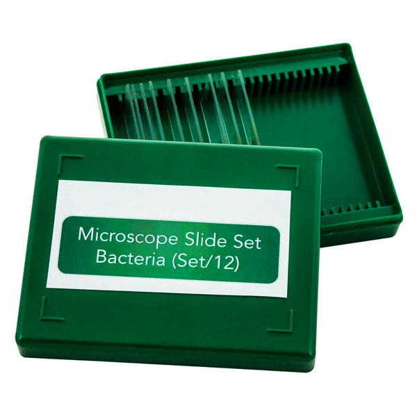
Bacteria slide Set of 12
Bacteria slide Set of 12 (Product Code: SLE/BL/11) is a set of 12 glass microscope slides of bacteria used to teach bacterial shapes, Gram staining and named species in biology and microbiology practicals. It ranges from mixed bacilli, cocci and spirillum to Rhizobium in a root nodule and Escherichia coli, and some samples need a 1000x magnification microscope. It is supplied by Jain Scientific Equipments Pvt Ltd (ScienceLab), Ambala, India, for microbiology teaching laboratories.
Specifications
| Parameter | Detail |
|---|---|
| Number of slides | 12 |
| Slide type | Glass microscope slides |
| Microscope requirement | 1000x magnification needed to view some samples |
| Bacterial shapes | Mixed bacteria – bacilli, cocci and spirillum |
| Gram reaction slides | Cocci, Gram positive and negative; Bacilli, Gram positive and negative |
| Named species | Bacillus megaterium; Bacillus subtilis; Escherichia coli; Streptococcus lactis; Staphylococcus epidermidis; Vibrio spirillum |
| Bacteria in host tissue | Rhizobium in root nodule; Mycobacterium tuberculosis in tissue |
| Product code | SLE/BL/11 |
| Supplied by | Jain Scientific Equipments Pvt Ltd (ScienceLab), Ambala, India |
The product listing names eleven slide subjects for this set of 12, so please confirm the complete slide list with us when you order.
What Is the Bacteria Slide Set Used For?
Teachers usually start with the mixed bacteria slide to show the three basic shapes, then move to the Gram-positive and Gram-negative cocci and bacilli, where the two staining reactions appear in contrasting colours. Named species such as Bacillus subtilis, Bacillus megaterium and Escherichia coli let students link a name to a shape and a staining result.
The Rhizobium root nodule slide connects microbiology with nitrogen fixation in plants, and the Mycobacterium tuberculosis tissue slide shows how bacteria appear within host tissue. Because some samples need 1000x magnification, plan to use an oil-immersion objective with immersion oil, and wipe the oil off each slide after use.
What to Specify When Ordering
- The number of sets needed for your practical groups.
- Whether your microscopes reach 1000x magnification, which some samples require.
- Whether immersion oil or a 1000x microscope should be quoted with the slides.
- Confirmation of the full 12-slide list, since eleven subjects are named in the listing.
Supplier
ScienceLab is the trading name of Jain Scientific Equipments Private Limited, based in Ambala, Haryana, India. The company supplies this bacteria slide set to schools, colleges, universities, microbiology teaching laboratories, dealers and importers in India and international markets, and can include microscopes and immersion oil in the same shipment. Company details are on the ScienceLab profile.
Related Prepared Slides
Items that complete a bacteria practical:
- Immersion Oil, 25ml — for oil-immersion viewing at high magnification.
- Monocular Microscope 1000x, LED light — a 1000x instrument for the samples that need it.
- Binocular Microscope, 1000x Mag LED — a binocular alternative at the same magnification.
- Microslide Helpful Bacteria — a lesson set on useful microbes.
- Microslide Harmful Bacteria — a lesson set on disease-causing bacteria.
Browse the full Prepared Slides range for more microbiology and biology slides.
Frequently Asked Questions
Which slides are included in the Bacteria slide Set of 12?
The Bacteria slide Set of 12 lists mixed bacteria, Gram-positive and Gram-negative cocci and bacilli, Bacillus megaterium, Rhizobium in root nodule, Mycobacterium tuberculosis in tissue, Escherichia coli, Bacillus subtilis, Vibrio spirillum, Streptococcus lactis and Staphylococcus epidermidis. Please confirm the remaining slide with us.
What magnification is needed for the Bacteria slide Set of 12?
A microscope with 1000x magnification is required to view some samples in the Bacteria slide Set of 12. At that power most microscopes use an oil-immersion objective, so keep immersion oil and lens tissue ready at the bench.
Does the Bacteria slide Set of 12 show Gram-positive and Gram-negative bacteria?
Yes, the Bacteria slide Set of 12 includes slides of Gram-positive and Gram-negative cocci and of Gram-positive and Gram-negative bacilli. Students can compare the two staining reactions side by side and relate them to the cell-wall differences taught in theory.
Which courses is the Bacteria slide Set of 12 suited to?
The Bacteria slide Set of 12 suits senior school biology, college microbiology and teacher-training practicals covering bacterial shapes, Gram staining and named species. Institutions with 1000x microscopes get the most from it, since some samples need that magnification.
Who supplies the Bacteria slide Set of 12?
The Bacteria slide Set of 12 (SLE/BL/11) is supplied by Jain Scientific Equipments Pvt Ltd (ScienceLab), Ambala, Haryana, India. The company ships slide sets to institutions, dealers and importers in India and international markets, alone or with microscopes.
What should a quotation request for the Bacteria slide Set of 12 include?
A quotation request for the Bacteria slide Set of 12 should state the product code SLE/BL/11, the number of sets and the delivery location. Add any microscopes or immersion oil you need so we can quote the complete practical set-up together.
Request a Quote
To get a price for the Bacteria slide Set of 12, add it to the enquiry cart or send your requirement through the contact page, listing the quantity and any related items.
Last updated: 24 September 2026

Unit System Slide Cabinet
Unit System Slide Cabinet (Product Code: SLE/PS/70) is a stackable storage cabinet used to keep up to five hundred 3 x 1 inch microscope slides flat and organised in biology departments and slide libraries. Its 25 aluminium trays give every slide its own compartment, and the door retracts into the top of the cabinet. It is supplied by Jain Scientific Equipments Pvt Ltd (ScienceLab), Ambala, India, for laboratories that need a dedicated lab storage cabinet for slides.
Specifications
| Parameter | Detail |
|---|---|
| Slide capacity | Five hundred 3 x 1 inch slides |
| Trays | 25 aluminium trays, horizontal |
| Slide position | Each slide lies flat in its own compartment |
| Door | Retracts into the top of the cabinet |
| Frame | Wood |
| Expansion | Additional units can be stacked on top |
| Size | 8 1/2 x 12 x 10 inch (H) |
| Product code | SLE/PS/70 |
| Supplied by | Jain Scientific Equipments Pvt Ltd (ScienceLab), Ambala, India |
What Is a Unit System Slide Cabinet Used For?
A department that builds up a prepared-slide collection needs storage that protects slides from dust, breakage and mix-ups. Keeping slides flat on horizontal trays helps mounted specimens and coverslips stay in position over the years, and giving each slide its own compartment shows at a glance which slide is out in a practical.
Arrange the trays by topic, for example plant slides on the upper trays and human histology lower down, and label each tray front so students return every slide to its place. The wood frame and aluminium trays give the collection a sturdy home, and when the collection grows a second unit can be stacked on top instead of finding new shelf space.
What to Specify When Ordering
- The number of cabinets, remembering that additional units stack on top of the first.
- Your slide format: the trays are made for 3 x 1 inch slides.
- The shelf or bench space available for a unit measuring 8 1/2 x 12 x 10 inch (H).
- Any prepared slide sets you want supplied to fill the cabinet.
Supplier
Jain Scientific Equipments Private Limited, which trades as ScienceLab, supplies the Unit System Slide Cabinet from Ambala, Haryana to schools, colleges, universities, research laboratories, dealers and importers in India and international markets. It can be shipped together with the prepared slide sets it is meant to hold. See the ScienceLab profile for company information.
Related Prepared Slides
Slides and storage that go with a department slide cabinet:
- Slide Tray Cabinet, Polypropylene — an alternative cabinet that holds separate slide trays.
- Slide Box, Wooden — a hinged box for a smaller collection or slides on loan.
- Basic Slide Set of 25 — prepared slides to start filling the trays.
- Human Histology Slide Set of 25 — a histology collection worth storing flat.
- Microscope Slide Case — a portable case for slides taken out for a practical.
All slide sets and storage items are listed under Prepared Slides.
Frequently Asked Questions
How many slides does the Unit System Slide Cabinet hold?
The Unit System Slide Cabinet holds five hundred 3 x 1 inch microscope slides on 25 aluminium trays. Each slide lies flat in its own compartment, so the cabinet suits a department collection rather than a single class set.
Can more Unit System Slide Cabinets be added later?
Yes, additional Unit System Slide Cabinet units can be stacked on top of the first one. A laboratory can therefore expand its slide storage in five-hundred-slide steps without taking up extra bench or floor area.
What are the dimensions of the Unit System Slide Cabinet?
The Unit System Slide Cabinet measures 8 1/2 x 12 x 10 inches, with 10 inches being the height. Check the space on your shelf or bench, and allow extra height if you plan to stack more units on top.
Why does the Unit System Slide Cabinet store slides flat?
The Unit System Slide Cabinet stores slides flat on horizontal trays because this keeps mounted specimens and coverslips in place over long periods. Slides stored on edge can let mounting media shift slowly, so flat storage suits long-term collections.
What is the Unit System Slide Cabinet made of?
The Unit System Slide Cabinet has a wood frame holding 25 aluminium trays. Its door retracts into the top of the cabinet, so it stays clear of the trays while slides are being selected or returned.
Who supplies the Unit System Slide Cabinet?
The Unit System Slide Cabinet (SLE/PS/70) is supplied by Jain Scientific Equipments Pvt Ltd (ScienceLab), Ambala, India. It can be sent to institutions, dealers and importers in India and international markets, on its own or with prepared slide sets.
Request a Quote
Add the Unit System Slide Cabinet to the enquiry cart or tell us through the contact page how many units and which slide sets you need. We will reply with a quotation for the complete storage order.
Last updated: 24 September 2026

Blue Box, for storing Microscope Slides
Blue Box, for storing Microscope Slides (Product Code: SLE/PS/71) is a compact slotted slide box used to store and carry microscope slides in school, college and research laboratories. Compared with conventional wooden boxes it is smaller, lighter and held to closer dimensional tolerances, and its slots take both 25 x 75 mm and 3 x 1 inch slides. It is supplied by Jain Scientific Equipments Pvt Ltd (ScienceLab), Ambala, India, in 2 sizes.
Specifications
| Parameter | Detail |
|---|---|
| Slide sizes accepted | 25 x 75 mm and 3 x 1 inch |
| Slide retention | Slides will not slip out of the slots once in place |
| Dust protection | Shoulder on each box excludes dust |
| Orientation | Shoulder identifies the top of the box |
| Compared with wooden boxes | Smaller, lighter and held to closer dimensional tolerances |
| Sizes available | 2 |
| Product code | SLE/PS/71 |
| Supplied by | Jain Scientific Equipments Pvt Ltd (ScienceLab), Ambala, India |
What Is the Blue Box for Microscope Slides Used For?
Students and technicians use the Blue Box to hold a working set of slides between sessions: a practical class’s slides, the sections for a project, or a batch of newly made mounts. Because slides grip in their slots, the box can be carried or tilted without slides sliding out and knocking together, and its lighter weight makes it easy to move between the preparation room and the microscope bench.
The shoulder does two jobs: it keeps dust away from the slides, and it shows at once which way up the box goes, so slides are not tipped out when the lid comes off. Number the slots and write a matching index on the lid, and the box becomes a simple catalogue of the slides inside.
What to Specify When Ordering
- Which of the 2 sizes you need; ask us for the slot count of each size.
- Your slide format, noting that both 25 x 75 mm and 3 x 1 inch slides fit.
- The number of boxes per student, group or research project.
Supplier
ScienceLab is the trading name of Jain Scientific Equipments Private Limited of Ambala, Haryana, which supplies the Blue Box together with slides, cover slips and prepared slide sets. Buyers who need vendor registration details can use CIN U29309HR2020PTC087597 and GSTIN 06AAECJ8618A1ZT for the supplying company. Schools, colleges, laboratories, dealers and importers in India and international markets can read more on the ScienceLab profile.
Related Prepared Slides
Slides and storage to use with the Blue Box:
- Microscope Glass Slides — plain slides to fill the box.
- Student-Grade Microscope Slides — plain slides for students’ own mounts.
- Glass Cover Slips — to finish the mounts before they are stored.
- Slide Box, Wooden — the conventional wooden option for comparison.
- Unit System Slide Cabinet — cabinet storage when the collection outgrows boxes.
More slide storage and slide sets are listed under Prepared Slides.
Frequently Asked Questions
Which slide sizes fit the Blue Box, for storing Microscope Slides?
Both 25 x 75 mm and 3 x 1 inch microscope slides fit the slots of the Blue Box, for storing Microscope Slides. Once a slide is in place it will not slip out, so metric and inch-sized slides can share one box.
How does the Blue Box compare with a wooden slide box?
Compared with conventional wooden boxes, the Blue Box, for storing Microscope Slides is smaller in size, lighter in weight and held to closer dimensional tolerances. That makes it easier to carry and gives a more consistent fit for the slides in each slot.
How many slides does each Blue Box hold?
The Blue Box, for storing Microscope Slides is available in 2 sizes, but the slot count of each size is not given in the product details. Please ask us through the contact page and we will confirm the capacity of each size in our quotation.
Does the Blue Box keep dust off the slides?
Yes, a shoulder on each Blue Box, for storing Microscope Slides excludes dust from the stored slides. The same shoulder identifies the top of the box, so it is opened the right way up and slides are not tipped out.
What material is the Blue Box made from?
The material of the Blue Box, for storing Microscope Slides is not stated in the product details. If your laboratory has material, cleaning or chemical-resistance requirements, please send them through our contact page before ordering so we can confirm suitability.
Who supplies the Blue Box, for storing Microscope Slides?
The Blue Box, for storing Microscope Slides (SLE/PS/71) is supplied by Jain Scientific Equipments Pvt Ltd (ScienceLab), Ambala, Haryana, India, to institutions, dealers and importers in India and international markets, alone or with slides and cover slips.
Request a Quote
Put the Blue Box in the enquiry cart or send us the size and quantity you need through the contact page. We will confirm the capacity of each size along with the price.
Last updated: 24 September 2026

Slide Tray Cabinet, Polypropylene
Slide Tray Cabinet, Polypropylene (Product Code: SLE/PS/72) is an autoclavable polypropylene cabinet used to store microscope slide trays horizontally in biology, microbiology and research laboratories. It holds 12 white styrene slide trays, or 240 slides, behind a vertically sliding clear acrylic door, and the cabinet can be autoclaved at 121 °C. It is supplied by Jain Scientific Equipments Pvt Ltd (ScienceLab), Ambala, India; the trays are not included.
Specifications
| Parameter | Detail |
|---|---|
| Cabinet material | Polypropylene |
| Capacity | 12 white styrene microscope slide trays, or 240 slides |
| Tray orientation | Horizontal |
| Door | Front, vertically sliding, clear acrylic |
| Autoclaving | Cabinet can be autoclaved at 121 °C |
| Size | 7 3/4 x 14 x 7 1/2 inch (H) |
| Trays | Not included |
| Product code | SLE/PS/72 |
| Supplied by | Jain Scientific Equipments Pvt Ltd (ScienceLab), Ambala, India |
What Is a Slide Tray Cabinet Used For?
Slide trays let a class or a research project keep its slides flat and in order; this cabinet then protects a stack of those trays from dust and knocks while leaving them visible through the clear door. With 12 trays loaded it keeps up to 240 slides in one compact unit on a bench or shelf.
Because the cabinet can be autoclaved at 121 °C, it is a practical piece of microbiology lab equipment where storage has to be decontaminated from time to time. Take out all trays and slides before autoclaving, let the cabinet cool and dry completely, then reload the trays in their usual order.
What to Specify When Ordering
- The number of cabinets required.
- Slide trays, which are not included: each cabinet takes 12 white styrene trays, so ask us to quote them.
- Slides to fill the trays, if you are setting up a new collection.
- Shelf space for a cabinet of 7 3/4 x 14 x 7 1/2 inch (H).
Supplier
Jain Scientific Equipments Private Limited trades as ScienceLab and supplies the polypropylene Slide Tray Cabinet from Ambala, Haryana, India. Laboratories, dealers and importers in India and international markets can ask for the cabinet together with slides and other microscopy accessories so the whole storage set-up arrives in one consignment. Details about the company are on the ScienceLab profile.
Related Prepared Slides
Items used with a slide tray cabinet in the same workflow:
- Unit System Slide Cabinet — a larger cabinet with its own built-in trays.
- Microscope Glass Slides — plain slides for the trays.
- Slide Drying Rack — dry stained or washed slides before they go into storage.
- Slide Box — box storage for a small set kept beside the microscope.
See the Prepared Slides category for more slide storage and slide sets.
Frequently Asked Questions
How many slides fit in the Slide Tray Cabinet, Polypropylene?
The Slide Tray Cabinet, Polypropylene holds 12 white styrene microscope slide trays horizontally, which equals 240 slides. The trays themselves are not included with the cabinet and need to be ordered separately.
Can the Slide Tray Cabinet, Polypropylene be autoclaved?
Yes, the Slide Tray Cabinet, Polypropylene can be autoclaved at 121 °C, which suits microbiology laboratories. Take out the slide trays and slides before autoclaving, and let the cabinet cool and dry completely before loading it again.
Are slide trays included with the Slide Tray Cabinet, Polypropylene?
No, trays are not included with the Slide Tray Cabinet, Polypropylene. The cabinet is designed for 12 white styrene microscope slide trays; ask us through the contact page about supplying suitable trays in the same order.
What kind of door does the Slide Tray Cabinet, Polypropylene have?
The Slide Tray Cabinet, Polypropylene has a vertically sliding, clear acrylic door on the front. Stored trays stay visible through the door, and because it moves vertically rather than swinging out, it needs no extra clearance in front of the cabinet.
What are the dimensions of the Slide Tray Cabinet, Polypropylene?
The Slide Tray Cabinet, Polypropylene measures 7 3/4 x 14 x 7 1/2 inches, the last figure being the height. Measure your shelf or bench before ordering, especially if several cabinets will stand side by side.
Who supplies the Slide Tray Cabinet, Polypropylene?
The Slide Tray Cabinet, Polypropylene (SLE/PS/72) is supplied by Jain Scientific Equipments Pvt Ltd (ScienceLab), Ambala, India. Institutions, dealers and importers in India and international markets can order it alone or with slides and trays.
Request a Quote
To price the Slide Tray Cabinet, Polypropylene, add it to the enquiry cart or contact us through the contact page. Tell us the number of cabinets and whether trays should be included in the quotation.
Last updated: 24 September 2026

Slide Box, Wooden
Slide Box, Wooden (Product Code: SLE/PS/73) is a hinged wooden box used to store one hundred 3 x 1 inch (25 x 75 mm) microscope slides in school, college and research laboratories. It has a light stain finish with brass hinges and a brass clasp, and measures 8 1/2 x 7 3/8 x 1 5/8 inches. It is supplied by Jain Scientific Equipments Pvt Ltd (ScienceLab), Ambala, India, as storage for prepared microscope slides.
Specifications
| Parameter | Detail |
|---|---|
| Capacity | One hundred slides |
| Slide size | 3 x 1 inch (25 x 75 mm) |
| Box size | 8 1/2 x 7 3/8 x 1 5/8 inch |
| Material | Wood with a light stain finish |
| Fittings | Brass hinges and clasp |
| Product code | SLE/PS/73 |
| Supplied by | Jain Scientific Equipments Pvt Ltd (ScienceLab), Ambala, India |
What Is a Wooden Slide Box Used For?
A hundred-slide box is a convenient size for a teacher’s demonstration collection, a student’s project slides, or several small prepared sets kept together. The hinged lid stays attached to the box and the clasp holds it shut, so the collection can be carried between the preparation room and the microscope bench without the lid coming loose.
Keep the box closed in a dry cupboard, as wood reacts to damp and heat, and a dry store protects both the box and the slides. Write an index of slide numbers on the inside of the lid so that each slide goes back to the same slot after a practical.
Supplier
Jain Scientific Equipments Private Limited, trading as ScienceLab from Ambala, Haryana, supplies this wooden slide box to schools, colleges, universities, dealers and importers in India and international markets. It can be packed together with prepared slide sets and blank slides in a single order. The ScienceLab profile has more about the company.
Related Prepared Slides
Other storage options and slides to keep in the box:
- Blue Box, for storing Microscope Slides — a lighter, more compact alternative to a wooden box.
- Slide Box — a small white-wood box with a shoulder cover.
- Wooden Slide Case — another wooden storage option from the Microscope range.
- Microscope Glass Slides — plain slides for new mounts.
- Human Histology Slide Set of 25 — a prepared set that fits comfortably in one box.
Browse all slide storage and slide sets in Prepared Slides.
Frequently Asked Questions
How many slides does the Slide Box, Wooden hold?
The Slide Box, Wooden holds one hundred microscope slides of 3 x 1 inch (25 x 75 mm) size. That capacity suits a teacher’s demonstration collection or several prepared slide sets stored together in one box.
What are the dimensions of the Slide Box, Wooden?
The Slide Box, Wooden measures 8 1/2 x 7 3/8 x 1 5/8 inches. Its slim depth lets several boxes stand together on a shelf or lie in a drawer in the preparation room without taking much space.
What fittings does the Slide Box, Wooden have?
The Slide Box, Wooden has brass hinges and a brass clasp. The hinged lid stays attached to the box, and the clasp keeps it closed so slides stay inside when the box is carried or moved.
What finish does the Slide Box, Wooden have?
The Slide Box, Wooden is made of wood with a light stain finish. Keep it in a dry cupboard away from direct heat and moisture, because wood responds to humidity and temperature changes over time.
Will 25 x 75 mm slides fit the Slide Box, Wooden?
Yes, the Slide Box, Wooden is sized for 3 x 1 inch slides, which correspond to 25 x 75 mm. Slides of other formats need a different box, so please ask us if your laboratory uses a non-standard size.
Who supplies the Slide Box, Wooden?
The Slide Box, Wooden (SLE/PS/73) is supplied by Jain Scientific Equipments Pvt Ltd (ScienceLab), Ambala, India. Schools, colleges, laboratories, dealers and importers in India and international markets can order it on its own or with slide sets.
Request a Quote
Add the Slide Box, Wooden to the enquiry cart or send the number of boxes you need through the contact page, and we will reply with a quotation.
Last updated: 24 September 2026

Slide Box
Slide Box (Product Code: SLE/PS/74) is a white-wood storage box with a shoulder-style cover, used to hold 25 x 75 mm microscope slides in teaching and research laboratories. Lifting off the top exposes the ends of the slides, so each one can be picked out without handling the others. It is supplied by Jain Scientific Equipments Pvt Ltd (ScienceLab), Ambala, India, and measures 6 7/8 x 3 1/2 x 1 1/8 inches.
Specifications
| Parameter | Detail |
|---|---|
| Slide size | 25 x 75 mm |
| Cover style | Shoulder cover |
| Material | White wood |
| Box size | 6 7/8 x 3 1/2 x 1 1/8 inch |
| Slide access | Removing the top exposes the slide ends so slides can be taken out |
| Product code | SLE/PS/74 |
| Supplied by | Jain Scientific Equipments Pvt Ltd (ScienceLab), Ambala, India |
What Is the Slide Box Used For?
A compact white-wood box suits small sets that are used often: the slides for one practical, the mounts a student is working on, or reference slides kept next to a microscope. At 6 7/8 inches long it is easy to hold in one hand, slip into a drawer or hand out to a group at the bench.
Because the shoulder cover lifts off to reveal the slide ends, students can grip a slide at its end and lift it out cleanly, which keeps fingerprints off the specimen area. Mark the end of each box with the set name or topic so boxes can be identified quickly on a shelf.
Supplier
ScienceLab, the trading name of Jain Scientific Equipments Private Limited in Ambala, Haryana, supplies this slide box to schools, colleges, universities, laboratories, dealers and importers in India and international markets. It can be ordered alongside blank slides and cover slips so students have a place to keep their own mounts. Visit the ScienceLab profile to learn about the company.
Related Prepared Slides
Slides, cover slips and tools used with a small slide box:
- Slide Box, Wooden — a larger hinged box for a bigger collection.
- Blue Box, for storing Microscope Slides — a slotted box that grips slides firmly.
- Student-Grade Microscope Slides — plain slides for student mounts.
- Glass Cover Slips, No. 1 — to cover specimens before storage.
- Microscope Slide Cover Forceps — for handling slides and cover slips without touching the glass.
Find more slide storage and slide sets under Prepared Slides.
Frequently Asked Questions
What slide size does the Slide Box take?
The Slide Box (SLE/PS/74) is made for 25 x 75 mm microscope slides, the common format also described as 3 x 1 inch. Check that your slides match this size before ordering, as larger formats will not fit.
How many slides does the Slide Box hold?
The slot count of the Slide Box is not stated in the product listing. Its outside size is 6 7/8 x 3 1/2 x 1 1/8 inches; please ask us through the contact page and we will confirm the capacity before you order.
How does the shoulder cover of the Slide Box work?
The Slide Box has a shoulder-style cover that lifts off the base. When the top is removed, the ends of the slides are exposed, so each slide can be gripped at its end and lifted out without touching the specimen area.
What is the Slide Box made of?
The Slide Box is made of white wood. Keep it in a dry place away from solvents and heat, which can mark or warp wood, and label each box so that slide sets are easy to find on a shelf.
Who supplies the Slide Box?
The Slide Box (SLE/PS/74) is supplied by Jain Scientific Equipments Pvt Ltd, trading as ScienceLab, Ambala, Haryana, India. Schools, colleges, dealers and importers in India and international markets can order it alone or with slides and cover slips.
What should a quotation request for the Slide Box include?
A quotation request for the Slide Box should give the product code SLE/PS/74, the number of boxes and your delivery location. If you need to know the slot count first, mention it and we will confirm it in the quotation.
Request a Quote
Add the Slide Box to the enquiry cart or send the quantity you need through the contact page. We will reply with a price and confirm the capacity.
Last updated: 24 September 2026

Histology Slide Scanner
Histology Slide Scanner (Product Code: SLE/PS/76) is a slide scanner used to digitise histology samples mounted on 75 x 25 mm slides with cover slips, for biology teaching and research laboratories. It scans an area of 24.3 x 36.5 mm at 10,000 dpi, 48-bit and 5 µm resolution, with a dynamic range of 3.8 OD. It is supplied by Jain Scientific Equipments Pvt Ltd (ScienceLab), Ambala, India, alongside its other biology lab equipment.
Specifications
| Parameter | Detail |
|---|---|
| Scanner model (as listed) | PrimeHisto XE |
| Slide format | 75 x 25 mm slides with cover slips |
| Scanning area | 24.3 mm (H) x 36.5 mm (W) |
| Resolution | 10,000 dpi; 5 µm |
| Bit depth | 48-bit |
| Dynamic range | 3.8 OD |
| Scan time | Digitises a sample within seconds (as described) |
| Intended users | Basic high school biology courses up to advanced college histology research |
| Product code | SLE/PS/76 |
| Supplied by | Jain Scientific Equipments Pvt Ltd (ScienceLab), Ambala, India |
What Is a Histology Slide Scanner Used For?
Instead of every student queuing at a microscope, a teacher can scan a prepared histology slide once and show or share the digital image with the whole class. Students can then zoom into tissue features on screen, annotate images for assignments, and return to the same section later for revision.
At college and research level, digitised slides make it simpler to archive a slide collection, compare sections side by side and include images in reports and presentations. For clean results, wipe the slide and cover slip before scanning and position the region of interest inside the 24.3 x 36.5 mm scanning area.
What to Specify When Ordering
- Your slide format, which should be 75 x 25 mm with cover slips.
- The computer and operating system you will use, so we can confirm connection and software details.
- Any prepared histology slide sets you want to digitise, quoted in the same order.
Supplier
Jain Scientific Equipments Private Limited, trading as ScienceLab from Ambala, Haryana, supplies this histology slide scanner to schools, colleges, universities and research laboratories, as well as dealers and importers in India and international markets. As it is an electronic instrument from another maker, ask us about the documentation, software and support that come with the unit you are quoted. The ScienceLab profile has company details.
Related Prepared Slides
Slides and imaging equipment for a digital histology workflow:
- Human Histology Slide Set of 25 — a varied tissue collection ready to digitise.
- Microslide Basic Human Histology — further histology material for class image banks.
- Glass Cover Slips — slides need cover slips before scanning.
- Research Trinocular Microscope — for live examination alongside scanned images.
- Microscope Digital Camera, 5MP — an alternative way to capture images through a microscope.
See more slide sets and accessories in the Prepared Slides category.
Frequently Asked Questions
What slides can the Histology Slide Scanner digitise?
The Histology Slide Scanner digitises samples mounted on 75 x 25 mm slides with cover slips. Slides should be clean and properly covered before scanning so that the image of the tissue is sharp and free of dust marks.
What resolution does the Histology Slide Scanner offer?
The Histology Slide Scanner offers 10,000 dpi resolution, equal to 5 µm, with 48-bit depth and a dynamic range of 3.8 OD. These figures are given for the PrimeHisto XE model named in the product listing.
How large is the scanning area of the Histology Slide Scanner?
The scanning area of the Histology Slide Scanner is 24.3 mm high by 36.5 mm wide. Position the region of interest within this area, because tissue outside it on the slide will not be captured in a single scan.
Who is the Histology Slide Scanner suitable for?
The Histology Slide Scanner is described as suitable from basic high school biology courses up to advanced college histology research. Schools use it to share slide images with a whole class, while colleges use it to archive and study sections digitally.
Which software and computer does the Histology Slide Scanner need?
Software, computer requirements and the connection interface of the Histology Slide Scanner are not listed in the product details. Please tell us your computer set-up through the contact page, and we will confirm what is supplied with the scanner in our quotation.
Who supplies the Histology Slide Scanner?
The Histology Slide Scanner (SLE/PS/76) is supplied by Jain Scientific Equipments Pvt Ltd (ScienceLab), Ambala, India. It can be sent to institutions, dealers and importers in India and international markets, together with prepared histology slides if required.
Request a Quote
To request a quotation for the Histology Slide Scanner, add it to the enquiry cart or write to us through the contact page. Include your computer set-up and any slide sets you want quoted with it.
Last updated: 24 September 2026

Student-Grade Microscope Slides
Student-Grade Microscope Slides (Product Code: SLE/PS/77) are economical plain glass slides used for making temporary slides in school and college biology practicals. Made of clear, noncorrosive glass with ground edges, they measure 3 x 1 inch, are 1.0 to 1.2 mm thick and come packed in boxes of 72 slides. They are supplied by Jain Scientific Equipments Pvt Ltd (ScienceLab), Ambala, India, and are not precleaned.
Specifications
| Parameter | Detail |
|---|---|
| Size | 3 x 1 inch |
| Thickness | 1.0 to 1.2 mm |
| Glass | Clear, noncorrosive |
| Edges | Ground |
| Cleaning | Not precleaned |
| Pack | Box of 72 slides |
| Grade | Student grade, economical |
| Product code | SLE/PS/77 |
| Supplied by | Jain Scientific Equipments Pvt Ltd (ScienceLab), Ambala, India |
What Are Student-Grade Microscope Slides Used For?
These are everyday slides for practical work: onion peel and cheek cell mounts, leaf epidermis peels, drops of pond water, pollen and simple stained preparations made by students. Ground edges make them more comfortable to handle in large classes, and the clear glass gives an unobstructed view of the specimen under the microscope.
Because the slides are not precleaned, wash them before use in warm water with a mild detergent, rinse well, and dry with a lint-free cloth so grease does not spoil the mount or the stain. With 72 slides in each box, it is easy to work out how many boxes a class needs for a term of practicals.
What to Specify When Ordering
- The number of boxes, remembering that each box holds 72 slides.
- Cover slips to go with the slides, if you need them quoted together.
- Whether precleaned slides are required instead, since these slides are not precleaned.
Supplier
ScienceLab is the trading name of Jain Scientific Equipments Private Limited, Ambala, Haryana, India, which supplies these student-grade slides by the box to schools, colleges, coaching institutes, dealers and importers in India and international markets. Boxes can be combined with cover slips, prepared slide sets and microscopes in one quotation. Company details are on the ScienceLab profile.
Related Prepared Slides
Items used with plain slides in a mounting practical:
- Glass Cover Slips — to cover each mount before viewing.
- Microscope Slide Cover Forceps — for lowering cover slips without trapping air bubbles.
- Slide Drying Rack — for drying slides after washing or staining.
- Microscope Glass Slides — another plain slide listing to compare.
- Student Microscope with Fixed Condenser — a class microscope for viewing student mounts.
See all slides and slide accessories in the Prepared Slides category.
Frequently Asked Questions
What size are the Student-Grade Microscope Slides?
Student-Grade Microscope Slides measure 3 x 1 inch and are 1.0 to 1.2 mm thick. This is the common slide format used on the stages of most student and laboratory microscopes.
Are the Student-Grade Microscope Slides precleaned?
No, Student-Grade Microscope Slides are not precleaned. Wash them in warm water with a mild detergent, rinse well and dry them with a lint-free cloth before use, so that grease or dust does not spoil the specimen or the stain.
How many Student-Grade Microscope Slides are in a box?
Student-Grade Microscope Slides are packaged in boxes of 72 slides. When ordering for a school, multiply the number of students or pairs by the slides each practical needs, then round up to whole boxes.
What kind of edges do the Student-Grade Microscope Slides have?
Student-Grade Microscope Slides have ground edges, which are smoother to hold than raw cut glass and reduce the chance of small cuts during handling. The glass is described as clear and noncorrosive.
Can Student-Grade Microscope Slides be reused?
Student-Grade Microscope Slides can be reused for temporary mounts if they are washed and dried carefully after each practical. Discard any slide that is chipped or deeply scratched, as damage makes mounts harder to view clearly.
Who supplies the Student-Grade Microscope Slides?
Student-Grade Microscope Slides (SLE/PS/77) are supplied by Jain Scientific Equipments Pvt Ltd (ScienceLab), Ambala, India, in boxes of 72. Schools, colleges, dealers and importers in India and international markets can order them in any number of boxes.
Request a Quote
Add Student-Grade Microscope Slides to the enquiry cart or send the number of boxes you need through the contact page. Let us know if cover slips should be quoted as well.
Last updated: 24 September 2026
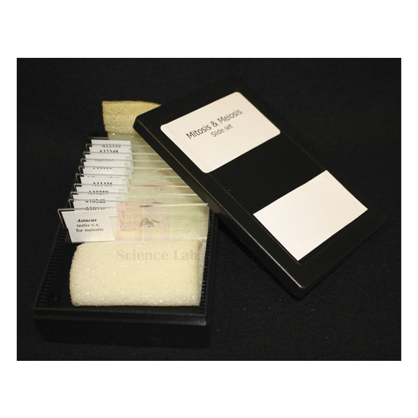
Mitosis & Meiosis Set, Microscope Slides
Mitosis & Meiosis Set, Microscope Slides (Product Code: SLE/PS/78) is a set of 10 prepared microscope slides used to compare mitosis and meiosis in plant and animal tissue during biology practicals. An onion root tip shows mitosis, five lily anther sections follow meiosis from early prophase to the second division, and crayfish and locust slides add animal material. It is supplied by Jain Scientific Equipments Pvt Ltd (ScienceLab), Ambala, India, as a microscope slides set for schools and colleges.
Specifications
| Parameter | Detail |
|---|---|
| Number of slides | 10 |
| Plant mitosis | Allium (onion) root tip LS |
| Plant meiosis | Lilium (lily) anthers TS – early prophase; late prophase; metaphase; late first division; second division |
| Animal testis | Astacus (crayfish) testis VS; Locust testis VS; Locust testis squash |
| Embryo | Locust embryo squash |
| Product code | SLE/PS/78 |
| Supplied by | Jain Scientific Equipments Pvt Ltd (ScienceLab), Ambala, India |
What Is a Mitosis and Meiosis Slide Set Used For?
Students first examine the onion root tip, where dividing cells show the stages of mitosis, then move to the five lily anther slides, which follow meiosis in sequence from early prophase to the second division. Seeing the stages in order lets them identify chromosome pairing, alignment and separation for themselves instead of relying only on textbook diagrams.
The animal slides widen the comparison: the crayfish and locust testis preparations show meiosis in developing sperm cells, while the locust embryo squash shows dividing cells in a growing animal. Ask students to draw each stage with labels and then list how the products of mitosis and meiosis differ in chromosome number.
Supplier
Jain Scientific Equipments Private Limited trades as ScienceLab from Ambala, Haryana, and supplies this ten-slide cell division set to schools, colleges, universities, dealers and importers in India and international markets. The set can be combined with other prepared slide collections, models and microscopes in a single order. Company details are on the ScienceLab profile.
Related Prepared Slides
Resources that support the same cell division lessons:
- Cell Division Slide Set of 9 — additional cell division slides for comparison.
- Microslide Meiosis — a lesson-based set focused on meiosis.
- Meiosis Model — a model to explain the stages before students look at slides.
- Basic Botany Slide (Set of 25) — wider plant material, including an onion root apex slide.
- Binocular Microscope 1000x mag — for viewing chromosomes at high magnification.
Explore all slide sets in the Prepared Slides category.
Frequently Asked Questions
Which slides are in the Mitosis & Meiosis Set, Microscope Slides?
The Mitosis & Meiosis Set, Microscope Slides contains 10 slides: Astacus testis VS, Allium root tip LS, five Lilium anther TS slides from early prophase to second division, Locust testis VS, Locust testis squash and Locust embryo squash.
Which slide in the set shows mitosis?
In the Mitosis & Meiosis Set, Microscope Slides, the Allium (onion) root tip LS is the main slide for mitosis, because the root tip is a region of rapid cell division. The Locust embryo squash also shows dividing cells in animal tissue.
Which stages of meiosis does the set show?
The five Lilium anther slides in the Mitosis & Meiosis Set, Microscope Slides show early prophase, late prophase, metaphase, late first division and the second division. Studying them in order lets students follow meiosis step by step.
What is a squash preparation?
A squash preparation, used for the locust testis and locust embryo slides in the Mitosis & Meiosis Set, Microscope Slides, is made by gently pressing tissue flat under a cover slip. This spreads the cells into a thin layer so chromosomes are easier to see.
Which courses is the Mitosis & Meiosis Set, Microscope Slides suited to?
The Mitosis & Meiosis Set, Microscope Slides suits senior school and undergraduate biology, where students compare the two types of cell division using plant and animal material. Teachers can also use it for class demonstrations at the microscope.
Who supplies the Mitosis & Meiosis Set, Microscope Slides?
The Mitosis & Meiosis Set, Microscope Slides (SLE/PS/78) is supplied by Jain Scientific Equipments Pvt Ltd (ScienceLab), Ambala, India. Institutions, dealers and importers in India and international markets can order single sets or several sets for parallel practical groups.
Request a Quote
Add the Mitosis & Meiosis Set, Microscope Slides to the enquiry cart or tell us through the contact page how many sets you need and where they should be delivered.
Last updated: 24 September 2026

Human Histology Slide Set of 25
Human Histology Slide Set of 25 (Product Code: SLE/PS/79) is a collection of 25 glass microscope slides of human tissues used to teach histology in college, allied health and senior school biology courses. It gives a varied cross-section of the body, from the cerebellum and spinal cord to the digestive tract, kidney and reproductive organs. It is supplied by Jain Scientific Equipments Pvt Ltd (ScienceLab), Ambala, India, among its prepared microscope slides.
Specifications
| Parameter | Detail |
|---|---|
| Number of slides | 25 |
| Slide type | Glass microscope slides |
| Coverage | A varied cross-section of human histology |
| Preparations | Sections; nerve in TS and LS, artery and vein in TS, scalp in VS |
| Product code | SLE/PS/79 |
| Supplied by | Jain Scientific Equipments Pvt Ltd (ScienceLab), Ambala, India |
Slides in the set, grouped by body system:
- Nervous system: Cerebellum, section; Spinal cord, section; Nerve, TS and LS.
- Circulation and lymphatic: Artery and vein, TS; Cardiac muscle, section; Lymph node, section; Spleen, section.
- Skin: Scalp, VS; Skin, heavily pigmented section.
- Muscle: Striated muscle, section.
- Digestive system: Oesophagus; Stomach, fundic portion; Duodenum; Ileum; Appendix; Colon; Liver; Pancreas (all sections).
- Endocrine and urinary: Thyroid gland, section; Kidney, section.
- Reproductive: Testis; Ovary; Uterus; Cervix; Mammary gland (all sections).
What Is a Human Histology Slide Set Used For?
The set lets students work system by system through human tissue types: nervous tissue in the cerebellum, spinal cord and nerve; the layered walls of artery and vein; the digestive tract from oesophagus to colon; and glandular organs such as the liver, pancreas and thyroid. Comparing striated muscle with cardiac muscle is a classic practical exercise that the set supports directly.
Scalp and heavily pigmented skin sections show the epidermis, hair follicles and pigment, while the testis, ovary, uterus, cervix and mammary gland slides support reproduction lessons. Locate each tissue layer at low power, switch to higher magnification to identify cell types, and record observations as labelled drawings.
Supplier
ScienceLab is the trading name of Jain Scientific Equipments Private Limited, Ambala, Haryana, India. The company supplies this human histology collection for teaching use to schools, colleges, universities, training institutes, dealers and importers in India and international markets, and can add microscopes or slide storage to the same order. More about the company is on the ScienceLab profile.
Related Prepared Slides
Slide sets and equipment that extend a histology course:
- Microslide Basic Human Histology — a lesson-based introduction to human tissues.
- Prepared Microscope Slide Set – Connective Tissues — adds connective tissue types.
- Prepared Microscope Slide Set – Digestion (Liver, Pancreas And Tongue) — a closer look at digestive organs.
- Histology Slide Scanner — digitise the sections for whole-class display.
- Unit System Slide Cabinet — flat, long-term storage for the collection.
Browse more collections in the Prepared Slides category.
Frequently Asked Questions
Which tissues are included in the Human Histology Slide Set of 25?
The Human Histology Slide Set of 25 covers nervous tissue, artery and vein, cardiac and striated muscle, lymph node, spleen, scalp and pigmented skin, the digestive tract from oesophagus to colon, liver, pancreas, thyroid, kidney, and reproductive organs including testis, ovary, uterus, cervix and mammary gland.
How are the slides in the Human Histology Slide Set of 25 prepared?
The Human Histology Slide Set of 25 consists of 25 glass microscope slides, mostly sections of a single organ. The nerve slide shows both transverse and longitudinal sections, the artery and vein slide is a transverse section, and the scalp is cut as a vertical section.
Which courses is the Human Histology Slide Set of 25 suited to?
The Human Histology Slide Set of 25 suits college biology, zoology and allied health teaching, as well as senior school courses covering human tissues. It gives a broad overview; departments needing more slides of one system can add topic sets from the same range.
What microscope is needed for the Human Histology Slide Set of 25?
The Human Histology Slide Set of 25 is examined with a compound light microscope, starting at low power to find the tissue and moving to high power for cell detail. The product details do not state a minimum magnification; ask us if you need a matching microscope.
How should the Human Histology Slide Set of 25 be stored?
Store the Human Histology Slide Set of 25 flat in a slide box or cabinet, away from direct sunlight and heat, which can fade stained sections over time. Wipe off any immersion oil with lens tissue after each use.
Who supplies the Human Histology Slide Set of 25?
The Human Histology Slide Set of 25 (SLE/PS/79) is supplied by Jain Scientific Equipments Pvt Ltd (ScienceLab), Ambala, India. Institutions, dealers and importers in India and international markets can order single sets or multiple sets for practical groups.
Request a Quote
Place the Human Histology Slide Set of 25 in the enquiry cart or send your requirement through the contact page, stating the number of sets and any microscopes or storage you want quoted.
Last updated: 24 September 2026

Cell Division Slide Set of 9
Cell Division Slide Set of 9 (Product Code: SLE/PS/80) is a set of nine prepared glass microscope slides used to show how chromosomes divide in plant and animal cells in school and college biology practicals. Teachers pick it because one box covers mitosis in Allium root tips, meiosis in Lilium and locust tissue, and an acetic-orcein squash of giant salivary-gland chromosomes. These prepared microscope slides are supplied by Jain Scientific Equipments Pvt Ltd (ScienceLab), Ambala, India.
Specifications
| Parameter | Detail |
|---|---|
| Slide type | Prepared glass microscope slides |
| Number of slides | 9 (listed below) |
| Topic | Chromosome division in animal and plant cells (mitosis and meiosis) |
| Plant material | Allium root apex and root tip; Lilium ovary and anther |
| Animal material | Locusta testis; Rana ova; insect salivary gland; mammal testis |
| Stain named in the set list | Acetic orcein (insect salivary gland squash) |
| Product code | SLE/PS/80 |
| Supplied by | Jain Scientific Equipments Pvt Ltd (ScienceLab), Ambala, India |
Slides in the set:
- Allium root apex LS, mitosis
- Allium root tip squash preparation
- Locusta testis VS
- Rana ova, various stages
- Lilium ovary, meiosis
- Lilium anther TS, three stages of development
- Insect salivary gland squash, acetic orcein, for giant chromosomes
- Mammal testis TS showing spermatogenesis
- Locusta testis squash showing meiosis
What Is the Cell Division Slide Set Used For?
A typical mitosis practical starts with the Allium root apex LS, where students scan the meristem just behind the root cap and count cells in prophase, metaphase, anaphase and telophase. The root tip squash then shows the same stages with the cells spread flat, which makes chromosome counting easier. For meiosis, the Lilium ovary and the three-stage Lilium anther section are compared with the locust testis squash, and the mammal testis TS links meiosis to sperm formation.
The Rana ova slide shows eggs at various stages, and the acetic-orcein salivary gland squash introduces giant chromosomes with visible banding, a useful bridge into genetics. As a microscope slide set for school and college labs, it gives the clearest results when students start on low power to locate the tissue and then move to higher power for chromosome detail.
Supplier
Jain Scientific Equipments Private Limited trades under the name ScienceLab and supplies this cell division set together with the microscopes, slide boxes and other prepared slide collections that a biology department orders for its practical course. The company works from Ambala, Haryana, and serves schools, colleges, training institutes and dealers in India and international markets. Background on the company is on the ScienceLab company profile page.
Related Prepared Slides
These items are often ordered with the Cell Division Slide Set of 9 for the same cell-division lessons:
- Mitosis & Meiosis Set, Microscope Slides — a second slide set on the two division types for larger practical groups.
- Compound Microscope — needed to view the root-tip and squash preparations at higher power.
- Slide Box — keeps the nine slides flat, labelled and protected between classes.
- Microslide Meiosis — photographic images of meiosis stages for pre-lab briefing or revision.
- Human Histology Slide Set of 25 — the next step once students move from cell division to tissues.
See the full range of Prepared Slides for other botany, zoology and histology sets.
Frequently Asked Questions
Which slides are included in the Cell Division Slide Set of 9?
The Cell Division Slide Set of 9 contains Allium root apex LS, Allium root tip squash, Locusta testis VS, Locusta testis squash, Rana ova at various stages, Lilium ovary, Lilium anther TS in three stages, an insect salivary gland squash and a mammal testis TS.
Does the Cell Division Slide Set of 9 cover both mitosis and meiosis?
Yes. The Cell Division Slide Set of 9 shows mitosis in the two Allium root preparations and meiosis in the Lilium ovary, the Lilium anther stages and the locust testis squash, while the mammal testis TS shows spermatogenesis.
What microscope is needed for the Cell Division Slide Set of 9?
Any compound microscope with low- and high-power objectives can be used with the Cell Division Slide Set of 9. Locate the dividing region on low power first, then switch to high power to study chromosome shape and arrangement in the squash preparations.
How should the Cell Division Slide Set of 9 be stored?
Store the Cell Division Slide Set of 9 flat in a slide box, away from heat, damp and direct sunlight. Hold slides by the edges, and wipe off immersion oil or fingerprints with lens tissue after each class so the preparations stay clear.
Who supplies the Cell Division Slide Set of 9?
The Cell Division Slide Set of 9 is supplied by Jain Scientific Equipments Pvt Ltd (ScienceLab), Ambala, Haryana, India. The company supplies prepared slides to schools, colleges, institutions and dealers in India and international markets.
What should a quotation request for the Cell Division Slide Set of 9 include?
For a quotation on the Cell Division Slide Set of 9, state the number of sets, the delivery city or country and any microscopes or slide boxes you need with it. Send these details through the contact page.
Request a Quote
To order the Cell Division Slide Set of 9 for your biology lab, send your quantity and delivery details through the contact page, or add it to the enquiry cart together with the other slides and microscopes on your list.
Last updated: 24 September 2026
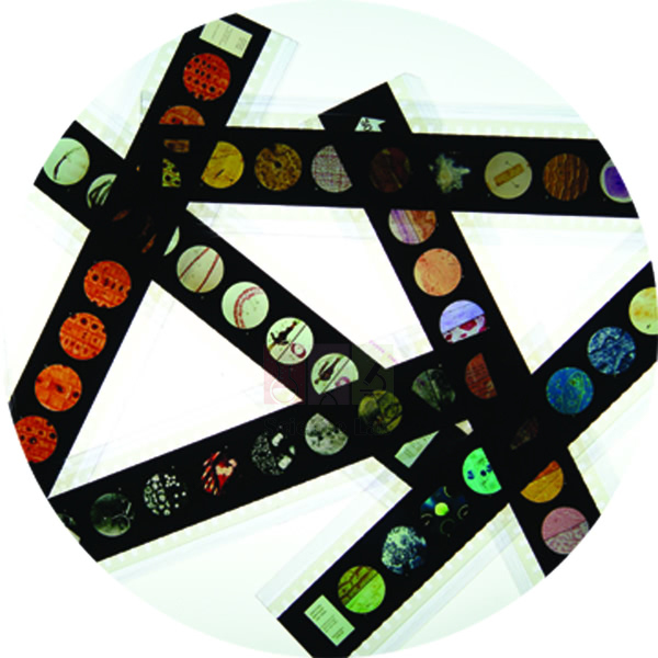
Microslide Animal Tissue Set 2 Muscle, Bone, Connective
Microslide Animal Tissue Set 2 Muscle, Bone, Connective (Product Code: SLE/PS/81) is a Microslide of 8 related 35mm images, photographed through a microscope, used to teach muscle, bone and connective tissue in school biology lessons. Students compare voluntary, involuntary and heart muscle alongside areolar tissue, bone, blood, fat and spinal cord, with arrows and callouts marking each key feature. This biology teaching aid is supplied by Jain Scientific Equipments Pvt Ltd (ScienceLab), Ambala, India.
Specifications
| Parameter | Detail |
|---|---|
| Format | Microslide: 8 related 35mm images photographed through a microscope |
| Viewed with | Microslide Viewer |
| Muscle images | Voluntary muscle cells, involuntary muscle cells, heart muscle |
| Connective and supporting tissue images | Areolar connective tissue, bone, blood, adipose tissue |
| Nervous tissue image | Spinal cord |
| Labelling | Arrows and callouts on the images |
| Accompanying material | Detailed text folder with a pocket for storing the Microslide |
| Product code | SLE/PS/81 |
| Supplied by | Jain Scientific Equipments Pvt Ltd (ScienceLab), Ambala, India |
What Is the Microslide Animal Tissue Set Used For?
The three muscle images set up the classic comparison in any tissue topic: long striated voluntary fibres, spindle-shaped involuntary cells and the branched fibres of heart muscle. Connective tissue then appears in four very different forms, from loose areolar tissue and fat-storing adipose tissue to the hard matrix of bone and the fluid tissue of blood. The spinal cord image rounds off the set with nervous tissue.
Because every image already carries arrows and callouts, teachers use this Microslide for a short briefing before students examine their own tissue slides, or for revision where microscope time is limited. The questions in the text folder work as a ready worksheet, which makes the set a practical biology teaching aid for large classes.
Supplier
ScienceLab is the trading name of Jain Scientific Equipments Private Limited, based in Ambala, Haryana, which supplies Microslide sets, viewers and prepared slides to schools, colleges and dealers in India and international markets. For vendor registration, the supplying company is listed under CIN U29309HR2020PTC087597 and GSTIN 06AAECJ8618A1ZT. More about the company is on the ScienceLab profile page.
Related Prepared Slides
Items that support the same tissue lessons as this Microslide:
- Microslide Viewer — the viewer these 35mm images are designed for.
- Microslide Basic Human Histology — adds cartilage, epithelium and nerve cells to the tissue unit.
- Microslide Cells of Body — looks at individual body cells rather than whole tissues.
- Prepared Microscope Slide Set – Connective Tissues — glass slides for students to examine under a microscope.
Browse all Prepared Slides for further Microslide topics and glass slide sets.
Frequently Asked Questions
Which images are in the Microslide Animal Tissue Set 2?
The Microslide Animal Tissue Set 2 Muscle, Bone, Connective has eight images: voluntary muscle cells, involuntary muscle cells, heart muscle, areolar connective tissue, bone, blood, adipose tissue and spinal cord, each marked with arrows and callouts.
Is the Microslide Animal Tissue Set 2 a glass microscope slide?
No. The Microslide Animal Tissue Set 2 is a set of 8 related 35mm images photographed through a microscope and viewed through a Microslide Viewer. For glass slides of similar tissues, see the connective tissues prepared slide set.
What comes with the Microslide Animal Tissue Set 2?
The Microslide Animal Tissue Set 2 comes with a detailed text folder written to inform students and question them on the topic. The folder has a pocket that holds the Microslide, so images and notes stay together.
Which muscle types does the Microslide Animal Tissue Set 2 show?
The Microslide Animal Tissue Set 2 shows all three muscle types: voluntary (skeletal) muscle cells, involuntary (smooth) muscle cells and heart muscle, so students can compare fibre shape and arrangement on one set.
Who supplies the Microslide Animal Tissue Set 2?
The Microslide Animal Tissue Set 2 Muscle, Bone, Connective is supplied by Jain Scientific Equipments Pvt Ltd (ScienceLab), Ambala, Haryana, India, to schools, colleges and dealers in India and international markets.
What information is needed to quote for the Microslide Animal Tissue Set 2?
To quote for the Microslide Animal Tissue Set 2, we need the number of sets, whether Microslide Viewers are also required, and the delivery location. Send these through the contact page.
Request a Quote
Ask for a quotation on the Microslide Animal Tissue Set 2 Muscle, Bone, Connective through the contact page, or place it in the enquiry cart with any viewers and other Microslide topics you need.
Last updated: 24 September 2026
220.jpg)
Microslide Basic Human Histology
Microslide Basic Human Histology (Product Code: SLE/PS/83) is a Microslide of 8 related 35mm photomicrographs used to introduce the basic tissues of the human body in school and early college biology. Its images run from a stained cross-section of bone and cartilage to ciliated epithelium, stained nerve cells, striated muscle and red and white blood cells, each marked with arrows and callouts. It is supplied by Jain Scientific Equipments Pvt Ltd (ScienceLab), Ambala, India.
Specifications
| Parameter | Detail |
|---|---|
| Format | 8 related 35mm images photographed through a microscope |
| Viewing device | Microslide Viewer |
| Supporting tissue images | Bone XS (stained), cartilage, fat cells |
| Epithelial image | Epithelial tissue / ciliated epithelial cell |
| Nerve and muscle images | Nerve cells (stained), striated muscle cells |
| Blood images | Red blood cells, white blood cells |
| Student notes | Detailed text folder with questions, plus a pocket that stores the Microslide |
| Product code | SLE/PS/83 |
| Supplied by | Jain Scientific Equipments Pvt Ltd (ScienceLab), Ambala, India |
What Is the Microslide Basic Human Histology Used For?
Most histology courses begin with four tissue families: epithelial, connective, muscle and nervous. This Microslide covers all four, so a teacher can move from the cilia on epithelial cells to the stained nerve cells, the banding of striated muscle and the supporting matrix of bone and cartilage. The two blood images let students compare red and white cells before they attempt a blood smear of their own.
Students can keep the images beside them while drawing, using the callouts to label their diagrams correctly. Because it needs only a Microslide Viewer rather than a microscope and stains, the set is an easy addition to school biology lab equipment for classes that are just starting human biology.
Supplier
Jain Scientific Equipments Private Limited, trading as ScienceLab, supplies this histology Microslide from Ambala, Haryana, as part of a wider list of prepared slides and biology teaching aids. Schools, colleges, teacher-training centres and distributors in India and international markets order through the company. Visit the company profile for more about ScienceLab.
Related Prepared Slides
Useful companions for a first human histology unit:
- Microslide Viewer — required to look at the 35mm images.
- Human Histology Slide Set of 25 — glass slides for microscope work once students know what to look for.
- Microslide Animal Tissue Set 2 Muscle, Bone, Connective — extends the unit with all three muscle types and more connective tissue.
- Human Blood Cell Types Model — a three-dimensional view of the blood cells seen in two of the images.
More topics are listed under Prepared Slides.
Frequently Asked Questions
Which tissues does the Microslide Basic Human Histology show?
The Microslide Basic Human Histology shows eight subjects: bone in cross-section, cartilage, fat cells, epithelial tissue with a ciliated epithelial cell, nerve cells, striated muscle cells, red blood cells and white blood cells.
Are the images in the Microslide Basic Human Histology stained?
Two images in the Microslide Basic Human Histology are identified as stained preparations: the bone cross-section and the nerve cells. The other six show cartilage, fat cells, ciliated epithelium, striated muscle, and red and white blood cells.
Is a microscope needed for the Microslide Basic Human Histology?
No microscope is needed. The Microslide Basic Human Histology holds 8 related 35mm images already photographed through a microscope, and it is designed to be viewed through a Microslide Viewer held up to the light.
What is the text folder supplied with the Microslide Basic Human Histology?
The Microslide Basic Human Histology comes with a detailed text folder that explains each image and asks students questions about it. A pocket in the folder holds the Microslide, so the notes and images are stored together.
Who supplies the Microslide Basic Human Histology?
Jain Scientific Equipments Pvt Ltd (ScienceLab), Ambala, Haryana, India, supplies the Microslide Basic Human Histology to schools, colleges, teacher-training centres and dealers in India and international markets.
How do I order the Microslide Basic Human Histology with other topics?
List the Microslide titles you want, the number of each and how many viewers you need, then send the list through the contact page. The Microslide Basic Human Histology can be quoted together with the rest of your order.
Request a Quote
Request pricing for the Microslide Basic Human Histology through the contact page, or add it to the enquiry cart alongside viewers and glass histology slides.
Last updated: 24 September 2026
846.jpg)
Microslide Cells Of Plants
Microslide Cells Of Plants (Product Code: SLE/PS/84) is a set of 8 related 35mm images, photographed through a microscope, used to teach plant cell structure in middle and secondary school biology. It moves from onion skin and green leaf cells to a woody stem shown at both 40x and 100x, cells of the anther and lily ovary, a Spirogyra cell and bacteria for comparison. It is supplied by Jain Scientific Equipments Pvt Ltd (ScienceLab), Ambala, India.
Specifications
| Parameter | Detail |
|---|---|
| Format | Microslide of 8 related 35mm images |
| For use with | Microslide Viewer |
| Leaf and epidermis images | Onion skin cells, green cells of a leaf |
| Stem images | Cells of a woody stem at 100x; cells of a woody stem at 40x |
| Reproductive tissue images | Cells of the anther, cells of the lily ovary |
| Comparison images | Spirogyra cell, bacteria |
| Labelling | Arrows and callouts point out the features being studied |
| Text folder | Detailed notes and questions, with a pocket to store the Microslide |
| Product code | SLE/PS/84 |
| Supplied by | Jain Scientific Equipments Pvt Ltd (ScienceLab), Ambala, India |
What Is the Microslide Cells Of Plants Used For?
Onion skin is the standard first plant-cell practical, and seeing a labelled image beforehand helps students recognise the cell wall and nucleus when they mount their own peel. The green leaf cells introduce chloroplasts, while the two woody stem images at 40x and 100x show how raising the magnification narrows the field of view but reveals more detail, a point students often miss.
Cells of the anther and the lily ovary link cell structure to plant reproduction, and the Spirogyra cell shows a simple filamentous alga with its spiral chloroplast. The bacteria image ends the set with a much smaller, simpler cell for contrast. Together they make a compact addition to plant biology lab equipment for schools that want students to prepare before microscope sessions.
Supplier
This plant-cell Microslide is supplied by Jain Scientific Equipments Private Limited, the Ambala company that trades as ScienceLab. It is sourced for botany and general science departments together with the matching viewer, plant cell models and glass slide sets, and is available to buyers in India and international markets. Find out more on the ScienceLab company profile.
Related Prepared Slides
Items that fit the same plant-cell lessons:
- Microslide Viewer — the viewer needed to see the 35mm images.
- Plant Cell Model — a three-dimensional model of the structures students look for in the images.
- Basic Plant Anatomy Slide Set — glass slides for the microscope follow-up.
- Microslide-The Cell Structure — places plant cells beside animal cells, bacteria and a virus.
- Microslide Photosynthesis — the natural next topic after green leaf cells.
See more in Prepared Slides.
Frequently Asked Questions
What images are in the Microslide Cells Of Plants?
The Microslide Cells Of Plants contains onion skin cells, green cells of a leaf, cells of a woody stem at 100x and at 40x, cells of the anther, cells of the lily ovary, a Spirogyra cell and bacteria.
Why does the Microslide Cells Of Plants show the woody stem twice?
The Microslide Cells Of Plants shows the same woody stem at 40x and at 100x so students can see how higher magnification shows a smaller area in more detail, which prepares them for switching objectives on a real microscope.
Why is a bacteria image included in the Microslide Cells Of Plants?
The bacteria image in the Microslide Cells Of Plants gives students a contrast: a much smaller cell without the large vacuole and chloroplasts seen in the plant images, which helps them explain what makes a plant cell different.
How is the Microslide Cells Of Plants viewed?
The Microslide Cells Of Plants is viewed through a Microslide Viewer, not a microscope. Its 8 images are already photographed through a microscope and carry arrows and callouts, so students can study them anywhere with light.
Who supplies the Microslide Cells Of Plants?
The Microslide Cells Of Plants is supplied by Jain Scientific Equipments Pvt Ltd (ScienceLab), Ambala, Haryana, India, to schools, colleges and distributors in India and international markets.
What should I include when asking for a quote on the Microslide Cells Of Plants?
When asking for a quote on the Microslide Cells Of Plants, include the number of sets, the number of viewers required and the delivery address. Use the contact page to send the details.
Request a Quote
Send your enquiry for the Microslide Cells Of Plants through the contact page, or add it to the enquiry cart with viewers and other plant biology items.
Last updated: 24 September 2026
158.jpg)
Microslide Cells of Body
Microslide Cells of Body (Product Code: SLE/PS/85) is a Microslide of 8 related 35mm images, photographed through a microscope, used to show how human body cells differ in shape and function in school biology classes. Starting from cheek cells, it continues through blood, lymph gland, bone, voluntary and involuntary muscle, nerve and gland cells, with arrows and callouts on each image. The set is supplied by Jain Scientific Equipments Pvt Ltd (ScienceLab), Ambala, India.
Specifications
| Parameter | Detail |
|---|---|
| Image format | 8 related 35mm images photographed through a microscope |
| Viewer required | Microslide Viewer |
| Lining and circulating cells | Cheek, blood, lymph gland |
| Support and movement | Bone, voluntary muscle, involuntary muscle |
| Control and secretion | Nerve, gland cells |
| On-image labels | Arrows and callouts |
| Folder | Detailed text folder that informs and questions students; pocket stores the Microslide |
| Product code | SLE/PS/85 |
| Supplied by | Jain Scientific Equipments Pvt Ltd (ScienceLab), Ambala, India |
What Is the Microslide Cells of Body Used For?
Cell specialisation is the idea this Microslide is built around. Students begin with the flat cheek cells they can collect from their own mouths, then see how blood, bone, muscle, nerve and gland cells are shaped for very different work. Placing voluntary and involuntary muscle on neighbouring images makes the link between structure and function easy to discuss.
Many teachers show it just before the cheek-cell practical, so students know what a stained human cell should look like before they prepare one. It also works as a revision station in human biology, sitting well alongside a human biology model set or a basic animal cell model.
Supplier
ScienceLab is the trade name used by Jain Scientific Equipments Private Limited of Ambala, Haryana, which supplies this Microslide and the viewer it needs to schools, colleges and distributors in India and international markets. Institutions and overseas buyers registering the company as a vendor can use its Importer-Exporter Code AAECJ8618A (DGFT) and GSTIN 06AAECJ8618A1ZT. Company background is on the ScienceLab profile.
Related Prepared Slides
Other items for a lesson on human cells:
- Microslide Viewer — lets students see the 35mm images without a microscope.
- Animal Cell Model — a generalised cell to compare with the specialised cells in the set.
- Microslide Ultra Structure of Animal Cells — moves from whole cells to the organelles inside them.
- Microslide Basic Human Histology — shows how these cells come together as tissues.
The full list is under Prepared Slides.
Frequently Asked Questions
Which cells does the Microslide Cells of Body show?
The Microslide Cells of Body shows eight kinds of human cells: cheek, blood, lymph gland, bone, voluntary muscle, involuntary muscle, nerve and gland cells, each labelled with arrows and callouts for students.
Does the Microslide Cells of Body help with the cheek cell practical?
Yes. The Microslide Cells of Body opens with a cheek cell image, so students can see the shape and nucleus they should look for before they scrape, stain and examine their own cheek cells under a microscope.
Is the Microslide Cells of Body a glass slide for a microscope?
No. The Microslide Cells of Body is a set of 8 related 35mm photographic images taken through a microscope. It is viewed through a Microslide Viewer, so no microscope, stain or slide preparation is needed.
What teaching material comes with the Microslide Cells of Body?
The Microslide Cells of Body comes with a detailed text folder that explains each image and asks students questions to check understanding. A pocket inside the folder keeps the Microslide safe between lessons.
Who supplies the Microslide Cells of Body?
The Microslide Cells of Body is supplied by Jain Scientific Equipments Pvt Ltd (ScienceLab) of Ambala, Haryana, India, to schools, colleges and distributors in India and international markets.
How many sets of the Microslide Cells of Body does a class need?
That depends on how you run the lesson. Many schools order the Microslide Cells of Body for small groups sharing one viewer; tell us your class size and number of viewers through the contact page for a quotation.
Request a Quote
For a quotation on the Microslide Cells of Body, write to us through the contact page or add it to the enquiry cart along with viewers and related Microslide titles.
Last updated: 24 September 2026
34.jpg)
Microslide Meiosis
Microslide Meiosis (Product Code: SLE/PS/86) is a set of 8 related 35mm images, photographed through a microscope, used to follow meiosis in the developing egg cell in secondary and college biology. The images trace the primary oocyte from tetrad formation to the first polar body, then the secondary oocyte, mature egg, pronucleus stage and a zygote ready for first cleavage, five of them at 750x. It is supplied by Jain Scientific Equipments Pvt Ltd (ScienceLab), Ambala, India.
Specifications
| Parameter | Detail |
|---|---|
| Format | Microslide – 8 related 35mm images |
| Viewing | Through a Microslide Viewer |
| Primary oocyte images | Tetrads formed; dyads separated; tetrads on spindle; first polar body forming (750x) |
| Later-stage images | Secondary oocyte – chromosomes formed (750x); mature egg (750x); pronucleus stage (750x); zygote ready for first cleavage (750x) |
| Magnification stated | 750x on five of the eight images |
| Labelling | Arrows and callouts locate the key features |
| Text folder | Detailed notes and questions; built-in pocket holds the Microslide |
| Product code | SLE/PS/86 |
| Supplied by | Jain Scientific Equipments Pvt Ltd (ScienceLab), Ambala, India |
What Is the Microslide Meiosis Used For?
Textbook meiosis diagrams usually show a generic cell; this set follows the process in a real egg cell, where the divisions are unequal and produce polar bodies. Students see tetrads, the paired homologous chromosomes, form and line up on the spindle, and then watch the first polar body pinch off before the secondary oocyte appears.
The final images carry the story beyond meiosis into fertilisation: the mature egg, the pronucleus stage and a zygote about to make its first cleavage. That makes this Microslide a useful visual biology teaching aid for linking the reproduction and genetics units, and for revising the stages before an examination.
Supplier
Jain Scientific Equipments Private Limited supplies the Microslide Meiosis under its ScienceLab trading name. From its base in Ambala, Haryana, the company puts together biology orders that combine Microslides, viewers, glass slide sets and models for schools, colleges and dealers in India and international markets. See the ScienceLab profile for company details.
Related Prepared Slides
Items that support the same cell-division lessons:
- Microslide Viewer — needed to view the eight images.
- Meiosis Model — an enlarged model that makes the chromosome movements easier to explain.
- Cell Division Slide Set of 9 — glass slides of meiosis in Lilium and locust tissue for microscope work.
- Mitosis & Meiosis Set, Microscope Slides — prepared slides that contrast the two types of division.
Explore more titles in Prepared Slides.
Frequently Asked Questions
Which stages does the Microslide Meiosis show?
The Microslide Meiosis shows the primary oocyte with tetrads formed, dyads separated, tetrads on the spindle and the first polar body forming, followed by the secondary oocyte, the mature egg, the pronucleus stage and a zygote ready for first cleavage.
At what magnification are the Microslide Meiosis images?
Five Microslide Meiosis images are marked 750x: first polar body forming, secondary oocyte, mature egg, pronucleus stage and zygote. No magnification is stated for the three earlier primary oocyte images; ask us if you need it confirmed.
Why does the Microslide Meiosis use egg cells?
The Microslide Meiosis follows meiosis in the developing egg because the divisions there are unequal and produce visible polar bodies, which lets students connect meiosis directly with egg formation and fertilisation.
Is the Microslide Meiosis used with a microscope?
No. The Microslide Meiosis holds 8 related 35mm images already photographed through a microscope, and students view them through a Microslide Viewer. It is often used before a slide practical on meiosis.
Does the Microslide Meiosis include teaching notes?
Yes. The Microslide Meiosis is accompanied by a detailed text folder that informs students about each stage and asks them questions. The folder has a pocket to store the Microslide.
Who supplies the Microslide Meiosis?
The Microslide Meiosis is supplied by Jain Scientific Equipments Pvt Ltd (ScienceLab), Ambala, Haryana, India. Schools, colleges and dealers in India and international markets can request a quotation through the contact page.
Request a Quote
Ask for pricing on the Microslide Meiosis through the contact page, or add it to the enquiry cart with a viewer and any meiosis models or slide sets you need.
Last updated: 24 September 2026
21.jpg)
Microslide Ultra Structure of Animal Cells
Microslide Ultra Structure of Animal Cells (Product Code: SLE/PS/87) is a Microslide of 8 related 35mm images, photographed through a microscope, used to teach the fine internal structure of animal cells in senior school and college biology. It focuses on cell parts such as the Golgi body and vacuole, a mitochondrion, a centriole, the nerve cell membrane, lampbrush chromosomes and muscle cell fibrils, alongside gland cells and a plasma cell. It is supplied by Jain Scientific Equipments Pvt Ltd (ScienceLab), Ambala, India.
Specifications
| Parameter | Detail |
|---|---|
| Format | Microslide, 8 related 35mm images photographed through a microscope |
| Viewed through | Microslide Viewer |
| Whole-cell images | Gland cells, plasma cell |
| Organelle and membrane images | Nerve cell membrane, Golgi body and vacuole, mitochondrion, centriole |
| Chromosome and fibril images | Lampbrush chromosomes, muscle cell fibrils |
| Labelling | Arrows and callouts on each image |
| Teaching notes | Detailed text folder with questions and a pocket for the Microslide |
| Product code | SLE/PS/87 |
| Supplied by | Jain Scientific Equipments Pvt Ltd (ScienceLab), Ambala, India |
What Is the Microslide Ultra Structure of Animal Cells Used For?
Once students can draw a basic animal cell, the next step is understanding what each organelle actually does. This set pairs structure with function: gland cells and a plasma cell are built for secretion, the Golgi body packages their products, the mitochondrion supplies energy and the centriole takes part in cell division.
Lampbrush chromosomes and muscle cell fibrils give extension material for genetics and physiology classes, while the nerve cell membrane leads into how signals travel. A good exercise is to build an organelle-and-function table with the class using the callouts. The Microslide complements the models found in a cell biology equipment collection.
Supplier
The company behind the ScienceLab name is Jain Scientific Equipments Private Limited, Ambala, Haryana. It supplies this cell-structure Microslide to biology departments that are building up a set of visual aids, and can combine it in one order with viewers, cell models and prepared slides for customers in India and international markets. Read about the company on the profile page.
Related Prepared Slides
Pair this Microslide with:
- Microslide Viewer — the viewer the eight images are made for.
- Mitochondrion Model — a three-dimensional look at one of the organelles pictured.
- Animal Cell Plaque — shows where each organelle sits within a whole cell.
- Microslide Cells of Body — whole specialised body cells before zooming in to organelles.
- Microslide-The Cell Structure — the introductory set on cell types and size.
Further titles are listed under Prepared Slides.
Frequently Asked Questions
Which cell structures does the Microslide Ultra Structure of Animal Cells show?
The Microslide Ultra Structure of Animal Cells shows gland cells, a plasma cell, the nerve cell membrane, the Golgi body and vacuole, a mitochondrion, a centriole, lampbrush chromosomes and muscle cell fibrils, each with arrows and callouts.
Which classes is the Microslide Ultra Structure of Animal Cells suited to?
The Microslide Ultra Structure of Animal Cells suits senior secondary and college biology, where students move from drawing a basic cell to studying organelles, membranes and chromosomes in more detail.
How is the Microslide Ultra Structure of Animal Cells different from Microslide-The Cell Structure?
Microslide-The Cell Structure compares whole cells from cork to a virus, while the Microslide Ultra Structure of Animal Cells looks inside animal cells at organelles such as the Golgi body, mitochondrion and centriole. Many schools use both in sequence.
What is needed to view the Microslide Ultra Structure of Animal Cells?
Only a Microslide Viewer is needed. The Microslide Ultra Structure of Animal Cells holds 8 related 35mm images already photographed through a microscope, so no microscope or slide preparation is involved.
Who supplies the Microslide Ultra Structure of Animal Cells?
Jain Scientific Equipments Pvt Ltd (ScienceLab), Ambala, Haryana, India, supplies the Microslide Ultra Structure of Animal Cells to schools, colleges and dealers in India and international markets.
What should a quotation request for the Microslide Ultra Structure of Animal Cells include?
Include the number of Microslide Ultra Structure of Animal Cells sets, any viewers or cell models you want with them, and the delivery location, then send the request through the contact page.
Request a Quote
Request a quotation for the Microslide Ultra Structure of Animal Cells through the contact page, or add it to the enquiry cart with viewers and cell biology models.
Last updated: 24 September 2026
412.jpg)
Microslide-The Cell Structure
Microslide-The Cell Structure (Product Code: SLE/PS/88) is a Microslide of 8 related 35mm images, photographed through a microscope, used to introduce cells and cell size in school biology. It runs from cork at 300x and onion skin at 200x through green leaf, cheek, blood and nerve cells to bacteria at 1500x and a virus at 50,000x, so students see both the variety and the scale of living things. It is supplied by Jain Scientific Equipments Pvt Ltd (ScienceLab), Ambala, India.
Specifications
| Parameter | Detail |
|---|---|
| Format | 8 related 35mm images for a Microslide Viewer |
| Plant images | Cork (300x), onion skin (200x), green leaf (300x) |
| Animal images | Cheek cells (900x), blood cells (900x), nerve cells (300x) |
| Microbe images | Bacteria (1500x), virus (50,000x) |
| Magnification range | 200x to 50,000x |
| Labelling | Arrows and callouts highlight the features to study |
| Folder | Detailed text folder with questions; pocket stores the Microslide |
| Product code | SLE/PS/88 |
| Supplied by | Jain Scientific Equipments Pvt Ltd (ScienceLab), Ambala, India |
What Is the Cell Structure Microslide Used For?
The cork image recalls the first cells ever described, the empty walls seen in a thin slice of cork, and makes a natural opening for a lesson on cell theory. Onion skin and green leaf follow with living plant cells, then cheek, blood and nerve cells show how differently animal cells can be shaped.
The magnification printed with each image turns the set into a lesson on scale: cheek and blood cells are shown at 900x, bacteria at 1500x and the virus at 50,000x. Students can rank the subjects by size and discuss why a virus is not usually counted as a cell, which makes this a strong first-lesson resource for any school biology lab.
Supplier
Jain Scientific Equipments Private Limited, known to customers as ScienceLab, supplies Microslide-The Cell Structure from Ambala, Haryana. Science departments often order it with the viewer, a compound microscope and other introductory Microslide titles, and the company handles these orders for schools, colleges and distributors in India and international markets. More background is on the ScienceLab profile page.
Related Prepared Slides
Items that extend an introductory cells lesson:
- Microslide Viewer — used to look at the eight images.
- Compound Microscope — for students to view their own onion skin and cheek cell preparations.
- Microslide Cells Of Plants — a closer look at plant cells, including a woody stem and Spirogyra.
- Microslide Cells of Body — more human cell types for the animal-cell part of the lesson.
- Prokaryotic Cell Model — shows the internal structure of the bacteria pictured in the set.
Browse the rest of the Prepared Slides range.
Frequently Asked Questions
What images are in Microslide-The Cell Structure?
Microslide-The Cell Structure contains cork (300x), onion skin (200x), green leaf (300x), cheek cells (900x), blood cells (900x), nerve cells (300x), bacteria (1500x) and a virus (50,000x), each labelled with arrows and callouts.
What magnifications does Microslide-The Cell Structure cover?
Microslide-The Cell Structure covers magnifications from 200x for onion skin up to 50,000x for the virus, with cells such as cheek and blood at 900x and bacteria at 1500x, which helps students compare sizes.
Why does Microslide-The Cell Structure begin with cork?
Microslide-The Cell Structure begins with cork because cork cells were the first cells ever described. Starting there lets teachers introduce the history of cell theory before moving on to living plant and animal cells.
Why is a virus included in Microslide-The Cell Structure?
The virus in Microslide-The Cell Structure gives a contrast with true cells. Seeing it at 50,000x beside bacteria at 1500x helps students grasp how small viruses are and why they are usually not classed as living cells.
How is Microslide-The Cell Structure viewed?
Microslide-The Cell Structure is viewed through a Microslide Viewer. The images were photographed through a microscope, so students can study them in class or at home without preparing slides.
Who supplies Microslide-The Cell Structure?
Microslide-The Cell Structure is supplied by Jain Scientific Equipments Pvt Ltd (ScienceLab), Ambala, Haryana, India. Schools and dealers in India and international markets can ask for a quotation through the contact page.
Request a Quote
To buy Microslide-The Cell Structure, send your quantity and delivery details through the contact page or use the enquiry cart to list it with viewers and microscopes.
Last updated: 24 September 2026
Microslide Helpful Bacteria
Microslide Helpful Bacteria (Product Code: SLE/PS/89) is a Microslide of 8 related 35mm images, photographed through a microscope, used to teach the useful roles of bacteria in school biology and environmental science. It covers bacteria of decay and fermentation at 2000x, nitrogen-fixing bacteria in soil, a root nodule and roots, and the bacteria behind vinegar, cheese and sour milk. It is supplied by Jain Scientific Equipments Pvt Ltd (ScienceLab), Ambala, India.
Specifications
| Parameter | Detail |
|---|---|
| Format | Microslide of 8 related 35mm images |
| Viewing | Microslide Viewer |
| Decomposition and fermentation | Bacteria of decay (2000x), bacteria of fermentation (2000x) |
| Nitrogen fixation | Nitrogen-fixing bacteria in soil, in a nodule and in roots |
| Food-related bacteria | Vinegar bacteria, cheese bacteria, sour milk bacteria |
| Labelling | Arrows and callouts mark the features being studied |
| Text folder | Detailed notes and questions, with a pocket for storing the Microslide |
| Product code | SLE/PS/89 |
| Supplied by | Jain Scientific Equipments Pvt Ltd (ScienceLab), Ambala, India |
What Is the Microslide Helpful Bacteria Used For?
Many students think of bacteria only as germs. This set balances that picture by showing bacteria that break down dead matter, ferment sugars and supply plants with nitrogen. The three nitrogen-fixing images follow the bacteria from the soil into a root nodule and the root itself, giving a clear visual thread through the nitrogen cycle.
The last three images bring the topic into the kitchen with vinegar, cheese and sour milk bacteria, a natural lead-in to a curd-making or fermentation activity. Schools putting together a microbiology teaching kit often pair this set with Microslide Harmful Bacteria so students weigh the benefits of bacteria against the diseases some of them cause.
Supplier
Microslide Helpful Bacteria is supplied by Jain Scientific Equipments Private Limited, which does business as ScienceLab from Ambala, Haryana. The company sources Microslide titles and viewers for biology and environmental science courses and delivers them with any other lab items on the same order to institutions and dealers in India and international markets. Company information is on the ScienceLab profile page.
Related Prepared Slides
These items fit the same microbiology lessons:
- Microslide Viewer — needed to see the images.
- Microslide Harmful Bacteria — the companion set on disease-causing bacteria.
- Bacteria slide Set of 12 — glass slides for examining bacteria under a microscope.
- Viruses & Bacteria Poster — a wall reference for the microbe topic.
- Microslide Five Kingdoms of Life — shows where bacteria sit in the classification of living things.
Find more in Prepared Slides.
Frequently Asked Questions
Which bacteria are shown in the Microslide Helpful Bacteria?
The Microslide Helpful Bacteria shows bacteria of decay, bacteria of fermentation, nitrogen-fixing bacteria in soil, in a nodule and in roots, and the bacteria associated with vinegar, cheese and sour milk.
Does the Microslide Helpful Bacteria cover the nitrogen cycle?
Yes. Three images in the Microslide Helpful Bacteria show nitrogen-fixing bacteria in soil, in a root nodule and in roots, so teachers can explain how these bacteria make nitrogen available to plants.
What magnification is stated for the Microslide Helpful Bacteria images?
The bacteria of decay and the bacteria of fermentation images in the Microslide Helpful Bacteria are marked 2000x. Magnification is not stated for the other six images; ask us if you need it confirmed.
Does the Microslide Helpful Bacteria contain live bacteria?
No. The Microslide Helpful Bacteria is a set of 8 photographic images taken through a microscope, viewed with a Microslide Viewer. There are no cultures to grow or dispose of, so it can be used in any classroom.
Who supplies the Microslide Helpful Bacteria?
The Microslide Helpful Bacteria is supplied by Jain Scientific Equipments Pvt Ltd (ScienceLab), Ambala, Haryana, India, to schools, colleges and dealers in India and international markets.
What details should I send for a Microslide Helpful Bacteria quotation?
For a Microslide Helpful Bacteria quotation, send the number of sets, the number of viewers you need and the delivery location through the contact page.
Request a Quote
Enquire about the Microslide Helpful Bacteria through the contact page, or add it to the enquiry cart along with the Microslide Harmful Bacteria and a viewer.
Last updated: 24 September 2026
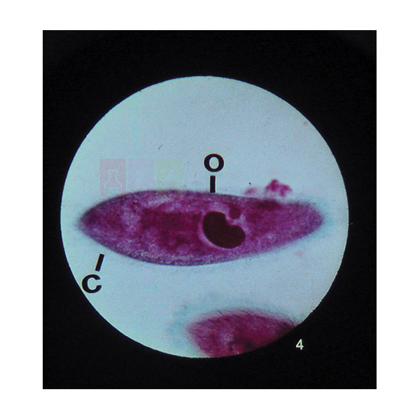
Microslide Five Kingdoms of Life
Microslide Five Kingdoms of Life (Product Code: SLE/PS/90) is a Microslide of 8 related 35mm images, photographed through a microscope, used to teach the five-kingdom classification of living things in school biology. It shows examples from Monera, Protista, Fungi, Plants and Animals, from nitrogen-fixing bacteria at 2000x and Gloeocapsa to Paramecium, Spirogyra, a mushroom, onion and Elodea, opening with Euglena as a classification puzzle. It is supplied by Jain Scientific Equipments Pvt Ltd (ScienceLab), Ambala, India.
Specifications
| Parameter | Detail |
|---|---|
| Format | 8 related 35mm images for a Microslide Viewer |
| Opening image | Euglena – a classification puzzle (400x) |
| Kingdom Monera | Nitrogen-fixing bacteria (2000x); Gloeocapsa (1000x) |
| Kingdom Protista | Paramecium (100x); Spirogyra (150x) |
| Fungus Kingdom | The mushroom (1/2x and 400x) |
| Plant Kingdom | Onion and Elodea (150x) |
| Animal Kingdom | Animal Kingdom image (750x) |
| Labelling | Arrows and callouts on each image |
| Folder | Detailed text folder with questions; pocket keeps the Microslide |
| Product code | SLE/PS/90 |
| Supplied by | Jain Scientific Equipments Pvt Ltd (ScienceLab), Ambala, India |
What Is the Microslide Five Kingdoms of Life Used For?
Classification lessons stick better when students see real organisms instead of names on a chart. The set opens with Euglena, which has chloroplasts like a plant yet swims like an animal, so the class has to argue where it belongs. From there it moves kingdom by kingdom, from the simplest cells in Monera to the Animal Kingdom.
The mushroom appears at both 1/2x and 400x, helping students connect a familiar fruiting body with its microscopic structure. Use the Microslide alongside a classification chart for biology class, asking groups to list the features that place each organism in its kingdom.
Supplier
ScienceLab, the trading name of Jain Scientific Equipments Private Limited in Ambala, Haryana, supplies Microslide Five Kingdoms of Life to schools that teach classification through images, models and posters. Biology departments, teacher-training colleges and dealers in India and international markets can order it with the rest of their classification resources. Details about the company are on the profile page.
Related Prepared Slides
Resources that support the same classification lessons:
- Microslide Viewer — for viewing the kingdom images.
- Classification of Living Things Poster — a wall summary of the groups shown in the set.
- Concepts Of Classification Kit — a hands-on sorting activity to follow the images.
- Classification of Living Things Lab Investigation — a structured investigation for older students.
- Euglena Model — an enlarged model of the organism that opens the set.
More titles are in Prepared Slides.
Frequently Asked Questions
Which organisms are shown in the Microslide Five Kingdoms of Life?
The Microslide Five Kingdoms of Life shows Euglena, nitrogen-fixing bacteria and Gloeocapsa (Monera), Paramecium and Spirogyra (Protista), a mushroom (Fungi), onion and Elodea (Plants) and an Animal Kingdom image.
Why is Euglena called a classification puzzle in the Microslide Five Kingdoms of Life?
Euglena opens the Microslide Five Kingdoms of Life as a classification puzzle because it makes food with chloroplasts like a plant but moves freely like an animal. Students debate its placement, which shows why classification systems change.
What magnifications are used in the Microslide Five Kingdoms of Life?
Magnifications in the Microslide Five Kingdoms of Life range from 100x for Paramecium to 2000x for nitrogen-fixing bacteria. The mushroom is shown at 1/2x and 400x, and the Animal Kingdom image at 750x.
How is the Microslide Five Kingdoms of Life used in class?
Students view the Microslide Five Kingdoms of Life through a Microslide Viewer and use the arrows and callouts with the text folder questions to identify each organism and the features that place it in its kingdom.
Who supplies the Microslide Five Kingdoms of Life?
The Microslide Five Kingdoms of Life is supplied by Jain Scientific Equipments Pvt Ltd (ScienceLab), Ambala, Haryana, India, to schools, colleges and dealers in India and international markets.
What should be included in a quotation request for the Microslide Five Kingdoms of Life?
For the Microslide Five Kingdoms of Life, state the number of sets, how many viewers you need and the delivery location, and add any classification kits or posters. Send it through the contact page.
Request a Quote
Ask for a quotation on the Microslide Five Kingdoms of Life through the contact page, or add it to the enquiry cart with viewers and classification resources.
Last updated: 24 September 2026
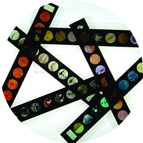
Microslide Harmful Bacteria
Microslide Harmful Bacteria (Product Code: SLE/PS/91) is a Microslide of 8 related 35mm images, photographed through a microscope, used to show bacteria that cause disease in people and crops in school biology and health education lessons. The listed subjects include the bacteria of diphtheria, typhoid fever, pneumonia, tuberculosis and food poisoning at 2000x, together with potato ring-rot and apple fire-blight bacteria. It is supplied by Jain Scientific Equipments Pvt Ltd (ScienceLab), Ambala, India.
Specifications
| Parameter | Detail |
|---|---|
| Format | Microslide – 8 related 35mm images photographed through a microscope |
| Viewing | Microslide Viewer |
| Human disease subjects | Diphtheria (2000x), typhoid fever bacteria (2000x), pneumonia bacteria (2000x), bacteria of tuberculosis (2000x), bacteria of food poisoning (2000x) |
| Plant disease subjects | Bacteria of potato ring-rot; bacteria of apple fire-blight (2000x) |
| Labelling | Arrows and callouts show the features to look for |
| Folder | Detailed text folder with questions; pocket holds the Microslide |
| Product code | SLE/PS/91 |
| Supplied by | Jain Scientific Equipments Pvt Ltd (ScienceLab), Ambala, India |
What Is the Microslide Harmful Bacteria Used For?
Typhoid, tuberculosis and food poisoning appear in most school health syllabuses, but the bacteria behind them should never be grown in a school lab. This Microslide lets students see those organisms as photographed images, with arrows and callouts pointing out their shape and arrangement, without anyone handling a culture.
Potato ring-rot and apple fire-blight widen the discussion to farming, showing that bacteria also damage crops and food supplies. Used with the text folder questions, the set supports lessons on hygiene, clean water and safe food handling, and it fits well into a health education kit for schools alongside Microslide Helpful Bacteria.
Supplier
Jain Scientific Equipments Private Limited supplies Microslide Harmful Bacteria through its ScienceLab brand, working from Ambala, Haryana. Schools and colleges usually request it together with the viewer and the other microbe and disease titles, and the company arranges supply for institutions and dealers in India and international markets. The ScienceLab profile has more about the company.
Related Prepared Slides
Items that support the same lessons on microbes and disease:
- Microslide Viewer — required to view the images.
- Microslide Helpful Bacteria — the useful side of bacteria, for a balanced lesson.
- Microslide Human Diseases — places bacterial disease beside viral, genetic and environmental illness.
- Microslide Body Defences Against Infection — how the body responds to the organisms shown here.
- Three Types of Bacteria, Stained — a stained preparation of three types of bacteria for comparison.
See all titles under Prepared Slides.
Frequently Asked Questions
Which bacteria are shown in the Microslide Harmful Bacteria?
The product details for the Microslide Harmful Bacteria list diphtheria, typhoid fever, pneumonia, tuberculosis and food poisoning bacteria, plus the bacteria of potato ring-rot and apple fire-blight. Ask us through the contact page if you need the full image list for all eight frames.
Is the Microslide Harmful Bacteria safe to use in a classroom?
Yes. The Microslide Harmful Bacteria contains photographic images only, taken through a microscope and viewed through a Microslide Viewer. There are no live bacteria or cultures, so nothing needs to be grown, handled or sterilised.
What magnification is used in the Microslide Harmful Bacteria?
Six of the listed subjects in the Microslide Harmful Bacteria are marked 2000x: the diphtheria, typhoid, pneumonia, tuberculosis, food-poisoning and apple fire-blight images. No magnification is stated for the potato ring-rot image.
Does the Microslide Harmful Bacteria cover plant diseases?
Yes. Besides human disease bacteria, the Microslide Harmful Bacteria includes the bacteria of potato ring-rot and apple fire-blight, which helps agriculture and environmental science students see how bacteria affect crops.
Who supplies the Microslide Harmful Bacteria?
The Microslide Harmful Bacteria is supplied by Jain Scientific Equipments Pvt Ltd (ScienceLab), Ambala, Haryana, India, to schools, colleges and dealers in India and international markets.
What is needed for a Microslide Harmful Bacteria quotation?
For a Microslide Harmful Bacteria quotation, tell us the number of sets, how many viewers you need and where the order is to be delivered. Send the details through the contact page.
Request a Quote
Ask for a quotation on the Microslide Harmful Bacteria through the contact page, or add it to the enquiry cart with viewers and related disease titles.
Last updated: 24 September 2026
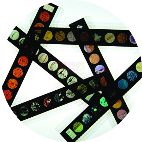
Microslide Nutrition
Microslide Nutrition (Product Code: SLE/PS/92) is a Microslide of 8 related 35mm images, photographed through a microscope, used to show how a shortage of vitamins and minerals changes body tissues in biology, home science and health education lessons. Each image pairs a nutrient with the tissue it affects, from vitamin A deficiency in the cornea at 200x to iodine deficiency in the thyroid at 400x and iron deficiency in blood at 300x. It is supplied by Jain Scientific Equipments Pvt Ltd (ScienceLab), Ambala, India.
Specifications
| Parameter | Detail |
|---|---|
| Format | Microslide of 8 related 35mm images |
| Viewed with | Microslide Viewer |
| Vitamin deficiency images | Vitamin A – cornea (200x); vitamin B1 – heart muscle (250x); vitamin B2 – skin (40x and 100x); vitamin B3 – skin (50x); vitamin D – bone (40x and 100x) |
| Mineral deficiency images | Iodine – thyroid (400x); potassium – heart muscle (100x); iron – blood (300x) |
| Labelling | Arrows and callouts identify the changes to look for |
| Teaching folder | Detailed text folder with questions; pocket stores the Microslide |
| Product code | SLE/PS/92 |
| Supplied by | Jain Scientific Equipments Pvt Ltd (ScienceLab), Ambala, India |
What Is the Microslide Nutrition Used For?
Food and nutrition chapters often present deficiency diseases as a list to memorise. This set shows what those deficiencies do at tissue level, in the cornea, skin, bone, thyroid, heart muscle and blood, which makes the link between diet and health far more concrete for students.
A practical approach is to give each group one nutrient, ask them to find foods rich in it, and then have them present the matching image using its callouts. The set connects naturally with the digestive system topic and with textbook conditions such as rickets, goitre and anaemia, making it a useful aid for health education classes.
Supplier
Jain Scientific Equipments Private Limited, which trades as ScienceLab, supplies Microslide Nutrition from Ambala, Haryana, to schools, colleges, home-science departments and dealers in India and international markets. Buyers who need vendor registration details can use the company's CIN U29309HR2020PTC087597 and GSTIN 06AAECJ8618A1ZT. See the ScienceLab profile for more about the company.
Related Prepared Slides
These items support the same nutrition and health lessons:
- Microslide Viewer — the viewer these 35mm images need.
- Microslide The Digestive System — covers how nutrients are taken into the body.
- Prepared Microscope Slide Set – Health And Disease — glass slides for microscope work on the same theme.
- Microslide Human Diseases — sets nutrition-related illness beside infectious and genetic disease.
The full range is under Prepared Slides.
Frequently Asked Questions
Which deficiencies are shown in the Microslide Nutrition?
The Microslide Nutrition shows deficiencies of vitamin A (cornea), vitamin B1 (heart muscle), vitamin B2 (skin), vitamin B3 (skin), vitamin D (bone), iodine (thyroid), potassium (heart muscle) and iron (blood).
What magnifications are used in the Microslide Nutrition?
The Microslide Nutrition images range from 40x to 400x: for example the cornea at 200x, the heart muscle at 100x and 250x, blood at 300x and the thyroid at 400x, with the skin and bone images at 40x and 100x.
Which subjects can use the Microslide Nutrition?
The Microslide Nutrition suits school biology, home science and health education, and any course that teaches how diet affects the body. Its text folder questions help students connect each nutrient with the tissue it affects.
Is the Microslide Nutrition meant for diagnosis?
No. The Microslide Nutrition is a classroom teaching aid made of photographed images. It illustrates tissue changes for learning purposes and is not intended for diagnosis or for giving medical or dietary advice.
Who supplies the Microslide Nutrition?
The Microslide Nutrition is supplied by Jain Scientific Equipments Pvt Ltd (ScienceLab), Ambala, Haryana, India, to schools, colleges and dealers in India and international markets.
How can I get a quotation for the Microslide Nutrition?
To get a quotation for the Microslide Nutrition, send the number of sets, the number of viewers and your delivery location through the contact page, or add the item to the enquiry cart.
Request a Quote
Send an enquiry for the Microslide Nutrition through the contact page, or use the enquiry cart to combine it with viewers and other health-topic Microslides.
Last updated: 24 September 2026

Microslide Lesson Set – Immunity
Microslide Lesson Set – Immunity (Product Code: SLE/PS/93) is a Microslide lesson set of related 35mm images, photographed through a microscope, used to teach how the immune system defends the body in senior school and college biology. Its listed subjects run from phagocytes, monocytes and macrophages to the bone marrow, lymph and thymus glands, T-cells with HIV budding, plasma cells, agglutinated red cells, eosinophils and basophils. It is supplied by Jain Scientific Equipments Pvt Ltd (ScienceLab), Ambala, India.
Specifications
| Parameter | Detail |
|---|---|
| Format | Microslide images; each Microslide is a set of 8 related 35mm images |
| Viewing | Microslide Viewer |
| Engulfing cells | Blood phagocyte, phagocytosis, monocyte, macrophage |
| Immune organs | Bone marrow, lymph gland, thymus gland |
| Lymphocytes and antibody cells | Normal T-cells, HIV budding from T-cell, plasma cell |
| Blood reactions and granulocytes | Normal red blood cells, agglutinated cells, eosinophil, basophil |
| Subjects listed | 14 |
| Labelling | Arrows and callouts on the images |
| Text folder | Detailed notes and questions, with a pocket to store the Microslides |
| Product code | SLE/PS/93 |
| Supplied by | Jain Scientific Equipments Pvt Ltd (ScienceLab), Ambala, India |
What Is the Microslide Immunity Lesson Set Used For?
Immunity stays abstract until students see the cells involved. The lesson set starts with phagocytes caught in the act of phagocytosis, then shows the monocytes and macrophages that clear away invaders, and the bone marrow, lymph gland and thymus where immune cells are produced and mature.
Normal T-cells beside HIV budding from a T-cell help explain why HIV weakens the immune system, while the plasma cell introduces antibody production. Normal and agglutinated red cells lead into blood grouping, and the eosinophil and basophil complete the white-cell family, making the set a strong resource for college biology and health education courses.
What to Specify When Ordering
- The number of Immunity lesson sets you need.
- Ask us to confirm how many Microslides and images the lesson set contains, since the listing names 14 subjects and each Microslide holds 8 images.
- How many Microslide Viewers are required for your groups.
- Any other Microslide titles, such as body defences or human diseases, to include on the same order.
Supplier
The Immunity lesson set is supplied by Jain Scientific Equipments Private Limited, the Ambala, Haryana company behind the ScienceLab name. It sources Microslide titles and viewers for human biology and health courses and supplies them to schools, colleges and dealers in India and international markets. You can read about the company on the ScienceLab profile page.
Related Prepared Slides
Items that support the same immunity lessons:
- Microslide Viewer — needed to view the images.
- Microslide Body Defences Against Infection — the wider picture of how the body keeps infection out.
- Human Blood Cell Types Model — an enlarged model of the blood cells shown in the set.
- Microslide Harmful Bacteria — the disease-causing organisms the immune system responds to.
- Microslide Human Diseases — connects immunity with infectious and non-infectious disease.
More titles are in Prepared Slides.
Frequently Asked Questions
What topics does the Microslide Lesson Set – Immunity cover?
The Microslide Lesson Set – Immunity lists blood phagocyte, phagocytosis, monocyte, macrophage, bone marrow, lymph gland, thymus gland, normal T-cells, HIV budding from a T-cell, plasma cell, normal red blood cells, agglutinated cells, eosinophil and basophil.
How many Microslides are in the Microslide Lesson Set – Immunity?
Each Microslide holds 8 related 35mm images, and the Microslide Lesson Set – Immunity lists 14 subjects. Please confirm the exact number of Microslides in the lesson set through the contact page before ordering.
Does the Microslide Lesson Set – Immunity cover HIV?
Yes. The Microslide Lesson Set – Immunity includes normal T-cells and an image of HIV budding from a T-cell, which teachers use to explain how the virus affects the cells that coordinate the immune response.
Does the Microslide Lesson Set – Immunity include teaching notes?
Yes. The Microslide Lesson Set – Immunity comes with a detailed text folder that informs students and asks them questions about each subject, and the folder has a pocket for storing the Microslides.
Which students is the Microslide Lesson Set – Immunity suitable for?
The Microslide Lesson Set – Immunity suits senior secondary and college students in biology, human biology and health education courses, where immune cells, antibodies and blood reactions are studied in detail.
Who supplies the Microslide Lesson Set – Immunity?
The Microslide Lesson Set – Immunity is supplied by Jain Scientific Equipments Pvt Ltd (ScienceLab), Ambala, Haryana, India, to schools, colleges and dealers in India and international markets.
Request a Quote
Ask for a quotation on the Microslide Lesson Set – Immunity through the contact page, or add it to the enquiry cart with viewers and related Microslide titles.
Last updated: 24 September 2026
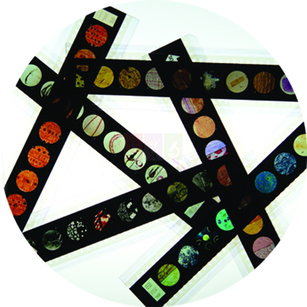
Microslide Human Diseases
Microslide Human Diseases (Product Code: SLE/PS/94) is a Microslide of 8 related 35mm images, photographed through a microscope, used to compare the different causes of human disease in school biology and health education. It moves from the influenza virus at 320,000x and tuberculosis to amoebic dysentery, the Trichina worm, sickle cell anaemia, a normal and a blackened lung, and atherosclerosis. It is supplied by Jain Scientific Equipments Pvt Ltd (ScienceLab), Ambala, India.
Specifications
| Parameter | Detail |
|---|---|
| Format | 8 related 35mm images for a Microslide Viewer |
| Viral disease | Influenza virus (320,000x) |
| Bacterial disease | Tuberculosis (100x and 1000x) |
| Protist disease | Amoebic dysentery (100x) |
| Parasitic worm | Trichina worm (65x) |
| Genetic disease | Sickle cell anaemia (400x) |
| Environmental illness | Normal lung (150x) and blackened lung (65x) |
| Multifactorial disease | Atherosclerosis (3x and 15x) |
| Labelling | Arrows and callouts guide students to key features |
| Folder | Detailed text folder with questions; pocket stores the Microslide |
| Product code | SLE/PS/94 |
| Supplied by | Jain Scientific Equipments Pvt Ltd (ScienceLab), Ambala, India |
What Is the Microslide Human Diseases Used For?
This set groups illness by cause rather than by body system: viral, bacterial, protist, parasitic, genetic, environmental and multifactorial. Students quickly see that not every disease is caught from someone else, because sickle cell anaemia is inherited, a blackened lung reflects what was breathed in and atherosclerosis builds up from several factors over time.
The normal and blackened lung pair is a strong starting point for discussing air pollution and smoking. The magnifications also teach scale, from atherosclerosis visible at just 3x and 15x to the influenza virus at 320,000x. Health education teachers often use the text folder questions as a group task before moving on to prevention.
Supplier
Microslide Human Diseases comes from Jain Scientific Equipments Private Limited, which supplies biology teaching material under the ScienceLab name from Ambala, Haryana. The company serves schools, colleges and distributors in India and international markets, and can quote this title together with the viewer and the related health Microslides. The company profile gives more background.
Related Prepared Slides
Other items for lessons on health and disease:
- Microslide Viewer — used to view the eight images.
- Microslide Smoking & Health — builds on the blackened lung image.
- Microslide Harmful Bacteria — a closer look at bacterial causes of disease.
- Microslide Lesson Set – Immunity — how the body fights the infections shown here.
- Prepared Microscope Slide Set – Health And Disease — glass slides for microscope study of the same theme.
Browse Prepared Slides for further titles.
Frequently Asked Questions
Which diseases are shown in the Microslide Human Diseases?
The Microslide Human Diseases shows the influenza virus, tuberculosis, amoebic dysentery, the Trichina worm, sickle cell anaemia, a normal lung beside a blackened lung, and atherosclerosis, each labelled with arrows and callouts.
What magnifications are used in the Microslide Human Diseases?
The Microslide Human Diseases spans 3x to 320,000x: atherosclerosis at 3x and 15x, the Trichina worm and blackened lung at 65x, tuberculosis at 100x and 1000x, sickle cell anaemia at 400x and the influenza virus at 320,000x.
Does the Microslide Human Diseases include non-infectious diseases?
Yes. Along with viral, bacterial, protist and parasitic infections, the Microslide Human Diseases covers a genetic disease (sickle cell anaemia), an environmental illness (blackened lung) and a multifactorial disease (atherosclerosis).
Is the Microslide Human Diseases a medical reference?
No. The Microslide Human Diseases is a teaching aid for school and college classes. Its photographed images and text folder explain disease causes for learning, and it is not intended for diagnosis or clinical use.
Who supplies the Microslide Human Diseases?
The Microslide Human Diseases is supplied by Jain Scientific Equipments Pvt Ltd (ScienceLab), Ambala, Haryana, India, to schools, colleges and distributors in India and international markets.
What should a quotation request for the Microslide Human Diseases include?
A quotation request for the Microslide Human Diseases should give the number of sets, the number of viewers and the delivery location, plus any related health titles. Send it through the contact page.
Request a Quote
Enquire about the Microslide Human Diseases through the contact page, or add it to the enquiry cart with a viewer and other health and disease titles.
Last updated: 24 September 2026
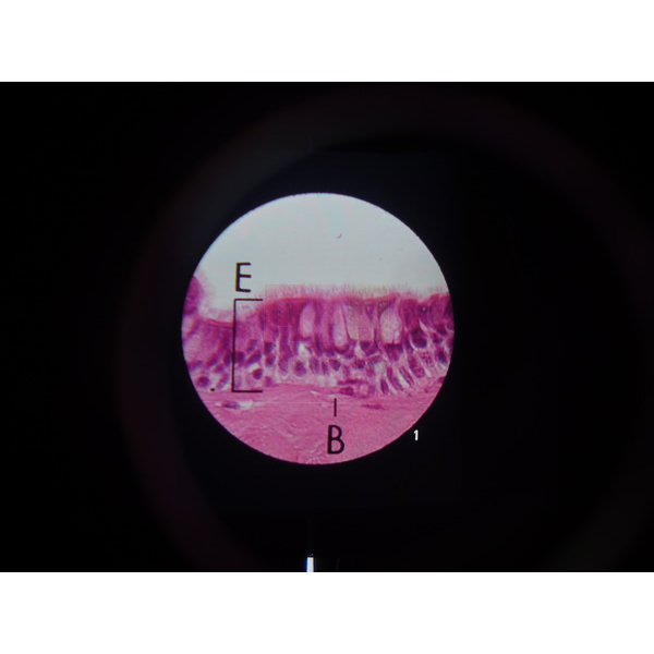
Microslide Smoking & Health
Microslide Smoking & Health (Product Code: SLE/PS/95) is a set of 8 related 35 mm images, photographed through a microscope, used to show how tobacco smoke changes the airways and lungs in school biology and health education lessons. Teachers use it to set normal bronchial epithelium and lung tissue beside thickened, cilia-damaged and blackened tissue, so students see the damage for themselves. This biology teaching aid is supplied by Jain Scientific Equipments Pvt Ltd (ScienceLab), Ambala, India.
Specifications
| Parameter | Detail |
|---|---|
| Format | Set of 8 related 35 mm images photographed through a microscope |
| Viewing | Designed to be viewed through a Microslide Viewer |
| Airway images | Normal Bronchial Epithelium (175x); Thickened Bronchial Epithelium (175x); Loss of Cilia – Bronchial Epithelium; Early Cancerous Invasion – Bronchial Epithelium |
| Lung images | Normal Lung Tissue; Blackened Lung Tissue (65x); Fibrosis and Emphysema – lung tissue |
| Micro-organism image | Alpha Virus on E. Coli (60,000x) |
| Labelling | Arrows and callouts point out the features being studied |
| Student notes | Detailed text folder that informs and questions students, with a pocket to store the Microslides |
| Product code | SLE/PS/95 |
| Supplied by | Jain Scientific Equipments Pvt Ltd (ScienceLab), Ambala, India |
What Is the Microslide Smoking & Health Used For?
The set works as a before-and-after comparison. Two views of bronchial epithelium at the same 175x magnification show how the airway lining thickens, and the next frames show cilia being lost and early cancerous invasion. The lung frames then move from normal tissue to blackened tissue at 65x and on to fibrosis and emphysema, helping students link what they see to breathlessness and poorer gas exchange.
Because every view carries arrows and callouts, pupils can work through the images in pairs using a Microslide Viewer and then answer the questions in the text folder. The last frame, Alpha Virus on E. Coli at 60,000x, is a useful prompt for discussing why some images need far higher magnification than a school microscope gives. Many teachers run it alongside a smoking model as part of a wider health education unit.
What to Specify When Ordering
- How many Microslide Smoking & Health sets you need (per class or per student group)
- Whether Microslide Viewers should be quoted with the sets
- Any other Microslide topics for the same health unit, such as Human Diseases
- Delivery city or country for the quotation
Supplier
Microslide Smoking & Health is supplied by Jain Scientific Equipments Private Limited, the company that trades as ScienceLab from Ambala, Haryana. We source this set for schools, colleges, dealers and importers in India and international markets, and we can place it on one quotation with viewers, models and other prepared slides. Read more about the company on our company profile.
Related Prepared Slides
These items support the same respiratory and health lessons:
- Microslide Viewer — needed to view the 35 mm images in the set
- Microslide Human Diseases — extends the lesson to other conditions
- Prepared Microscope Slide Set – Health And Disease — glass slides including lung of smoking and emphysema for microscope work
- Effects of Smoking Model — a three-dimensional companion for class demonstration
- Smoking Experiment — a hands-on kit for the same topic
See the full range of Prepared Slides.
Frequently Asked Questions
What images are in Microslide Smoking & Health?
Microslide Smoking & Health contains 8 images: normal and thickened bronchial epithelium (both 175x), loss of cilia, early cancerous invasion, normal lung tissue, blackened lung tissue (65x), fibrosis and emphysema, and Alpha Virus on E. Coli (60,000x).
Do I need a viewer for Microslide Smoking & Health?
Yes. The Microslide Smoking & Health images are designed to be seen through a Microslide Viewer, which is listed as a separate product. Mention in your enquiry whether you need viewers and how many, and we will quote them together with the sets.
What does the text folder with Microslide Smoking & Health contain?
The Microslide Smoking & Health text folder gives detailed notes written to inform students and question them about smoking and the lungs. It also has a pocket that holds the Microslides, so images and notes stay together between lessons.
Which classes is Microslide Smoking & Health suitable for?
Microslide Smoking & Health suits secondary school biology and health education lessons on breathing, the lungs and the effects of tobacco. It is useful wherever students should see tissue changes without preparing or staining their own specimens.
Who supplies Microslide Smoking & Health?
Microslide Smoking & Health is supplied by Jain Scientific Equipments Pvt Ltd (ScienceLab) of Ambala, Haryana, India. The company serves schools, colleges, dealers and importers and can combine the set with viewers and models on one quotation.
What should a quotation request for Microslide Smoking & Health include?
For a Microslide Smoking & Health quotation, give the product code SLE/PS/95, the number of sets, any Microslide Viewers required and the delivery city or country. Send these details through our contact page and we will reply with availability and terms.
Request a Quote
To order Microslide Smoking & Health, send your quantity and delivery details through our contact page, or add it to the enquiry cart with the other prepared slides you need.
Last updated: 24 September 2026
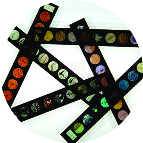
Microslide Body Defences Against Infection
Microslide Body Defences Against Infection (Product Code: SLE/PS/96) is a set of 8 related 35 mm photomicrographs used to teach how the human body keeps out and fights infection in secondary biology classes. It moves from barrier tissues such as skin and the airway lining to white blood corpuscles, phagocytosis, lymphatic organs and a plasma cell, at magnifications from 7x to 37,000x. It is supplied by Jain Scientific Equipments Pvt Ltd (ScienceLab), Ambala, India.
Specifications
| Parameter | Detail |
|---|---|
| Set contents | 8 related 35 mm images, each photographed through a microscope |
| First-line barriers | The Skin (200x); Epithelial Lining of Respiratory Tract (175x) / Isolated Cell from Wind Pipe (1400x); Pyloric Stomach (100x) / Eyelid (30x) |
| Blood cells | White Blood Corpuscles (500x) / Phagocytosis (25,000x); Plasma Cell (37,000x) |
| Organs | Lymph Gland (40x); Tonsils and Adenoids (7x); The Liver (20x) |
| Guidance on the images | Arrows and callouts mark the key features |
| Accompanying material | Detailed text folder that informs and questions students; built-in pocket stores the Microslides |
| Viewing | Microslide Viewer |
| Product code | SLE/PS/96 |
| Supplied by | Jain Scientific Equipments Pvt Ltd (ScienceLab), Ambala, India |
What Is the Microslide Body Defences Against Infection Used For?
This set lets a teacher build an immunity lesson layer by layer. Students begin with the skin, the lining of the respiratory tract and a single cell from the windpipe, the stomach and the eyelid, and discuss how each surface blocks or traps microbes. They then move inward to white blood corpuscles and phagocytosis at 25,000x, where a cell engulfs foreign material.
The later frames show where defence cells gather: a lymph gland, the tonsils and adenoids, and the liver, ending with a plasma cell at 37,000x as a lead-in to antibodies. With the callouts guiding them, pupils can study the images at their own pace in a Microslide Viewer and use the text folder questions for homework or revision. It fits well among biology teaching aids for immunity and disease units.
Supplier
Jain Scientific Equipments Private Limited, trading as ScienceLab, supplies Microslide Body Defences Against Infection from Ambala, Haryana, to schools, teacher-training colleges and distributors in India and international markets. For vendor registration, institutions can record the supplying company under GSTIN 06AAECJ8618A1ZT and LEI 3358004HRD1JIVXKYX49. More about the company is on our profile page.
Related Prepared Slides
Buyers planning an immunity or disease unit often add:
- Microslide Viewer — for looking at the eight images
- Microslide Lesson Set – Immunity — continues the topic of the immune response
- Microslide Harmful Bacteria — shows the micro-organisms the body defends against
- Microslide Cells of Body — background on the cell types students meet in this set
Compare other sets in our Prepared Slides category.
Frequently Asked Questions
Which tissues are shown in Microslide Body Defences Against Infection?
Microslide Body Defences Against Infection shows the skin, respiratory tract lining with an isolated windpipe cell, pyloric stomach with eyelid, white blood corpuscles with phagocytosis, a lymph gland, tonsils and adenoids, the liver and a plasma cell.
What magnifications does Microslide Body Defences Against Infection cover?
Microslide Body Defences Against Infection spans 7x for the tonsils and adenoids up to 37,000x for the plasma cell. Other stated values are 20x, 30x, 40x, 100x, 175x, 200x, 500x, 1400x and 25,000x.
How do students view Microslide Body Defences Against Infection?
Students view Microslide Body Defences Against Infection through a Microslide Viewer, not a standard microscope, because the images are 35 mm photographs. The viewer is a separate listing, so include it in your enquiry if your school does not already have one.
Is there written material with Microslide Body Defences Against Infection?
Yes. Microslide Body Defences Against Infection includes a detailed text folder with notes and questions on the topic, and the folder has a pocket in which the Microslides are stored after use.
Who supplies Microslide Body Defences Against Infection?
Microslide Body Defences Against Infection is supplied by Jain Scientific Equipments Pvt Ltd (ScienceLab), Ambala, Haryana, India, to schools, colleges and dealers in India and abroad. Vendor details such as GSTIN and LEI are available for institutional registration.
Can Microslide Body Defences Against Infection be ordered in bulk or for export?
Yes, Microslide Body Defences Against Infection can be quoted in multiple sets for a school network or a dealer, and for delivery outside India. State the quantity and destination on our contact page so the quotation reflects your order.
Request a Quote
Ask for a quotation for Microslide Body Defences Against Infection through our contact page, or place it in the enquiry cart along with any viewers or related Microslides.
Last updated: 24 September 2026
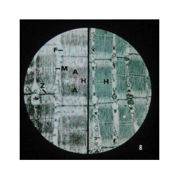
Microslide Nerve & Muscle Action
Microslide Nerve & Muscle Action (Product Code: SLE/PS/97) is a set of 8 related 35 mm images taken through a microscope, used to follow a nerve impulse from a single nerve cell to a contracting muscle in biology and physiology lessons. Its distinguishing frames show sarcomeres in both the relaxed and contracted state, with synapses, synaptic knobs and the nerve-muscle junction along the way. The set is supplied by Jain Scientific Equipments Pvt Ltd (ScienceLab), Ambala, India.
Specifications
| Parameter | Detail |
|---|---|
| What you get | 8 related 35 mm images photographed through a microscope |
| Nerve images | Single Cell; Synapse; Axons; Myelinated Axons; Synaptic Knobs |
| Junction image | Nerve-Muscle Junction |
| Muscle images | Striated Muscle Cell; Sarcomeres – relaxed and contracted |
| On-image help | Arrows and callouts locate the important structures |
| Text folder | Detailed folder that informs and questions students, with a handy pocket for the Microslides |
| Viewed with | Microslide Viewer |
| Product code | SLE/PS/97 |
| Supplied by | Jain Scientific Equipments Pvt Ltd (ScienceLab), Ambala, India |
What Is the Microslide Nerve & Muscle Action Used For?
Teachers use the set to tell one continuous story. Students start with a whole nerve cell, compare plain axons with myelinated axons, and then look closely at a synapse and the synaptic knobs where the signal passes from one cell to the next. The nerve-muscle junction image shows where that signal reaches a muscle fibre.
The last frames switch to the muscle itself: a striated muscle cell, followed by sarcomeres shown relaxed and contracted, which makes the shortening of the sarcomere during contraction easy to explain. With the callouts as a guide and the text folder questions to answer, this set suits both class teaching and self-study at senior secondary and early college level, and pairs well with a synapse model on the demonstration bench.
Supplier
ScienceLab is the trading name of Jain Scientific Equipments Private Limited, Ambala, Haryana, which supplies Microslide Nerve & Muscle Action to schools, colleges and distributors. Shipments outside India are handled under Importer-Exporter Code AAECJ8618A (DGFT), and the company is GST-registered under GSTIN 06AAECJ8618A1ZT. Background on the business is on our company profile.
Related Prepared Slides
Items that help students connect these images to three-dimensional structures:
- Microslide Viewer — the viewer these 35 mm images are made for
- Human Nerve Ending and Synapse Model — a model version of the synapse frames
- Skeletal Muscle Fiber Model — enlarges the striated muscle structure seen in the set
- Microslide Animal Tissue Set 2 Muscle, Bone, Connective — more muscle and tissue images for the same unit
More topic sets are listed under Prepared Slides.
Frequently Asked Questions
What does Microslide Nerve & Muscle Action show?
Microslide Nerve & Muscle Action shows 8 views: a single nerve cell, a synapse, axons, myelinated axons, the nerve-muscle junction, synaptic knobs, a striated muscle cell, and sarcomeres in relaxed and contracted states.
Are magnifications stated for Microslide Nerve & Muscle Action?
No magnification values are given in our listing for Microslide Nerve & Muscle Action. If your syllabus or worksheet needs them, ask us through the contact page and we will check the details for the sets we quote.
Can Microslide Nerve & Muscle Action be used with a normal microscope?
Microslide Nerve & Muscle Action is made for a Microslide Viewer rather than a compound microscope, because the images are 35 mm photographs taken through a microscope. Add viewers to your enquiry if needed.
How is Microslide Nerve & Muscle Action stored?
Microslide Nerve & Muscle Action comes with a detailed text folder that has a pocket for the Microslides, so the images are kept inside the folder with their notes and questions between lessons.
Who supplies Microslide Nerve & Muscle Action?
Microslide Nerve & Muscle Action is supplied by Jain Scientific Equipments Pvt Ltd (ScienceLab) in Ambala, Haryana, India, for schools, colleges and dealers in India and international markets, with IEC and GSTIN details for export paperwork.
What information speeds up a quotation for Microslide Nerve & Muscle Action?
For a Microslide Nerve & Muscle Action quotation, quote code SLE/PS/97, the number of sets, whether viewers or models are needed, and the delivery location. We reply with availability and terms once we have these details.
Request a Quote
Send an enquiry for Microslide Nerve & Muscle Action through our contact page, or add it to the enquiry cart with any viewers and models for your physiology lessons.
Last updated: 24 September 2026
Microslide The Digestive System
Microslide The Digestive System (Product Code: SLE/PS/98) is a set of 8 related 35 mm microscope photographs used to show the structure of the human gut, from the mouth to the small intestine, in school biology classes. Students follow food past the salivary gland and taste buds, down the gullet, into the stomach lining with its gastric glands and on to the intestinal villi. It is supplied by Jain Scientific Equipments Pvt Ltd (ScienceLab), Ambala, India.
Specifications
| Parameter | Detail |
|---|---|
| Number of images | 8 related 35 mm images, photographed through a microscope |
| Mouth | Salivary gland; Taste buds |
| Gullet | Gullet (Oesophagus); Smooth muscle; Junction of gullet and stomach |
| Stomach | Stomach lining and gastric glands |
| Small intestine | Small intestine; Intestinal glands and villi |
| Teaching features | Arrows and callouts on the images; detailed text folder with a pocket to store the Microslides |
| Viewer | Made to be viewed through a Microslide Viewer |
| Product code | SLE/PS/98 |
| Supplied by | Jain Scientific Equipments Pvt Ltd (ScienceLab), Ambala, India |
What Is the Microslide The Digestive System Used For?
The images are arranged in the order food travels, which makes the set a natural companion to a lesson on digestion and absorption. Students can relate each structure to its job: saliva from the salivary gland, taste detection in the taste buds, the smooth muscle that moves food along the oesophagus, and the change in lining where the gullet joins the stomach.
In the stomach and small intestine frames, the callouts draw attention to gastric glands, intestinal glands and villi, so learners can explain how the folded lining increases the surface for absorption. Working in a Microslide Viewer, a group can annotate a worksheet directly from the images, then use the text folder questions to check understanding. It is a compact addition to school biology lab equipment where time for sectioning and staining is limited.
Supplier
We supply Microslide The Digestive System as part of the biology teaching range offered by Jain Scientific Equipments Private Limited, known by its trading name ScienceLab, from Ambala in Haryana. Schools and dealers in India and international markets can combine it with models and glass slide sets on a single enquiry. Our profile page explains more about the company.
Related Prepared Slides
For a complete digestion unit, buyers also look at:
- Microslide Viewer — required to see the images
- Prepared Microscope Slide Set – Digestion — glass slides of gut tissue for microscope practicals
- Human Digestive System Model — shows where each tissue sits in the body
- Microslide Nutrition — links what we eat to how it is digested
- Kit for Digestion Experiments — practical work on the same topic
Browse the rest of our Prepared Slides.
Frequently Asked Questions
What parts of the gut appear in Microslide The Digestive System?
Microslide The Digestive System covers the salivary gland, taste buds, gullet (oesophagus), smooth muscle, the junction of gullet and stomach, stomach lining with gastric glands, the small intestine, and intestinal glands with villi.
Does Microslide The Digestive System include the liver, pancreas or large intestine?
No, Microslide The Digestive System does not list the liver, pancreas or large intestine; its images end at the small intestine and villi. For liver, pancreas and tongue, see our separate glass slide set on digestion.
What is supplied with Microslide The Digestive System?
Microslide The Digestive System comes as 8 related 35 mm images with a detailed text folder of notes and questions. The folder has a handy pocket, so the Microslides are stored with it after class.
How are the images in Microslide The Digestive System labelled?
Microslide The Digestive System uses arrows and callouts on the images to help students find the important features, such as the gastric glands and villi, without needing a teacher to point them out on each view.
Who supplies Microslide The Digestive System?
Microslide The Digestive System is supplied by Jain Scientific Equipments Pvt Ltd (ScienceLab), based in Ambala, Haryana, India. The company supplies schools, colleges, dealers and importers in India and in international markets.
Can I order Microslide The Digestive System for several schools?
Yes, Microslide The Digestive System can be quoted in the number of sets you need for several schools or a dealer order. Give the product code SLE/PS/98, quantity and delivery location through our contact page.
Request a Quote
For pricing and availability of Microslide The Digestive System, write to us on the contact page or add it to your enquiry cart together with any viewers or models.
Last updated: 24 September 2026
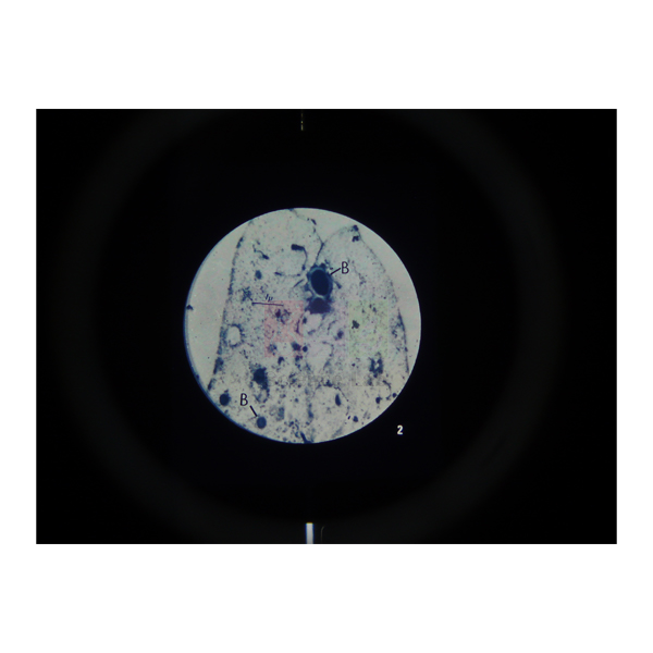
Microslide The Circulatory System
Microslide The Circulatory System (Product Code: SLE/PS/99) is a set of 8 related 35 mm photographs taken through a microscope, used to teach the blood, blood vessels, lymph vessels and heart in secondary biology. Alongside human blood, a phagocyte and an artery with its vein, it includes two views of a rat heart at 4x and 6x and a close-up of the valve at the aorta. It is supplied by Jain Scientific Equipments Pvt Ltd (ScienceLab), Ambala, India.
Specifications
| Parameter | Detail |
|---|---|
| Images per set | 8 related 35 mm images photographed through a microscope |
| Blood | Human Blood; Phagocyte |
| Vessels | Artery and vein; Lung capillaries; Lymph vessel and valves |
| Heart | Rat Heart (4x); Rat heart (6x); Valve at aorta |
| Callouts | Arrows and callouts guide students to key features |
| Folder | Detailed text folder to inform and question students; handy pocket stores the Microslides |
| For use in | Microslide Viewer |
| Product code | SLE/PS/99 |
| Supplied by | Jain Scientific Equipments Pvt Ltd (ScienceLab), Ambala, India |
What Is the Microslide The Circulatory System Used For?
The set gives a class a microscope-level view of the transport system without cutting or staining tissue. A typical sequence starts with human blood and a phagocyte, then compares the thick wall of an artery with its matching vein, and moves to the thin capillaries of the lung where exchange with the air takes place.
The lymph vessel with its valves shows the second, one-way return route for tissue fluid, and the rat heart images at 4x and 6x, followed by the valve at the aorta, help students see why valves stop blood flowing backwards. Viewed in a Microslide Viewer, with callouts on each frame, the set is a practical way to back up a heart model or a glass slide practical.
What to Specify When Ordering
- The number of Microslide The Circulatory System sets
- Whether you also need Microslide Viewers
- Whether a glass slide set of circulatory tissue should be quoted for microscope practicals
Supplier
Microslide The Circulatory System reaches schools, colleges and dealers through Jain Scientific Equipments Private Limited, which operates under the ScienceLab name in Ambala, Haryana. We supply it within a wider range of biology teaching resources for buyers in India and international markets. Visit our company profile for more about who we are.
Related Prepared Slides
These pair well with the set in circulation lessons:
- Microslide Viewer — the viewer the 35 mm images are designed for
- Prepared Microscope Slide Set – Circulatory System — glass slides of artery, vein, cardiac muscle and blood for microscope work
- Human Circulatory System Model — places the vessels in the whole body
- Human Lymphatic System Model — expands on the lymph vessel image
All topic sets are grouped in Prepared Slides.
Frequently Asked Questions
Which images are included in Microslide The Circulatory System?
Microslide The Circulatory System includes human blood, a phagocyte, artery and vein, lung capillaries, a lymph vessel with valves, a rat heart at 4x, a rat heart at 6x, and the valve at the aorta.
How is the heart shown in Microslide The Circulatory System?
Microslide The Circulatory System shows the heart in two rat heart images, at 4x and 6x, so students can study heart structure at low magnification, and adds a separate frame of the valve at the aorta for a closer look at a heart valve.
Does Microslide The Circulatory System replace glass microscope slides?
Microslide The Circulatory System complements rather than replaces glass slides. Its 35 mm images are viewed in a Microslide Viewer with callouts, while glass slides are for students practising their own microscope work.
What student notes come with Microslide The Circulatory System?
Microslide The Circulatory System includes a detailed text folder designed to stimulate, inform and question students about circulation, with a pocket in the folder for storing the Microslides.
Who supplies Microslide The Circulatory System?
Microslide The Circulatory System is supplied by Jain Scientific Equipments Pvt Ltd (ScienceLab), Ambala, Haryana, India, to schools, colleges, dealers and importers, and can be quoted together with models and slide sets.
Request a Quote
Request a quotation for Microslide The Circulatory System on our contact page, giving the quantity and delivery location, or use the enquiry cart to list it with the other items for your lab.
Last updated: 24 September 2026
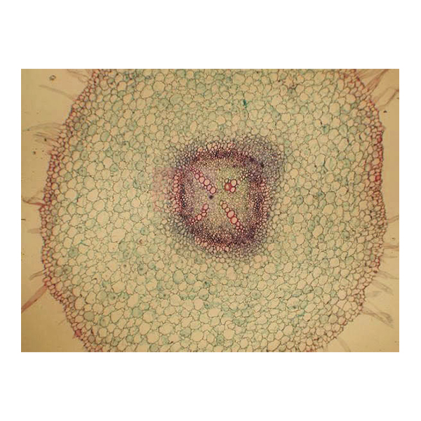
Basic Plant Anatomy Slide Set
Basic Plant Anatomy Slide Set (Product Code: SLE/PS/100) is a set of 20 glass microscope slides used to study the internal structure of vascular plants in school and college botany practicals. The prepared microscope slides cover roots, stems, leaves, flowers and seeds of plants such as Vicia, Zea, Lilium, Helianthus and Tilia, so one box supports a whole plant anatomy course. It is supplied by Jain Scientific Equipments Pvt Ltd (ScienceLab), Ambala, India.
Specifications
| Parameter | Detail |
|---|---|
| Slides in the set | 20 glass microscope slides |
| Theme | Major aspects of vascular plant structure |
| Roots | Vicia, root apex LS; Vicia (bean), young root TS; Helianthus (sunflower), young and old root TS |
| Stems | Zea (maize), stem TS and LS; Dicot, stem, apex LS; Cucurbita (marrow), stem TS and LS; Helianthus, stem, bundles TS; Helianthus, old stem TS and LS; Tilia (lime), young and old stem TS; Tilia, old stem RLS |
| Leaves and epidermis | Prunus (cherry), leaf TS; Ammophila (marram grass), leaf TS; Monocot, epidermis, for stomata E |
| Flowers and pollen | Lilium, anther TS; Lilium, ovary TS and LS; Lilium, ovary, various stages TS; Stigma with pollen tubes; Pollen grains |
| Seeds and embryo | Zea, seed LS; Capsella, mature cotyledon stage LS |
| Product code | SLE/PS/100 |
| Supplied by | Jain Scientific Equipments Pvt Ltd (ScienceLab), Ambala, India |
Label abbreviations: TS = transverse section, LS = longitudinal section, RLS = radial longitudinal section, E = entire (whole mount).
What Is the Basic Plant Anatomy Slide Set Used For?
Students use the set under a compound microscope to compare tissues across plant organs. Paired sections such as Zea and Cucurbita stems in both TS and LS show how vascular bundles look when cut across and along, while young and old stems of Tilia and Helianthus reveal secondary growth. The Vicia root apex and young root sections show the growing tip and the arrangement of root tissues.
For reproduction and adaptation topics, the Lilium anther and ovary slides, stigma with pollen tubes and pollen grains cover the flower, the Capsella slide shows an embryo at the mature cotyledon stage, and the marram grass leaf is a classic example of a leaf adapted to dry, windy sites. A slide box keeps the set in order, and the slides serve well as reference material in any botany lab equipment list for practical examinations.
What to Specify When Ordering
- Number of Basic Plant Anatomy Slide Sets (one per bench or one per class)
- Whether storage such as a slide box should be quoted with the sets
- Whether you also need microscopes for the practical
Supplier
Jain Scientific Equipments Private Limited supplies the Basic Plant Anatomy Slide Set under its ScienceLab trading name from Ambala, Haryana, serving schools, universities, dealers and importers in India and international markets. For tender and vendor forms, the supplying company's details are Importer-Exporter Code AAECJ8618A (DGFT) and GSTIN 06AAECJ8618A1ZT. See our company profile for more.
Related Prepared Slides
Useful additions for a plant anatomy practical:
- Prepared Microscope Slide Set – Transport In Plants — multiple copies of stem, root and leaf sections for a whole class
- Basic Botany Slide Set — covers algae, fungi, mosses and ferns alongside flowering plants
- Cell Division Slide Set of 9 — adds mitosis and meiosis to the plant course
- Slide Box — storage for the 20 slides
- Monocular Biological Microscope 400x, LED — a student microscope for viewing the sections
View all Prepared Slides.
Frequently Asked Questions
How many slides are in the Basic Plant Anatomy Slide Set?
The Basic Plant Anatomy Slide Set contains 20 glass microscope slides showing the major aspects of vascular plant structure, covering roots, stems, leaves, flowers, pollen, a seed and an embryo.
Which plants are used in the Basic Plant Anatomy Slide Set?
The Basic Plant Anatomy Slide Set uses Vicia (bean), Zea (maize), Lilium, Tilia (lime), Prunus (cherry), Cucurbita (marrow), Helianthus (sunflower), Capsella, Ammophila (marram grass) and a monocot epidermis prepared for viewing stomata.
What do TS, LS, RLS and E mean on the Basic Plant Anatomy Slide Set?
On the Basic Plant Anatomy Slide Set, TS means transverse section, LS longitudinal section, RLS radial longitudinal section and E an entire, or whole-mount, preparation. Several slides carry both TS and LS cuts of the same organ.
What microscope is needed for the Basic Plant Anatomy Slide Set?
The Basic Plant Anatomy Slide Set is viewed with an ordinary compound microscope of the kind used in school and college biology labs. Tell us if you need microscopes as well, and we will include them in the same quotation.
Who supplies the Basic Plant Anatomy Slide Set?
The Basic Plant Anatomy Slide Set is supplied by Jain Scientific Equipments Pvt Ltd (ScienceLab), Ambala, Haryana, India. The company provides IEC and GSTIN details to institutions and importers that need them for registration or export paperwork.
How is the Basic Plant Anatomy Slide Set different from the Basic Botany Slide Set?
The Basic Plant Anatomy Slide Set focuses on the internal structure of flowering and other vascular plants in 20 slides, whereas the Basic Botany Slide Set spreads its slides across the major plant groups, including algae, fungi, mosses and ferns.
Request a Quote
Ask for a quotation for the Basic Plant Anatomy Slide Set through our contact page, or add it to the enquiry cart with any microscopes or storage you need.
Last updated: 24 September 2026
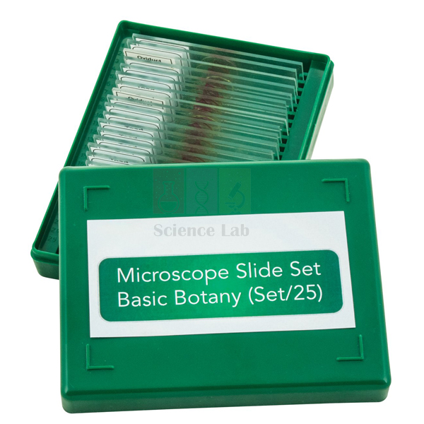
Basic Botany Slide Set
Basic Botany Slide Set (Product Code: SLE/PS/101) is a set of 25 glass microscope slides of botanical specimens used to survey the major plant groups in school and college botany classes. It runs from single-celled Chlamydomonas and Spirogyra through fungi, liverworts, mosses and ferns to pine and flowering plants, making it a ready collection of permanent slides for biology classification work. It is supplied by Jain Scientific Equipments Pvt Ltd (ScienceLab), Ambala, India.
Specifications
| Parameter | Detail |
|---|---|
| Quantity | 25 glass microscope slides |
| Algae | Chlamydomonas E; Spirogyra, vegetative E; Spirogyra, conjugation E; Fucus, dioecious antheridial and oogonial conceptacles |
| Fungi and yeast | Mucor, conjugation; Saccharomyces (yeast) budding; Penicillium, mycelium and conidia |
| Liverworts and mosses | Marchantia (liverwort) thallus, antheridia VS; Marchantia, thallus, archegonia VS; Marchantia, developing sporogonium VS; Mnium, archegonial head VS; Mnium (moss) antheridial head VS |
| Ferns | Dryopteris (fern), pinnule with sori TS; Dryopteris prothallus with antheridia and archegonia E |
| Conifer | Pinus (pine) leaf TS |
| Flowering plants | Zea (maize) root TS; Zea stem TS; Helianthus stem TS; Helianthus (sunflower) young root TS; Prunus (cherry) leaf TS; Tilia (lime) young and old stem TS; Cucurbita (marrow) stem, for sieve tubes LS; Ranunculus (buttercup) flower bud LS; Pollen grains, wind and insect distribution E; Allium (onion) root apex, mitosis LS |
| Product code | SLE/PS/101 |
| Supplied by | Jain Scientific Equipments Pvt Ltd (ScienceLab), Ambala, India |
What Is the Basic Botany Slide Set Used For?
The set is built for comparing groups rather than studying one plant in depth. A class can place the Marchantia, Mnium and Dryopteris slides side by side to see antheridia and archegonia in a liverwort, a moss and a fern, then contrast them with the Ranunculus flower bud and pollen grains adapted for wind and insect distribution. The algae and fungi slides show conjugation in Spirogyra and Mucor, budding yeast and Penicillium conidia.
The flowering-plant slides add everyday anatomy practice with root, stem and leaf sections, while the Cucurbita LS is chosen for sieve tubes and the Allium root apex for stages of mitosis. Teachers often keep the set as a reference collection in the botany lab and bring out the relevant slides as each plant group is taught.
Supplier
The Basic Botany Slide Set is supplied by Jain Scientific Equipments Private Limited, the Ambala, Haryana company that trades as ScienceLab. We source prepared slides for schools, colleges, laboratories and dealers in India and international markets and can quote them alongside microscopes and slide storage. Find out more on our company profile.
Related Prepared Slides
Other items used with a plant diversity course:
- Basic Plant Anatomy Slide Set — 20 slides for deeper study of vascular plant structure
- Cell Division Slide Set of 9 — builds on the Allium mitosis slide
- Basic Slide Set of 25 — a mixed plant and animal set for general biology
- Slide Box, Wooden — keeps the 25 slides organised
- Binocular Biological Microscope — for examining the specimens
Explore our Prepared Slides category for more sets.
Frequently Asked Questions
Which plant groups does the Basic Botany Slide Set cover?
The Basic Botany Slide Set covers algae, fungi and yeast, liverworts, mosses, ferns, a conifer (pine) and flowering plants, using 25 glass microscope slides that represent the major plant groups studied in botany.
Does the Basic Botany Slide Set show plant reproduction?
Yes. The Basic Botany Slide Set includes antheridia and archegonia of Marchantia, Mnium and a Dryopteris prothallus, a developing Marchantia sporogonium, fern sori, Fucus conceptacles, conjugation in Spirogyra and Mucor, a Ranunculus flower bud and pollen grains.
What do the letters E, VS, TS and LS mean on the Basic Botany Slide Set?
On the Basic Botany Slide Set, E marks an entire (whole-mount) specimen, VS a vertical section, TS a transverse section and LS a longitudinal section, telling students how each specimen was cut before mounting.
Is the Basic Botany Slide Set suitable for class practicals?
The Basic Botany Slide Set suits teacher demonstrations and small-group practicals because it holds one slide of each of 25 specimens. For whole-class work on a single topic, schools usually order extra sets or a multi-copy set.
Who supplies the Basic Botany Slide Set?
The Basic Botany Slide Set is supplied by Jain Scientific Equipments Pvt Ltd (ScienceLab), Ambala, Haryana, India, to schools, colleges and dealers in India and international markets.
What should I include in a quotation request for the Basic Botany Slide Set?
For the Basic Botany Slide Set, give product code SLE/PS/101, the number of sets, any microscopes or slide boxes required and the delivery address or country. Send them via our contact page for a prompt reply.
Request a Quote
Enquire about the Basic Botany Slide Set on our contact page, or add it to the enquiry cart with the other slides and equipment for your botany lab.
Last updated: 24 September 2026
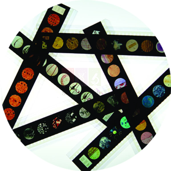
Microslide The Root Of a Flowering Plant
Microslide The Root Of a Flowering Plant (Product Code: SLE/PS/102) is a Microslide set of related 35 mm images photographed through a microscope, used to teach root structure and root growth in plant biology lessons. Its named views follow a buttercup root from the central cylinder, epidermis and cortex down through the root cap and the zones of growth, elongation and maturation to the root hairs. It is supplied by Jain Scientific Equipments Pvt Ltd (ScienceLab), Ambala, India.
Key Details
| Parameter | Detail |
|---|---|
| Format | Microslides are sets of 8 related 35 mm images photographed through a microscope |
| Root structure views | Buttercup Root; Central Cylinder; Epidermis and Cortex |
| Root growth views | Root Cap and Zone of Growth; Zone of Elongation; Zone of Maturation; Root Tip – Root hairs |
| Annotations | Arrows and callouts help students locate the features being studied |
| Text folder | Detailed folder to stimulate, inform and question students, with a pocket for storing the Microslides |
| Viewing device | Microslide Viewer |
| Product code | SLE/PS/102 |
| Supplied by | Jain Scientific Equipments Pvt Ltd (ScienceLab), Ambala, India |
What Is the Microslide The Root Of a Flowering Plant Used For?
The set helps students connect what a root looks like inside with how it grows. The buttercup root views show the outer epidermis, the wide cortex and the central cylinder that carries water and food, which is the standard dicot root pattern examined in school syllabuses.
The growth sequence then moves along the tip: the root cap protecting the zone of growth, cells lengthening in the zone of elongation, and tissues specialising in the zone of maturation, where root hairs increase the surface for taking up water. Because the callouts point to each zone, pupils can label a root-tip diagram straight from the Microslide Viewer before trying a root section under the microscope. It is a simple piece of plant biology lab equipment for revision and for classes without a slide-preparation facility.
Supplier
Jain Scientific Equipments Private Limited, which does business as ScienceLab in Ambala, Haryana, supplies Microslide The Root Of a Flowering Plant to schools, colleges and education suppliers. We handle orders for India and international markets and can add viewers or glass slide sets to the same quotation. Company background is available on our profile page.
Related Prepared Slides
Items that extend a lesson on roots:
- Microslide Viewer — needed to look at the images
- Basic Plant Anatomy Slide Set — glass slides including Vicia and sunflower root sections
- Microslide Photosynthesis — links root hairs and vascular tissue to leaf function
- Kit for Demonstrating Root Pressure — a practical on water uptake by roots
See every set in Prepared Slides.
Frequently Asked Questions
What views are named in Microslide The Root Of a Flowering Plant?
Microslide The Root Of a Flowering Plant names seven views: buttercup root, central cylinder, epidermis and cortex, root cap and zone of growth, zone of elongation, zone of maturation, and root tip with root hairs. Ask us for the complete image list.
How many images does Microslide The Root Of a Flowering Plant contain?
Microslides are described as sets of 8 related 35 mm images, while our listing for Microslide The Root Of a Flowering Plant names seven views. Please confirm the full contents with us through the contact page before ordering.
Which plant is shown in Microslide The Root Of a Flowering Plant?
Microslide The Root Of a Flowering Plant uses the buttercup root for its structural views, a commonly studied example of a flowering plant root, together with root-tip views of the growth zones and root hairs.
Is a text folder included with Microslide The Root Of a Flowering Plant?
Yes. Microslide The Root Of a Flowering Plant is accompanied by a detailed text folder that informs and questions students, and the folder has a handy pocket to keep the Microslides safe.
Who supplies Microslide The Root Of a Flowering Plant?
Microslide The Root Of a Flowering Plant is supplied by Jain Scientific Equipments Pvt Ltd (ScienceLab), Ambala, Haryana, India, which serves schools, colleges and dealers in India and international markets.
Request a Quote
To receive a quotation for Microslide The Root Of a Flowering Plant, use our contact page or add the set to the enquiry cart with any viewers you need.
Last updated: 24 September 2026
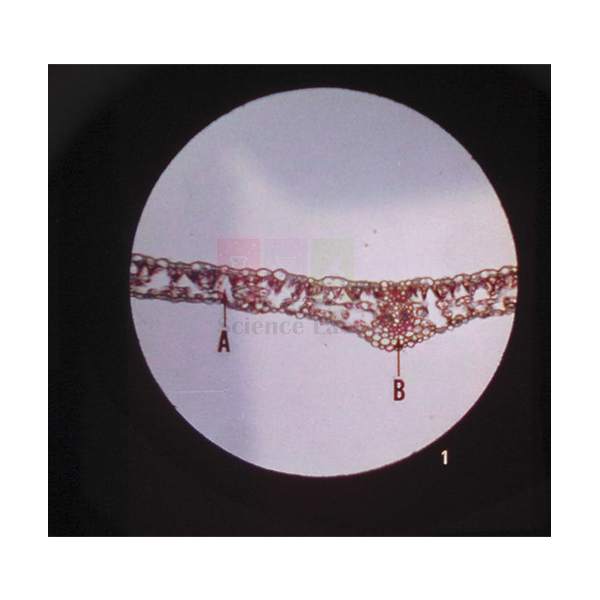
Microslide Photosynthesis
Microslide Photosynthesis (Product Code: SLE/PS/103) is a set of 8 related 35 mm images, photographed through a microscope, used to show where and how plants make and store food in school biology lessons on photosynthesis. It pairs green leaf structure at 110x and 350x with images of a leaf kept with and without sunlight, a variegated leaf, starch grains and a bean seed. The set is supplied by Jain Scientific Equipments Pvt Ltd (ScienceLab), Ambala, India.
Specifications
| Parameter | Detail |
|---|---|
| Contents | 8 related 35 mm images photographed through a microscope |
| Leaf structure | Green leaf (110x); Green leaf (350x) |
| Supply of raw materials | Root hair; Vascular bundles |
| Conditions for photosynthesis | Leaf with and without sunlight; Leaf – Variegated |
| Food storage | Starch grains; Bean seed |
| Student aids | Arrows and callouts on the images; detailed text folder with a pocket that holds the Microslides |
| Viewed in | Microslide Viewer |
| Product code | SLE/PS/103 |
| Supplied by | Jain Scientific Equipments Pvt Ltd (ScienceLab), Ambala, India |
What Is the Microslide Photosynthesis Used For?
Teachers use the set to turn photosynthesis from an equation into something students can see. The two green leaf images, at 110x and then 350x, let a class move from the overall layout of the leaf to a closer look at its cells. The root hair and vascular bundle frames show how water reaches the leaf.
The comparison of a leaf with and without sunlight and the variegated leaf image support the classic discussion of why light and chlorophyll are needed, and the starch grains and bean seed show where the products end up in storage. Students can study the callouts in a Microslide Viewer before or after running a starch test with a photosynthesis experiment kit, using the text folder questions to explain their results.
Supplier
Microslide Photosynthesis is sourced and supplied by Jain Scientific Equipments Private Limited, whose trading name is ScienceLab, from Ambala in Haryana, India. Our customers include schools, colleges and dealers in India and international markets who want slides, apparatus and kits quoted together. Read about the company on our company profile.
Related Prepared Slides
Pair the set with these photosynthesis resources:
- Microslide Viewer — for viewing the eight images
- Prepared Microscope Slide Set – Photosynthesis and Adaptations — glass slides for microscope practicals on leaves
- Photosynthesis Apparatus — demonstrates the process in a working experiment
- Kit for Photosynthesis Experiments — hands-on tests to match the images
- Microslide The Root Of a Flowering Plant — more detail on roots and root hairs
Browse more in Prepared Slides.
Frequently Asked Questions
What images are in Microslide Photosynthesis?
Microslide Photosynthesis contains green leaf at 110x, green leaf at 350x, root hair, vascular bundles, a leaf with and without sunlight, a variegated leaf, starch grains and a bean seed, all photographed through a microscope.
Why does Microslide Photosynthesis include a variegated leaf?
Microslide Photosynthesis includes a variegated leaf because its green and pale areas let students discuss the role of chlorophyll, alongside the leaf shown with and without sunlight, which relates to the need for light.
Can Microslide Photosynthesis be used without a microscope?
Yes. Microslide Photosynthesis is viewed in a Microslide Viewer rather than a microscope, because its images were already photographed through a microscope. Order viewers with the set if your school does not have them.
What written material comes with Microslide Photosynthesis?
Microslide Photosynthesis comes with a detailed text folder designed to stimulate, inform and question students, with a handy pocket where the Microslides are kept between lessons.
Who supplies Microslide Photosynthesis?
Microslide Photosynthesis is supplied by Jain Scientific Equipments Pvt Ltd (ScienceLab), Ambala, Haryana, India. The company quotes it for schools, colleges and dealers in India and international markets, together with related apparatus if needed.
What details should a Microslide Photosynthesis enquiry include?
A Microslide Photosynthesis enquiry should give code SLE/PS/103, the number of sets, whether viewers or photosynthesis apparatus are wanted, and the delivery location. Our contact page is the quickest way to send them.
Request a Quote
Request pricing for Microslide Photosynthesis via our contact page, or add it to the enquiry cart alongside other photosynthesis resources.
Last updated: 24 September 2026

Glass Cover Slips
Glass Cover Slips (Product Code: SLE/PS/104) are thin, square, pre-cleaned cover glasses used to cover specimens mounted on microscope slides in school, college and research laboratories. Each slip measures 22 x 22 mm with a thickness of 0.13–0.17 mm, and a box holds approximately 100 pieces (10 g), a convenient pack for student practicals. They are supplied by Jain Scientific Equipments Pvt Ltd (ScienceLab), Ambala, India.
Specifications
| Parameter | Detail |
|---|---|
| Type | Pre-cleaned cover glasses |
| Shape | Square |
| Dimensions | 22 x 22 mm |
| Thickness | 0.13–0.17 mm |
| Pack | Approximately 100 pieces (10 g) per box |
| Product code | SLE/PS/104 |
| Supplied by | Jain Scientific Equipments Pvt Ltd (ScienceLab), Ambala, India |
What Are Glass Cover Slips Used For?
A cover slip is placed over a drop of mountant, water or stain on a microscope slide to flatten the specimen, keep it from drying out and protect the objective lens from contact with the sample. In school biology practicals they are used for temporary mounts such as onion epidermis, cheek cells, pond water and pollen, and in colleges for stained smears and sections.
To avoid trapping air bubbles, touch one edge of the slip to the liquid and lower it slowly with a needle or forceps. Handle the slips by their edges, as the 0.13–0.17 mm glass is thin and breaks easily, and put broken pieces in a glass waste container. Their thickness range includes 0.17 mm, the cover-glass thickness many microscope objectives are designed for. As a biology lab equipment supplier in India, we can quote them together with plain slides for new practical kits.
What to Specify When Ordering
- Number of boxes (each approximately 100 pieces)
- Confirmation that the 22 x 22 mm square size suits your slides and mounts
- Whether plain microscope slides or cover-slip forceps should be added
Supplier
Jain Scientific Equipments Private Limited, which trades as ScienceLab from Ambala, Haryana, supplies Glass Cover Slips as a routine consumable to schools, colleges, laboratories and dealers in India and international markets. For vendor registration, the supplying company is registered under CIN U29309HR2020PTC087597 and GSTIN 06AAECJ8618A1ZT. More information is on our company profile.
Related Prepared Slides
Items used with cover slips when making slides:
- Microscope Glass Slides — the plain slides the cover slips sit on
- Glass Cover Slips, No. 1 — an alternative cover-slip listing to compare
- Microscope Slide Cover Forceps — for picking up and lowering thin slips
- Slide Box — storage for finished slides
Find more slides and accessories in Prepared Slides.
Frequently Asked Questions
What size are these Glass Cover Slips?
These Glass Cover Slips are square, measuring 22 x 22 mm, with a thickness between 0.13 and 0.17 mm. The size covers a typical specimen area on a standard microscope slide used in school and college practicals.
How many Glass Cover Slips are in a box?
Each box of Glass Cover Slips (SLE/PS/104) holds approximately 100 pieces, about 10 g of glass. Order the number of boxes you need for the term, since slips are used once for temporary mounts and break easily.
Do the Glass Cover Slips need cleaning before use?
The Glass Cover Slips are supplied pre-cleaned. Keep the box closed and handle slips by their edges so that fingerprints and dust do not spoil the view; if a slip is marked, wipe it gently with lens tissue.
Can Glass Cover Slips be used with high-power objectives?
Glass Cover Slips at 0.13–0.17 mm are thin enough for the high-power objectives on school and college compound microscopes, and 0.17 mm is the thickness many objectives are designed around. Check your microscope manual if you use specialised objectives.
Who supplies these Glass Cover Slips?
These Glass Cover Slips are supplied by Jain Scientific Equipments Pvt Ltd (ScienceLab), Ambala, Haryana, India, to schools, colleges, laboratories and dealers. CIN and GSTIN details are available for vendor registration.
What should I include in a Glass Cover Slips enquiry?
For Glass Cover Slips, give code SLE/PS/104, the number of boxes, any slides or forceps you want quoted together, and the delivery city or country. Bulk and export quantities can be requested through our contact page.
Request a Quote
Order Glass Cover Slips by sending your quantity through our contact page, or add them to the enquiry cart with slides and other consumables.
Last updated: 24 September 2026

Prepared Microscope Slide Set – Health And Disease
Prepared Microscope Slide Set – Health And Disease (Product Code: SLE/PS/105) is a set of prepared glass microscope slides used to compare healthy and diseased human-body tissue in biology, health science and introductory pathology classes. It covers eight preparations with 2 slides of each, from lymph node and spleen to emphysema, thrombus, lung of smoking and atherosclerosis of the main and coronary arteries. It is supplied by Jain Scientific Equipments Pvt Ltd (ScienceLab), Ambala, India.
Specifications
| Parameter | Detail |
|---|---|
| Lymph node section | 2 slides |
| Spleen section | 2 slides |
| Spleen TS | 2 slides |
| Emphysema | 2 slides |
| Lung of smoking | 2 slides |
| Thrombus | 2 slides |
| Main artery atherosclerosis | 2 slides |
| Coronary artery atherosclerosis | 2 slides |
| Product code | SLE/PS/105 |
| Supplied by | Jain Scientific Equipments Pvt Ltd (ScienceLab), Ambala, India |
What Is the Prepared Microscope Slide Set – Health And Disease Used For?
The set brings disease topics from the textbook to the microscope. Lymph node and spleen slides show the organs where immune cells gather, giving students a reference for normal lymphoid tissue. The lung slides let them compare emphysema and the lung of a smoker, a strong visual link to lessons on smoking and gas exchange.
The cardiovascular slides cover a thrombus and atherosclerosis in both a main artery and a coronary artery, so a class can see how deposits narrow the vessel and discuss heart disease risk factors. With two copies of each preparation, one slide can stay on the demonstration microscope while the second passes round a small group. These prepared microscope slides also suit revision sessions before practical examinations.
What to Specify When Ordering
- Number of sets, bearing in mind each holds 2 slides per preparation
- Whether normal-tissue sets (such as circulatory or histology slides) should be quoted for comparison
- Storage and microscopes, if the lab needs them
Supplier
This slide set is supplied by Jain Scientific Equipments Private Limited, the company behind the ScienceLab name in Ambala, Haryana, for schools, colleges, training institutes and dealers in India and international markets. Institutions completing vendor forms can use the company's GSTIN 06AAECJ8618A1ZT and its Legal Entity Identifier 3358004HRD1JIVXKYX49. Visit our profile page to learn more.
Related Prepared Slides
Good companions for a health and disease unit:
- Prepared Microscope Slide Set – Circulatory System — healthy artery, vein and cardiac muscle for comparison with the diseased vessels
- Microslide Smoking & Health — annotated images of smoke-damaged airways and lungs
- Human Histology Slide Set of 25 — a wider set of normal human tissues
- Microscope Slide Case — protects the slides between classes
- Binocular Microscope, 1000x Mag LED — a microscope for detailed tissue work
More sets are listed under Prepared Slides.
Frequently Asked Questions
What slides are in the Prepared Microscope Slide Set – Health And Disease?
The Prepared Microscope Slide Set – Health And Disease contains lymph node section, spleen section, spleen TS, emphysema, thrombus, lung of smoking, main artery atherosclerosis and coronary artery atherosclerosis, with 2 slides of each preparation.
Why does the Health And Disease slide set include two of each slide?
The Prepared Microscope Slide Set – Health And Disease lists 2 slides per preparation, which lets a teacher keep one on a demonstration microscope while students examine the other, or keep a spare if a slide is damaged.
Is the Health And Disease slide set meant for diagnosis?
No. The Prepared Microscope Slide Set – Health And Disease is a teaching resource for showing students what diseased tissue looks like under a microscope. It is not intended for clinical or diagnostic use.
What microscope suits the Health And Disease slide set?
The Prepared Microscope Slide Set – Health And Disease can be examined with a standard compound microscope; higher magnification helps with cell detail in the lymph node, spleen and artery wall. We can quote microscopes on the same enquiry.
Who supplies the Prepared Microscope Slide Set – Health And Disease?
The Prepared Microscope Slide Set – Health And Disease is supplied by Jain Scientific Equipments Pvt Ltd (ScienceLab), Ambala, Haryana, India, which provides GSTIN and LEI details to institutions and importers on request.
What should an enquiry for the Health And Disease slide set include?
For the Prepared Microscope Slide Set – Health And Disease, send product code SLE/PS/105, the number of sets, any companion slide sets or microscopes, and the delivery location through our contact page.
Request a Quote
Request a quotation for the Prepared Microscope Slide Set – Health And Disease through our contact page, or add it to the enquiry cart with any other slide sets you are ordering.
Last updated: 24 September 2026

Prepared Microscope Slide Set – Exchange Surfaces and Epithelia
Prepared Microscope Slide Set – Exchange Surfaces and Epithelia (Product Code: SLE/PS/106) is a class set of prepared glass microscope slides used to study epithelial linings and the supporting tissues of gas exchange surfaces in biology practicals. It provides 10 slides each of ciliated epithelium, squamous epithelium, loose connective tissue, lung cartilage and hyaline cartilage, enough for groups to work on the same tissue at once. It is supplied by Jain Scientific Equipments Pvt Ltd (ScienceLab), Ambala, India.
Specifications
| Parameter | Detail |
|---|---|
| Ciliated epithelium | 10 slides |
| Squamous epithelium | 10 slides |
| Loose connective tissue | 10 slides |
| Lung cartilage | 10 slides |
| Hyaline cartilage | 10 slides |
| Slide type | Prepared microscope slides |
| Product code | SLE/PS/106 |
| Supplied by | Jain Scientific Equipments Pvt Ltd (ScienceLab), Ambala, India |
What Is the Prepared Microscope Slide Set – Exchange Surfaces and Epithelia Used For?
Exchange surfaces depend on thin linings and firm support, and this set lets students examine both. Squamous epithelium shows the flattened cells that keep diffusion distances short, while ciliated epithelium shows the hair-like cilia that sweep mucus along the airways. Loose connective tissue shows the packing layer that sits beneath such linings.
The lung cartilage and hyaline cartilage slides explain how the airways are held open, so students can relate structure to function when they draw and label the tissues. Because the set holds 10 of every preparation, a teacher can run one tissue per lesson with the whole class, or rotate the five preparations round stations. It is a practical microscope slide set for school and junior college biology labs preparing students for practical examinations.
What to Specify When Ordering
- Number of sets, based on 10 slides per preparation
- Whether linked sets such as Circulatory System or Connective Tissues should be quoted as well
- Microscopes or slide storage needed for the practical room
Supplier
ScienceLab, the trading name of Jain Scientific Equipments Private Limited in Ambala, Haryana, supplies this class set of epithelium and cartilage slides to schools, colleges and education distributors in India and international markets. We can combine it with other topic sets so a department receives one consolidated quotation. For company details, see our company profile.
Related Prepared Slides
Sets and equipment often ordered alongside:
- Prepared Microscope Slide Set – Connective Tissues — bone, adipose and areolar tissue to extend the tissue topic
- Prepared Microscope Slide Set – Circulatory System — the blood side of gas exchange
- Prepared Microscope Slide Set – Health And Disease — shows emphysema, where the exchange surface is damaged
- Monocular Biological Microscope 400x, LED — a student microscope for the practical
View the complete Prepared Slides range.
Frequently Asked Questions
What tissues are in the Exchange Surfaces and Epithelia slide set?
The Prepared Microscope Slide Set – Exchange Surfaces and Epithelia contains ciliated epithelium, squamous epithelium, loose connective tissue, lung cartilage and hyaline cartilage, with 10 slides of each preparation.
Is the Exchange Surfaces and Epithelia slide set enough for a whole class?
The Prepared Microscope Slide Set – Exchange Surfaces and Epithelia holds 10 slides of each tissue, so ten students or pairs can examine the same preparation together. Larger classes can order additional sets or rotate the slides between groups.
Why does the Exchange Surfaces and Epithelia set include cartilage?
The Prepared Microscope Slide Set – Exchange Surfaces and Epithelia includes lung and hyaline cartilage because cartilage supports the airways that carry air to the gas exchange surface, letting students connect lining tissues with the structures that keep air passages open.
What magnification is needed for the Exchange Surfaces and Epithelia slides?
The Prepared Microscope Slide Set – Exchange Surfaces and Epithelia is examined with a standard compound microscope; low power finds the tissue, and high power shows cell shape and cilia. We can quote suitable microscopes with the set.
Who supplies the Prepared Microscope Slide Set – Exchange Surfaces and Epithelia?
The Prepared Microscope Slide Set – Exchange Surfaces and Epithelia is supplied by Jain Scientific Equipments Pvt Ltd (ScienceLab), Ambala, Haryana, India, to schools, colleges and dealers in India and international markets.
Request a Quote
For a quotation on the Prepared Microscope Slide Set – Exchange Surfaces and Epithelia, contact us through our contact page or add it to the enquiry cart with the other sets your department needs.
Last updated: 24 September 2026

Prepared Microscope Slide Set – Circulatory System
Prepared Microscope Slide Set – Circulatory System (Product Code: SLE/PS/107) is a set of prepared glass microscope slides used for hands-on microscope practicals on blood, blood vessels, heart muscle and lymphatics. It contains 10 slides each of artery and vein, cardiac muscle, cardiac muscle (LS), human blood thin film and lymphatic (TS), so a full class can examine one preparation together. It is supplied by Jain Scientific Equipments Pvt Ltd (ScienceLab), Ambala, India.
Specifications
| Parameter | Detail |
|---|---|
| Artery and vein | 10 slides |
| Cardiac muscle | 10 slides |
| Cardiac muscle (LS) | 10 slides |
| Human blood thin film | 10 slides |
| Lymphatic (TS) | 10 slides |
| Product code | SLE/PS/107 |
| Supplied by | Jain Scientific Equipments Pvt Ltd (ScienceLab), Ambala, India |
What Is the Prepared Microscope Slide Set – Circulatory System Used For?
This set is designed for students to find structures themselves under the microscope. The artery and vein slide is the standard exercise for comparing wall thickness and lumen shape, and it usually leads to a labelled drawing. The blood thin film lets students tell red cells from the fewer white cells and practise counting fields of view.
Two cardiac muscle preparations, one of them a longitudinal section, give a class the chance to look for the striped, branching fibres of heart muscle, while the lymphatic transverse section completes the picture of how tissue fluid returns to the blood. With 10 copies of each slide, the preparations can rotate round the lab in one or two periods, which suits school biology lab equipment used for board practical preparation.
Supplier
The Circulatory System slide set comes from Jain Scientific Equipments Private Limited, trading as ScienceLab, of Ambala, Haryana. We supply prepared slide sets like this to schools, colleges and dealers across India and in international markets, and we can list them on the same quotation as microscopes and heart models. The company profile gives more background.
Related Prepared Slides
Resources that support a circulation practical:
- Microslide The Circulatory System — annotated images to preview before the practical
- Prepared Microscope Slide Set – Health And Disease — atherosclerosis and thrombus slides for comparison with healthy vessels
- Human Circulatory System Model — shows the route blood takes around the body
- Binocular Microscope, 1000x Mag LED — for close study of the blood film
- Immersion Oil, 25ml — for oil-immersion viewing of blood cells
Our Prepared Slides category lists the other sets.
Frequently Asked Questions
What does the Prepared Microscope Slide Set – Circulatory System contain?
The Prepared Microscope Slide Set – Circulatory System contains 10 slides each of five preparations: artery and vein, cardiac muscle, cardiac muscle (LS), human blood thin film, and lymphatic (TS).
How do the two cardiac muscle slides in the Circulatory System set differ?
In the Prepared Microscope Slide Set – Circulatory System, one cardiac muscle preparation is listed as LS, a longitudinal section, and the other simply as cardiac muscle. Ask us through the contact page if you need the orientation of the second one confirmed.
What magnification suits the blood film in the Circulatory System slide set?
The human blood thin film in the Prepared Microscope Slide Set – Circulatory System can be scanned at low and high power; many teachers use an oil-immersion objective with immersion oil to show white cell nuclei in detail.
How is the Circulatory System slide set different from the Microslide on the same topic?
The Prepared Microscope Slide Set – Circulatory System is made of glass slides for students to examine under a microscope, whereas Microslide The Circulatory System provides annotated photographic images that are looked at in a Microslide Viewer.
Who supplies the Prepared Microscope Slide Set – Circulatory System?
The Prepared Microscope Slide Set – Circulatory System is supplied by Jain Scientific Equipments Pvt Ltd (ScienceLab) in Ambala, Haryana, India, to schools, colleges and dealers in India and international markets.
What is needed for a quotation on the Circulatory System slide set?
For the Prepared Microscope Slide Set – Circulatory System, send code SLE/PS/107, the number of sets, any microscopes or immersion oil you want added, and the delivery location through our contact page.
Request a Quote
Get a quotation for the Prepared Microscope Slide Set – Circulatory System via our contact page, or add it to the enquiry cart together with microscopes and other slide sets.
Last updated: 24 September 2026

Prepared Microscope Slide Set – Transport In Plants
Prepared Microscope Slide Set – Transport In Plants (Product Code: SLE/PS/108) is a set of prepared glass microscope slides used to trace the path of water and food through roots, stems and leaves in plant biology practicals. It includes 5 slides each of eight preparations, from Vicia root hairs and sunflower roots to Helianthus, Zea and Tilia stems and the rolled leaf of marram grass. The set is supplied by Jain Scientific Equipments Pvt Ltd (ScienceLab), Ambala, India.
Specifications
| Parameter | Detail |
|---|---|
| Vicia (root apex) | 5 slides |
| Vicia young root hairs | 5 slides |
| Helianthus (sunflower), young and old root TS | 5 slides |
| Helianthus, stem, bundles TS | 5 slides |
| Helianthus, old stem, TS and LS | 5 slides |
| Zea stem TS and LS | 5 slides |
| Tilia old stem (RLS) | 5 slides |
| Ammophila (marram grass), leaf TS | 5 slides |
| Product code | SLE/PS/108 |
| Supplied by | Jain Scientific Equipments Pvt Ltd (ScienceLab), Ambala, India |
TS = transverse section; LS = longitudinal section; RLS = radial longitudinal section.
What Is the Prepared Microscope Slide Set – Transport In Plants Used For?
The slides follow water upward through the plant. Students begin at the Vicia root apex and young root hairs, where water enters, then examine young and old sunflower roots to locate the central xylem and phloem. Stem sections of Helianthus and Zea compare the ring of vascular bundles in a dicot with the scattered bundles of a monocot, and the TS and LS pairs show the same tissues cut two ways.
The old Helianthus and Tilia stems show how vascular tissue builds up as a stem ages, and the marram grass leaf, adapted to dry sand dunes, links transport to water loss and transpiration. Five copies of each slide allow small groups to share preparations, and teachers often run the set alongside a transpiration experiment so observation and measurement come together. It is a focused addition to any plant biology lab equipment list.
What to Specify When Ordering
- Number of sets for your class size (5 slides of each preparation per set)
- Whether single-copy reference sets, such as the Basic Plant Anatomy Slide Set, are also needed
- Any transpiration or root-pressure apparatus to include in the same quotation
Supplier
Jain Scientific Equipments Private Limited supplies the Transport In Plants slide set through its ScienceLab business in Ambala, Haryana. Schools, colleges and dealers in India and international markets order prepared slides from us together with botany apparatus, which keeps a department's purchasing on one quotation. Learn about the company on our profile page.
Related Prepared Slides
Items that complement a plant transport practical:
- Basic Plant Anatomy Slide Set — one-of-each reference slides of the same plants
- Kit for Demonstration of Transpiration in Plants — measures the water loss the leaf slides explain
- Kit for Demonstrating Root Pressure — shows water being pushed up from the roots
- Microslide The Root Of a Flowering Plant — annotated images of root zones and root hairs
- Monocular Biological Microscope 400x — a microscope for student groups
See more in Prepared Slides.
Frequently Asked Questions
Which slides are in the Prepared Microscope Slide Set – Transport In Plants?
The Prepared Microscope Slide Set – Transport In Plants has 5 slides each of Vicia root apex, Vicia young root hairs, sunflower young and old root TS, Helianthus stem bundles TS, Helianthus old stem TS and LS, Zea stem TS and LS, Tilia old stem RLS and marram grass leaf TS.
Does the Transport In Plants slide set show both monocot and dicot stems?
Yes. The Prepared Microscope Slide Set – Transport In Plants includes Zea (maize), a monocot, and Helianthus (sunflower) and Tilia (lime), both dicots, so students can compare how vascular bundles are arranged in each group.
Why is marram grass included in the Transport In Plants slide set?
The Prepared Microscope Slide Set – Transport In Plants includes an Ammophila (marram grass) leaf TS because this dune grass is a standard example of a leaf adapted to reduce water loss, connecting transport with transpiration.
How many students can use one Transport In Plants slide set?
Each Prepared Microscope Slide Set – Transport In Plants carries 5 slides of every preparation, so five students or groups can examine the same slide type at once. Order more sets for larger classes working simultaneously.
Who supplies the Prepared Microscope Slide Set – Transport In Plants?
The Prepared Microscope Slide Set – Transport In Plants is supplied by Jain Scientific Equipments Pvt Ltd (ScienceLab), Ambala, Haryana, India, for schools, colleges and dealers in India and international markets.
Request a Quote
Send an enquiry for the Prepared Microscope Slide Set – Transport In Plants on our contact page, or add it to the enquiry cart with any apparatus for the same practical.
Last updated: 24 September 2026

Prepared Microscope Slide Set – Connective Tissues
Prepared Microscope Slide Set – Connective Tissues (Product Code: SLE/PS/109) is a set of prepared glass microscope slides used to identify connective tissue, the three muscle types and related tissues in animal histology practicals. It supplies 5 slides each of eight preparations, including compact bone for Haversian canals and lamellae, adipose and areolar tissue, cardiac, striated and non-striated muscle, ciliated epithelium and spinal cord. It is supplied by Jain Scientific Equipments Pvt Ltd (ScienceLab), Ambala, India.
Specifications
| Parameter | Detail |
|---|---|
| Compact bone, for Haversian canals and lamellae | 5 slides |
| Adipose tissue, section | 5 slides |
| Areolar tissue, stained for fibres and cells E | 5 slides |
| Cardiac muscle showing the nuclei, striations and intercalated discs LS | 5 slides |
| Striated muscle, showing striations and nuclei LS | 5 slides |
| Non-striated (involuntary) muscle E | 5 slides |
| Pseudo-stratified ciliated epithelium TS | 5 slides |
| Spinal cord TS | 5 slides |
| Product code | SLE/PS/109 |
| Supplied by | Jain Scientific Equipments Pvt Ltd (ScienceLab), Ambala, India |
E = entire (whole mount); LS = longitudinal section; TS = transverse section.
What Is the Prepared Microscope Slide Set – Connective Tissues Used For?
The set supports the classic tissue identification practical. Students see how compact bone is organised into Haversian systems with rings of lamellae, how adipose tissue is packed with fat cells, and how areolar tissue, stained for fibres and cells, holds other tissues together. These three slides show why connective tissues look so different despite sharing a common role.
The muscle slides let a class compare the three types side by side: striated muscle with its striations and nuclei, cardiac muscle with intercalated discs, and non-striated (involuntary) muscle. Pseudo-stratified ciliated epithelium and a spinal cord section round out a survey of the main animal tissue types. With 5 slides of each, small groups can move through the preparations and record labelled drawings for their practical files.
Supplier
Jain Scientific Equipments Private Limited, which trades as ScienceLab in Ambala, Haryana, supplies the Connective Tissues slide set to schools, colleges, teaching laboratories and dealers in India and international markets. Buyers registering a new vendor can note the supplying company's Corporate Identification Number U29309HR2020PTC087597 and GSTIN 06AAECJ8618A1ZT. More on the company is on our profile page.
Related Prepared Slides
Other sets and tools used in histology practicals:
- Prepared Microscope Slide Set – Exchange Surfaces and Epithelia — more epithelium and cartilage slides
- Prepared Microscope Slide Set – Circulatory System — extra cardiac muscle and blood vessel slides
- Human Histology Slide Set of 25 — a broader single-copy reference set
- Binocular Biological Microscope — for examining tissue detail
Browse all Prepared Slides.
Frequently Asked Questions
What preparations are in the Prepared Microscope Slide Set – Connective Tissues?
The Prepared Microscope Slide Set – Connective Tissues contains compact bone, adipose tissue, areolar tissue, cardiac muscle LS, striated muscle LS, non-striated muscle, pseudo-stratified ciliated epithelium TS and spinal cord TS, with 5 slides of each.
Does the Connective Tissues slide set include muscle slides?
Yes. Besides bone, adipose and areolar tissue, the Prepared Microscope Slide Set – Connective Tissues includes cardiac, striated and non-striated muscle, plus ciliated epithelium and spinal cord, so all main animal tissue types can be compared.
What features does the compact bone slide in the Connective Tissues set show?
The compact bone slide in the Prepared Microscope Slide Set – Connective Tissues is prepared to show Haversian canals and lamellae, the concentric rings that make up the structural units of compact bone.
How many learners can share one Connective Tissues slide set?
One Prepared Microscope Slide Set – Connective Tissues provides 5 slides of each preparation, enough for five groups to view the same tissue together. Additional sets can be quoted for larger practical classes.
Who supplies the Prepared Microscope Slide Set – Connective Tissues?
The Prepared Microscope Slide Set – Connective Tissues is supplied by Jain Scientific Equipments Pvt Ltd (ScienceLab), Ambala, Haryana, India. CIN and GSTIN details are available to institutions that need them for vendor registration.
What should I send to get a quotation for the Connective Tissues slide set?
For the Prepared Microscope Slide Set – Connective Tissues, send code SLE/PS/109, the number of sets, any microscopes or companion slide sets, and your delivery city or country through our contact page.
Request a Quote
Request a quote for the Prepared Microscope Slide Set – Connective Tissues through our contact page, or add it to the enquiry cart along with other histology sets.
Last updated: 24 September 2026

Prepared Microscope Slide Set – Skin and Excretion
Prepared Microscope Slide Set – Skin and Excretion (Product Code: SLE/PS/110) is a group of ready-mounted microscope slides used to study skin structure and the organs of excretion in school and college biology practicals. It brings together two skin sections, a bladder-wall section and two kidney sections, so a class can follow how the body removes water, salts and wastes. These prepared microscope slides are supplied by Jain Scientific Equipments Pvt Ltd (ScienceLab), Ambala, India.
Key Details
| Parameter | Detail |
|---|---|
| Slide 1 | Skin, showing hair insertion and sebaceous glands, VS |
| Slide 2 | Skin, sweat glands, VS |
| Slide 3 | Transitional epithelium, bladder wall, TS |
| Slide 4 | Kidney, injected, thick section |
| Slide 5 | Kidney, section |
| Figure listed against each preparation | 10 (the listing does not say what this counts; the number of slides per preparation is confirmed in your quotation) |
| Label abbreviations | VS = vertical section; TS = transverse section |
| Product code | SLE/PS/110 |
| Supplied by | Jain Scientific Equipments Pvt Ltd (ScienceLab), Ambala, India |
What Is the Skin and Excretion Slide Set Used For?
Teachers use this set for practical lessons on excretion and temperature control. Students locate the hair follicle and its sebaceous gland on the first skin slide, then compare it with the coiled sweat glands on the second, which leads naturally into how sweating removes water and salts. The two kidney slides let the class pick out cortex and medulla, and the injected preparation, in which blood vessels are filled with a coloured medium, makes the kidney's rich blood supply easier to trace.
The bladder-wall slide shows transitional epithelium, the stretchable lining that allows the bladder to fill and empty. Begin every slide at low power to find the region of interest before moving to a higher objective. As part of school biology lab equipment, the set pairs well with a kidney model so students connect what they see under the lens with whole-organ anatomy.
What to Specify When Ordering
- The number of sets required, for example one per practical group or one for the teacher's demonstration.
- Confirmation of how many slides of each preparation you expect (the listing shows the figure 10 against each).
- Any microscopes, slide boxes or organ models to be quoted in the same enquiry.
Supplier
ScienceLab is the trading name of Jain Scientific Equipments Private Limited, based in Ambala, Haryana. The company supplies prepared slide sets like this one to schools, colleges, dealers and importers, and can combine them with microscopes and anatomy models so a department receives one consolidated quotation. More about the company is on the ScienceLab company profile.
Related Prepared Slides
These items are often ordered with the skin and excretion slides for the same practical sessions:
- Prepared Microscope Slide Set – Exchange Surfaces and Epithelia — other epithelium types to compare with the bladder lining.
- Human Kidney, Nephron, and Glomerulus Model Set — whole-organ and nephron view to match the kidney sections.
- Human Skin Section Model — enlarged 3D view of the glands seen on the skin slides.
- Monocular Biological Microscope 400x, LED — a classroom microscope for viewing the set.
- Slide Box — keeps the slides flat and protected between lessons.
See the full range in Prepared Slides.
Frequently Asked Questions
What slides are in the Prepared Microscope Slide Set – Skin and Excretion?
The Prepared Microscope Slide Set – Skin and Excretion lists five preparations: skin showing hair insertion and sebaceous glands (VS), skin sweat glands (VS), transitional epithelium of the bladder wall (TS), an injected thick kidney section and a plain kidney section.
How many slides of each preparation are supplied?
Our listing for the Skin and Excretion slide set shows the figure 10 beside each preparation but does not state what it counts. We therefore confirm the exact number of slides per preparation in your quotation; please ask through the contact page before you order.
What do VS and TS mean on the slide labels?
On the Skin and Excretion slide labels, VS means vertical section, cut at right angles to the skin surface, and TS means transverse section, cut straight across the organ. Knowing the plane of the cut helps students interpret the layers and glands they see.
What microscope is needed to view these slides?
A standard compound microscope with low- and high-power objectives is suitable for the Skin and Excretion prepared slides. Start at low power to find the gland or kidney region, then change to a higher objective; a school microscope can be quoted in the same enquiry.
How should the skin and excretion slides be stored?
Store the Skin and Excretion prepared slides flat in a slide box or tray, away from heat and direct sunlight, and hold them by the edges. Wipe fingerprints off gently with lens tissue and never let the objective lens press on the coverslip.
Who supplies the Skin and Excretion slide set?
Jain Scientific Equipments Pvt Ltd (ScienceLab), Ambala, India supplies the Prepared Microscope Slide Set – Skin and Excretion to schools, colleges, dealers and importers, handling quotations and orders for customers in India and international markets.
What information does a quotation request need?
To quote the Prepared Microscope Slide Set – Skin and Excretion, send product code SLE/PS/110, the number of sets, your delivery location and any microscopes or storage boxes you want added. The contact page and the enquiry cart both reach our sales team.
Request a Quote
Send your requirement for this slide set through the ScienceLab contact page, or add it to the enquiry cart with other prepared slides and microscopes for one combined quotation.
Last updated: 24 September 2026

Prepared Microscope Slide Set – Photosynthesis and Adaptations
Prepared Microscope Slide Set – Photosynthesis and Adaptations (Product Code: SLE/PS/111) is a collection of eight prepared plant slides used to show how leaves, stomata, pollen and root hairs are built for their jobs in secondary and college botany classes. Its mix of ordinary leaf sections with marram grass and fig preparations lets students compare typical leaves with plants adapted to harsher conditions. The set is supplied by Jain Scientific Equipments Pvt Ltd (ScienceLab), Ambala, India.
Key Details
| Parameter | Detail |
|---|---|
| Leaf sections | Dicot leaf, TS; Ligustrum leaf, TS; Prunus (cherry) leaf, TS |
| Stomata preparations | Monocot epidermis, for stomata, E; Ficus (fig), sunken stomata |
| Adaptation preparation | Ammophila (marram grass) |
| Reproduction and roots | Pine, mature pollen grain; Vicia, young root hairs |
| Preparations listed | 8 |
| Figure listed against each preparation | 5 (meaning not stated in the listing; slides per preparation confirmed at quotation) |
| Label abbreviations | TS = transverse section; E = entire (whole mount) |
| Product code | SLE/PS/111 |
| Supplied by | Jain Scientific Equipments Pvt Ltd (ScienceLab), Ambala, India |
What Is the Photosynthesis and Adaptations Slide Set Used For?
The three leaf sections are the core of a photosynthesis lesson: students identify the upper epidermis, the tightly packed palisade layer, the looser spongy tissue and the veins, then explain how each helps capture light and exchange gases. The monocot epidermis preparation shows stomata and guard cells from above, so learners can count pores or sketch their shape.
The remaining slides move the lesson towards adaptation. Marram grass is a sand-dune plant, and the fig preparation shows sunken stomata, both useful for discussing how plants limit water loss. Pine pollen introduces wind pollination, while the Vicia root-hair slide shows how roots increase their surface for absorbing water. Used with a photosynthesis experiment kit, the set links structure seen under the microscope with results measured at the bench, making it a practical piece of botany lab equipment.
What to Specify When Ordering
- How many sets you need for your practical groups.
- The number of slides of each preparation you expect, since the listing shows the figure 5 without explaining it.
- Whether photosynthesis apparatus or a microscope should be quoted alongside.
Supplier
Jain Scientific Equipments Private Limited, known to customers as ScienceLab, supplies this botany slide set from Ambala, Haryana. Botany departments, schools and resellers can place it on the same order as practical apparatus and other slide collections, which keeps paperwork and delivery simple. Company background is available on the ScienceLab profile page.
Related Prepared Slides
Items that support the same plant-biology practicals:
- Photosynthesis Apparatus — bench experiment to pair with the leaf sections.
- Prepared Microscope Slide Set – Transport In Plants — continues the topic into xylem and phloem.
- Microslide Photosynthesis — a viewer-based lesson on the same theme.
- Basic Plant Anatomy Slide Set — stems and roots to complete plant structure.
- Binocular Microscope 400x, LED Illumination — comfortable two-eye viewing of leaf tissues.
Browse all slide sets under Prepared Slides.
Frequently Asked Questions
Which plant preparations are in the Photosynthesis and Adaptations set?
The Prepared Microscope Slide Set – Photosynthesis and Adaptations lists eight preparations: dicot leaf TS, monocot epidermis for stomata, Ligustrum leaf TS, marram grass, mature pine pollen grain, fig sunken stomata, cherry leaf TS and Vicia young root hairs.
How many slides of each preparation does the set contain?
The Photosynthesis and Adaptations slide set listing shows the figure 5 beside each preparation without saying what it counts. We confirm the number of slides per preparation in writing with your quotation, so please raise this through the contact page when you enquire.
What does the letter E mean on some slide labels?
In the Photosynthesis and Adaptations set, E marks an entire or whole-mount preparation, such as the monocot epidermis peeled and mounted flat, whereas TS marks a thin transverse section cut across a leaf. Whole mounts show surface features like stomata in plan view.
Which slides illustrate adaptation to dry conditions?
In the Photosynthesis and Adaptations slide set, the marram grass and fig sunken-stomata preparations are the ones teachers usually use for water-saving adaptations. Comparing them with the ordinary dicot, privet and cherry leaf sections helps students explain why stomata position and leaf form differ.
Can these slides support a photosynthesis practical?
Yes, the Photosynthesis and Adaptations slide set supports the theory side of a photosynthesis practical by showing leaf tissues and stomata. It does not replace bench apparatus, so many schools order it together with photosynthesis apparatus to link observation with measured results.
Who supplies this slide set?
Jain Scientific Equipments Pvt Ltd (ScienceLab) of Ambala, India supplies the Prepared Microscope Slide Set – Photosynthesis and Adaptations to schools, colleges, laboratories and trade buyers in India and international markets, with quotations issued on request.
Request a Quote
For a quotation on code SLE/PS/111, write to us via the contact page or place the set in the enquiry cart with any apparatus or microscopes you need.
Last updated: 24 September 2026

Prepared Microscope Slide Set – Digestion
Prepared Microscope Slide Set – Digestion (Product Code: SLE/PS/112) is a set of prepared gut-tissue slides used to trace the alimentary canal from stomach to colon in biology and physiology practicals. Six preparations cover two regions of the stomach wall, two duodenum sections, the ileum with villi and goblet cells, and the colon, so learners can see how the gut lining changes along its length. It is supplied by Jain Scientific Equipments Pvt Ltd (ScienceLab), Ambala, India.
Key Details
| Parameter | Detail |
|---|---|
| Stomach | Stomach wall, fundic, VS; Stomach wall, pyloric region, VS |
| Small intestine | Duodenum, section; Duodenum showing Brunner's glands; Ileum showing villi and goblet cells, TS |
| Large intestine | Colon, section |
| Preparations listed | 6 |
| Figure listed against each preparation | 10 (not explained in the listing; slide count per preparation confirmed with your quote) |
| Label abbreviations | VS = vertical section; TS = transverse section |
| Product code | SLE/PS/112 |
| Supplied by | Jain Scientific Equipments Pvt Ltd (ScienceLab), Ambala, India |
What Is the Digestion Slide Set Used For?
The set is designed around a simple question students can answer at the microscope: how does the lining of the gut change as its job changes? The fundic stomach section shows deep gastric glands, while the pyloric section lets the class compare the region that opens into the small intestine. In the duodenum, Brunner's glands in the wall are a distinctive landmark, and the ileum slide displays finger-like villi with mucus-secreting goblet cells, the classic example of a surface built for absorption.
Finishing with the colon, where villi are absent, gives a clear contrast for written answers and drawings. Teachers often give each group one region, then rotate the slides so everyone sketches the full sequence. As a microscope slides set for school and college histology, it works well beside a digestive system model that shows where each tissue sits in the body.
What to Specify When Ordering
- Number of sets, based on how many groups will rotate through the six preparations.
- The slide count per preparation you expect, as the figure 10 in the listing is not explained.
- Whether you also want the companion liver, pancreas and tongue slide set.
Supplier
This histology set comes from Jain Scientific Equipments Private Limited, which trades as ScienceLab in Ambala, Haryana. The company supplies prepared slides to teaching laboratories, colleges and distributors, and can quote them together with organ models and microscopes. You can read about the business on our company profile.
Related Prepared Slides
Useful companions for a digestive-system practical:
- Prepared Microscope Slide Set – Digestion (Liver, Pancreas And Tongue) — adds the accessory organs, tongue and oesophagus.
- Human Digestive System Model — shows where each sectioned region lies.
- Human Stomach Model — whole-organ view of the fundus and pylorus.
- Microslide The Digestive System — viewer-based lesson on the same topic.
- Monocular Microscope 1000x, LED light — higher magnification for goblet cells.
More sets are listed in Prepared Slides.
Frequently Asked Questions
Which parts of the gut does the Digestion slide set cover?
The Prepared Microscope Slide Set – Digestion covers the stomach wall (fundic and pyloric regions, both VS), the duodenum (a general section and one showing Brunner's glands), the ileum with villi and goblet cells (TS) and a section of the colon.
How many slides of each preparation are included?
The Digestion slide set listing shows the figure 10 next to each preparation, but it does not state what that figure counts. We confirm the number of slides per preparation in your quotation, so please mention your expected quantity when you contact us.
Does the Digestion set include liver or pancreas slides?
No, the Prepared Microscope Slide Set – Digestion covers the stomach, duodenum, ileum and colon only. Liver, pancreas, islets of Langerhans, tongue and oesophagus are in the separate Digestion (Liver, Pancreas And Tongue) set, which can be quoted together with this one.
Which magnification suits villi and goblet cells?
On the Digestion slide set, villi are easy to find at low power, while goblet cells in the ileum lining are clearer at a higher objective. A compound microscope with several objectives lets students move from the overall wall layers to individual cells.
Who supplies the Prepared Microscope Slide Set – Digestion?
Jain Scientific Equipments Pvt Ltd (ScienceLab), Ambala, India supplies the Prepared Microscope Slide Set – Digestion to schools, colleges and trade customers, and quotes it for delivery within India and to international markets.
What should I include in a quotation request?
For the Prepared Microscope Slide Set – Digestion, include code SLE/PS/112, the number of sets, your delivery address and any companion slide sets, models or microscopes. Send the details through the contact page or add the items to the enquiry cart.
Request a Quote
Ask for a quotation via the ScienceLab contact page, or collect this set and its companion items in the enquiry cart and submit them together.
Last updated: 24 September 2026
334.jpg)
Prepared Microscope Slide Set – Digestion (Liver, Pancreas And Tongue)
Prepared Microscope Slide Set – Digestion (Liver, Pancreas And Tongue) (Product Code: SLE/PS/113) is a set of prepared tissue slides used to examine the accessory digestive organs, the tongue and the oesophagus in biology and anatomy practicals. Its highlight is a transverse section of pancreas showing the islets of Langerhans, alongside liver, tongue taste-bud and oesophagus preparations. The set is supplied by Jain Scientific Equipments Pvt Ltd (ScienceLab), Ambala, India, for biology labs in India and international markets.
Key Details
| Parameter | Detail |
|---|---|
| Liver | Liver, section |
| Pancreas | Pancreas, section; Pancreas showing islets of Langerhans, TS |
| Mouth and gullet | Tongue showing taste buds, VS; Oesophagus, section |
| Preparations listed | 5 |
| Figure listed against each preparation | 10 (the listing does not define it; we confirm slides per preparation when quoting) |
| Label abbreviations | TS = transverse section; VS = vertical section |
| Product code | SLE/PS/113 |
| Supplied by | Jain Scientific Equipments Pvt Ltd (ScienceLab), Ambala, India |
What Is the Liver, Pancreas and Tongue Slide Set Used For?
This set covers the parts of digestion that happen outside the main tube of the gut. On the liver slide, students look for the repeating lobules and the rows of liver cells around central veins. The two pancreas slides work well side by side: the general section shows the enzyme-producing tissue, and the islets section picks out the pale clusters of hormone-secreting cells, a useful bridge from digestion to blood-sugar control.
The tongue section shows taste buds set into the surface, which ties the practical to the senses, and the oesophagus slide shows the layered lining and muscular wall that move food to the stomach. For zoology and human biology courses, the set works as a compact histology unit and fits neatly beside liver and tongue models on the same bench.
What to Specify When Ordering
- The number of sets for your class or department.
- The number of slides per preparation you require, since the figure 10 in the listing is not defined.
- Whether the companion Digestion set (stomach to colon) should be quoted as well.
Supplier
Jain Scientific Equipments Private Limited trades as ScienceLab from Ambala, Haryana, and supplies this slide set to colleges, schools and resellers serving human histology teaching. Buyers who need vendor details for registration can note that the supplying company holds Importer-Exporter Code AAECJ8618A (DGFT) and GSTIN 06AAECJ8618A1ZT. Further company information is on the ScienceLab profile.
Related Prepared Slides
Pair these items with the set for a complete digestive-organs lesson:
- Prepared Microscope Slide Set – Digestion — stomach, duodenum, ileum and colon sections.
- Human Liver With Gall Bladder Model — whole-organ view to match the liver section.
- The Liver Model with the Spleen bile the Pancreas and Duodenum — shows how liver and pancreas connect to the gut.
- Microstructure of the Human Tongue Model — enlarged taste-bud structure.
- Binocular Biological Microscope — for detailed study of islets and liver cells.
See every slide set in Prepared Slides.
Frequently Asked Questions
What is included in the Liver, Pancreas and Tongue slide set?
The Prepared Microscope Slide Set – Digestion (Liver, Pancreas And Tongue) lists five preparations: a liver section, a pancreas section, pancreas showing islets of Langerhans (TS), tongue showing taste buds (VS) and an oesophagus section.
How many slides of each preparation come with the set?
The listing for the Liver, Pancreas and Tongue slide set places the figure 10 against every preparation but does not say what it counts. We confirm the number of slides per preparation in your written quotation; please state the quantity you need when enquiring.
Why does the set have two pancreas slides?
The Liver, Pancreas and Tongue slide set includes a general pancreas section and a transverse section showing the islets of Langerhans, so students can compare the enzyme-producing tissue with the hormone-producing islet cells and discuss both roles of the organ.
Is this set different from the main Digestion slide set?
Yes, the Digestion (Liver, Pancreas And Tongue) set covers the liver, pancreas, tongue and oesophagus, while the separate Prepared Microscope Slide Set – Digestion covers stomach, duodenum, ileum and colon. Ordering both gives a full alimentary-system series.
Who supplies this slide set and can it be exported?
Jain Scientific Equipments Pvt Ltd (ScienceLab), Ambala, India supplies the Prepared Microscope Slide Set – Digestion (Liver, Pancreas And Tongue) and handles orders for customers in India and international markets. Send your destination with the enquiry so the quotation covers delivery.
How should these prepared slides be handled in class?
Hold the Liver, Pancreas and Tongue slides by their edges, focus first at low power, and raise the objective before changing slides so the lens never touches the coverslip. Return them flat to a slide box after the lesson.
Request a Quote
Request a quotation for code SLE/PS/113 on the contact page, or add it to the enquiry cart together with the companion Digestion set and any models.
Last updated: 24 September 2026

Prepared Microscope Slide Set – Key Stage 4 Biology
Prepared Microscope Slide Set – Key Stage 4 Biology (Product Code: SLE/PS/114) is a core set of prepared slides used to cover cells, cell division, tissues, blood and the nervous system in upper-secondary biology practicals. Its six preparations span both plant and animal material, from onion root-tip mitosis and moss leaf cells to a human blood smear and spinal cord. The set is supplied by Jain Scientific Equipments Pvt Ltd (ScienceLab), Ambala, India.
Key Details
| Parameter | Detail |
|---|---|
| Plant preparations | Allium (onion) root tip, mitosis, LS; Moss leaf, for cells |
| Circulation | Artery and vein, TS; Human blood smear |
| Tissues and nervous system | Squamous epithelium; Spinal cord, TS |
| Preparations listed | 6 |
| Figure listed against each preparation | 10 (meaning not given in the listing; the slide count per preparation is confirmed on your quotation) |
| Label abbreviations | LS = longitudinal section; TS = transverse section |
| Product code | SLE/PS/114 |
| Supplied by | Jain Scientific Equipments Pvt Ltd (ScienceLab), Ambala, India |
What Is the Key Stage 4 Biology Slide Set Used For?
Key Stage 4 is the curriculum stage covering the last two years of compulsory secondary school, at GCSE level, and this set gathers the slides most often needed for that course in one order. The moss leaf gives a clear first look at plant cells with cell walls and chloroplasts, while the onion root tip lets students find cells in different stages of mitosis and estimate how many are dividing.
On the animal side, the artery and vein section shows why arteries have thicker, more muscular walls, the blood smear lets students tell red cells from the larger white cells, squamous epithelium shows flat lining cells, and the spinal cord section introduces grey and white matter. As a microscope slides set for school biology, it suits both practical lessons and revision sessions before exams.
What to Specify When Ordering
- The number of sets, often one per pair or small group at the microscope bench.
- Your expected number of slides per preparation, because the listing's figure of 10 is not explained.
- Any microscopes or extra cell-division slides to be included in the same quotation.
Supplier
ScienceLab, the trading name of Jain Scientific Equipments Private Limited in Ambala, Haryana, supplies this curriculum slide set to secondary schools, colleges and educational distributors. Schools equipping several labs can request it together with microscopes and models on a single quotation. The ScienceLab company profile gives more background.
Related Prepared Slides
These items extend the same secondary-biology topics:
- Mitosis & Meiosis Set, Microscope Slides — more cell-division stages beyond the root tip.
- Prepared Microscope Slide Set – Circulatory System — builds on the artery, vein and blood slides.
- Human Blood Cell Types Model — 3D reference for the cells in the smear.
- Cell Division Slide Set of 9 — a wider cell-division series.
- Monocular Microscope, 400x, LED — a student microscope for the practicals.
Explore more sets in Prepared Slides.
Frequently Asked Questions
Which slides are in the Key Stage 4 Biology set?
The Prepared Microscope Slide Set – Key Stage 4 Biology lists six preparations: Allium root tip showing mitosis (LS), artery and vein (TS), human blood smear, moss leaf for cells, spinal cord (TS) and squamous epithelium.
How many slides of each preparation are in the set?
The Key Stage 4 Biology slide set listing shows the figure 10 for each preparation without explaining it. We confirm the exact number of slides per preparation in your quotation, so please tell us the quantity your classes need when you enquire.
Is the set only for the Key Stage 4 curriculum?
No, the Key Stage 4 Biology slide set covers topics common to most secondary and pre-university biology courses, including mitosis, plant cells, blood, blood vessels, epithelium and the spinal cord, so schools following other syllabuses can use it equally well.
What can students observe on the onion root-tip slide?
On the Key Stage 4 Biology set's Allium root-tip slide, students can find cells at different stages of mitosis, such as condensed chromosomes lining up and separating, and can count dividing cells against non-dividing ones to discuss how fast the root grows.
Who supplies the Key Stage 4 Biology slide set?
Jain Scientific Equipments Pvt Ltd (ScienceLab), Ambala, India supplies the Prepared Microscope Slide Set – Key Stage 4 Biology to schools, colleges and distributors, and quotes orders for delivery across India and to international markets.
What details should a quotation request include?
For the Prepared Microscope Slide Set – Key Stage 4 Biology, send code SLE/PS/114, the number of sets, the delivery location and any microscopes or extra slide sets. Use the contact page or the enquiry cart to submit it.
Request a Quote
To order this set, send your enquiry through the contact page or add it to the enquiry cart alongside microscopes and other slide sets.
Last updated: 24 September 2026

Insect Anatomy Slide Set of 9
Insect Anatomy Slide Set of 9 (Product Code: SLE/PS/115) is a set of nine prepared microscope slides used to compare the body parts of common insects in zoology and general biology classes. It covers the honey bee, house fly, cabbage-white type butterfly (Pieris) and cockroach, so students can see how legs, wings, mouthparts, antennae and breathing structures differ between insects. The set is supplied by Jain Scientific Equipments Pvt Ltd (ScienceLab), Ambala, India.
Key Details
| Parameter | Detail |
|---|---|
| Number of slides | 9 microscope slides showing anatomical parts |
| Apis (bee) | Legs, E; Spiracles, E; Head and eyes, VS; Wing, E |
| Musca (house fly) | Imago, proboscis, E |
| Pieris (butterfly) | Imago, proboscis, E; Imago, antenna, E |
| Periplaneta (cockroach) | Trachea, E; Mouthparts, disarticulated, E |
| Label abbreviations | E = entire (whole mount); VS = vertical section; imago = adult insect |
| Product code | SLE/PS/115 |
| Supplied by | Jain Scientific Equipments Pvt Ltd (ScienceLab), Ambala, India |
What Is the Insect Anatomy Slide Set Used For?
The strongest lesson with this set is on feeding: the cockroach's separated chewing mouthparts, the house fly's sponging proboscis and the butterfly's long sucking proboscis show three very different ways insects take in food. The bee slides then add locomotion and senses, with legs, a membranous wing and a section through the head and compound eyes.
For respiration, the bee spiracle and cockroach trachea mounts show the openings and air tubes that carry oxygen straight to the tissues, a useful contrast with lungs. Most of the slides are whole mounts, so they are easiest to study at low magnification first. The set is practical zoology lab equipment for schools and colleges and works well alongside an insect collection kit for field work.
Supplier
Jain Scientific Equipments Private Limited supplies this zoology slide set under its ScienceLab trading name from Ambala, Haryana. Colleges, school biology departments and distributors can order it along with insect kits, magnifiers and microscopes, and the company prepares one combined quotation for the whole list. See the ScienceLab profile for company details.
Related Prepared Slides
Other items for insect and invertebrate studies:
- Insect Lab Kit — equipment for collecting and examining whole insects.
- Insect Mount Set — mounted specimens to compare with the slide details.
- Bee, Hind Leg — a single-subject slide that extends the bee leg study.
- Earthworm Slide Set of 6 — another invertebrate series for comparison.
- Stereo Binocular Microscope — low-power 3D viewing of whole mounts and specimens.
The complete range is in Prepared Slides.
Frequently Asked Questions
Which slides are in the Insect Anatomy Slide Set of 9?
The Insect Anatomy Slide Set of 9 contains bee legs, bee spiracles, bee head and eyes (VS), bee wing, house fly proboscis, butterfly proboscis, butterfly antenna, cockroach trachea and disarticulated cockroach mouthparts, all but the head section being whole mounts.
What do imago and disarticulated mean on the labels?
In the Insect Anatomy Slide Set of 9, imago means the adult insect, so the fly and butterfly slides come from adults. Disarticulated means the cockroach mouthparts have been separated and spread out, letting students identify each part individually.
What magnification suits these insect slides?
Most slides in the Insect Anatomy Slide Set of 9 are whole mounts, so start with the lowest objective of a compound microscope to see the whole structure, then increase magnification for details such as wing veins, hairs on the legs or the spiral lining of the trachea.
Does the set compare mouthparts of different insects?
Yes, the Insect Anatomy Slide Set of 9 includes three feeding structures, the cockroach's chewing mouthparts, the house fly's proboscis and the butterfly's proboscis, which makes it well suited to lessons on how insect mouthparts match their diets.
Who supplies the Insect Anatomy Slide Set of 9?
Jain Scientific Equipments Pvt Ltd (ScienceLab), Ambala, India supplies the Insect Anatomy Slide Set of 9 to schools, colleges and trade buyers, and can quote single sets or larger quantities for customers in India and international markets.
How do I request a quotation for this set?
To get a quotation for the Insect Anatomy Slide Set of 9, send code SLE/PS/115 with the number of sets and your delivery location through the contact page, or add it to the enquiry cart with any insect kits or microscopes.
Request a Quote
Enquire about this nine-slide set on the contact page, or place it in the enquiry cart with related zoology items for a single quote.
Last updated: 24 September 2026
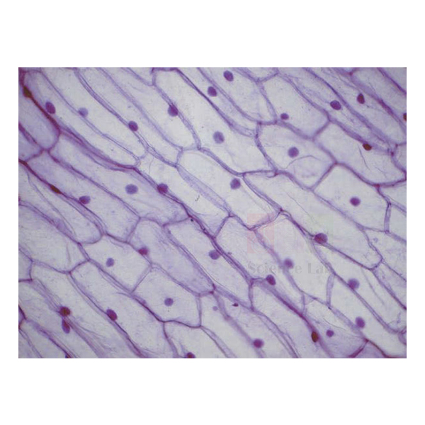
Beginners Microscope Slide Set
Beginners Microscope Slide Set (Product Code: SLE/PS/116) is a set of 12 glass microscope slides used to introduce young and inexperienced microscopists to focusing and observation in schools and science clubs. The subjects were chosen because they are easy to recognise, ranging from a printed letter e and paper fibres to onion skin, cork, a water flea and butterfly wing scales. The set is supplied by Jain Scientific Equipments Pvt Ltd (ScienceLab), Ambala, India, as a simple microscopy starter kit for school use.
Key Details
| Parameter | Detail |
|---|---|
| Number of slides | 12 glass microscope slides |
| Intended users | Young and inexperienced microscopists |
| Everyday materials | Letter e; Paper fibres; Cotton material |
| Plant material | Onion skin; Cork section; Twig section |
| Insect parts | Housefly wing; Housefly leg; Butterfly wing scales |
| Water life | Hydra; Water flea; Water organisms |
| Product code | SLE/PS/116 |
| Supplied by | Jain Scientific Equipments Pvt Ltd (ScienceLab), Ambala, India |
What Is the Beginners Microscope Slide Set Used For?
A good first lesson starts with the letter e slide: students see that the image appears upside down and reversed, and that moving the slide one way makes the image travel the other way. Paper and cotton fibres then build confidence with focusing, before moving on to living-world subjects such as onion skin cells and the tiny empty boxes in cork that gave cells their name.
The insect and water-life slides reward closer looking, with overlapping scales on the butterfly wing, hairs on the housefly leg and the transparent body of the water flea. Because every subject is familiar, children can link what they see to real objects and draw it with confidence. Teachers of middle-school science and hobby clubs use the set before moving pupils on to sectioned tissue slides.
Supplier
The Beginners Microscope Slide Set is supplied by Jain Scientific Equipments Private Limited, the Ambala company behind the ScienceLab name. It is offered to schools, activity centres, science clubs and retailers, and can be bundled on one quotation with a student microscope and blank slides for pupils' own mounts. The company profile has more about ScienceLab.
Related Prepared Slides
Useful next steps and companions for new microscope users:
- Monocular Mirror Microscope, 400x — a simple microscope for first observations.
- Basic Slide Set of 25 — the next step, with tissue sections for syllabus work.
- Microscope Glass Slides — blank slides for pupils to make their own mounts.
- Glass Cover Slips, No. 1 — to cover home-made onion or hair mounts.
- Minibeast Magnifier — hand-lens viewing of small creatures before the microscope.
Find more sets in Prepared Slides.
Frequently Asked Questions
What slides are in the Beginners Microscope Slide Set?
The Beginners Microscope Slide Set contains 12 glass slides: cork section, cotton material, housefly wing, butterfly wing scales, letter e, housefly leg, hydra, water organisms, onion skin, paper fibres, water flea and twig section.
Who is the Beginners Microscope Slide Set designed for?
The Beginners Microscope Slide Set was selected for young and inexperienced microscopists, so it suits school pupils in their first microscope lessons, science clubs and teachers introducing focusing, magnification and careful drawing before moving on to stained tissue sections.
Why does the set include a letter e slide?
The letter e slide in the Beginners Microscope Slide Set shows beginners that a microscope image is inverted and reversed, and that moving the slide left makes the image move right. It is a quick way to teach correct slide handling.
Which microscope works with these slides?
The Beginners Microscope Slide Set fits any standard compound microscope that takes glass slides. Children should start with the lowest-power objective, focus upwards away from the slide, and switch to higher power only once the subject is centred and sharp.
How should children handle the glass slides?
Pupils using the Beginners Microscope Slide Set should hold each glass slide by its edges, keep it flat, and return it to its box after use. An adult should supervise, and any chipped or cracked slide should be set aside safely.
Who supplies the set and how do I order?
Jain Scientific Equipments Pvt Ltd (ScienceLab), Ambala, India supplies the Beginners Microscope Slide Set. Send code SLE/PS/116, the number of sets and your delivery location through the contact page or enquiry cart for a quotation.
Request a Quote
Ask for a quote on the contact page, or add the set to the enquiry cart together with a student microscope and blank slides.
Last updated: 24 September 2026
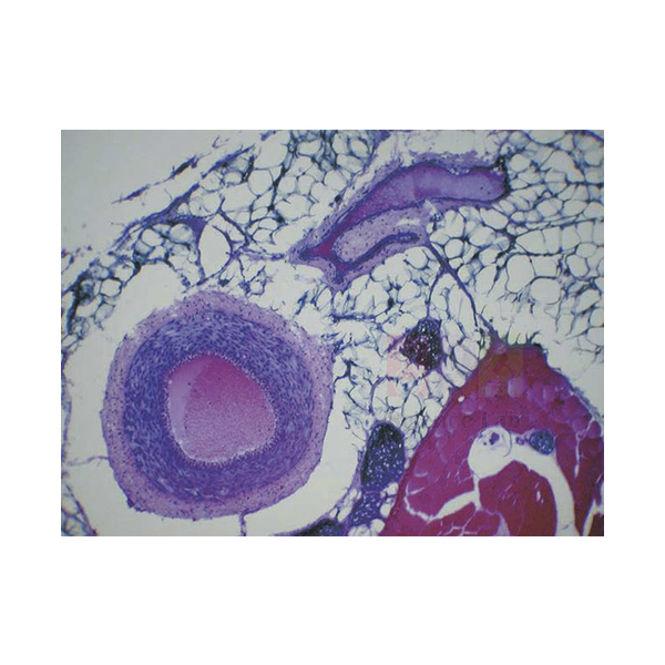
Basic Slide Set of 25
Basic Slide Set of 25 (Product Code: SLE/PS/118) is a collection of 25 glass prepared microscope slides used to cover the core plant, fungal and animal preparations of a biology syllabus in school and college labs. Its breadth is the main point: one box moves from onion root-tip mitosis and dicot stems to yeast, Mucor, blood, kidney, lung and reproductive organs. The set is supplied by Jain Scientific Equipments Pvt Ltd (ScienceLab), Ambala, India.
Specifications
| Parameter | Detail |
|---|---|
| Number of slides | 25 glass microscope slides, as specified for a biology syllabus |
| Cell division | Allium root tip, mitosis, LS; Allium root tip, squash; Lily anther tetrads, TS |
| Plant structure | Dicot stem apex, LS; Dicot leaf, TS; Dicot stem old, TS; Dicot stem separate bundles, TS; Monocot (Zea) root, TS; Monocot leaf epidermis, E; Moss leaf, for cells |
| Fungi | Yeast (Saccharomyces), E; Mucor asexual, E |
| Blood and circulation | Human blood smear; Artery and vein, TS |
| Excretion and breathing | Kidney, TS; Kidney injected, VS; Lung, VS; Lung injected, VS |
| Skin and tissues | Skin, sweat glands, VS; Skin, hair insertion, VS; Squamous epithelium; Spinal cord, TS; Pancreas, TS |
| Reproduction | Ovary, VS; Testis, TS |
| Label abbreviations | TS = transverse section; LS = longitudinal section; VS = vertical section; E = entire (whole mount) |
| Product code | SLE/PS/118 |
| Supplied by | Jain Scientific Equipments Pvt Ltd (ScienceLab), Ambala, India |
What Is the Basic Slide Set of 25 Used For?
Departments buy this set when they want one slide collection that serves a whole year of practical work. Plant lessons use the four dicot slides to compare young and old stems, vascular bundles and leaf layers, while the maize root and monocot epidermis show how monocots differ. The two onion root-tip slides, one sectioned and one squashed, give two views of mitosis, and the lily anther tetrads bring in meiosis.
Animal and human lessons draw on blood, blood vessels, kidney, lung, skin, spinal cord, pancreas and the reproductive organs, and the plain and injected kidney and lung slides let students compare tissue layout with blood supply. Yeast and Mucor round off a microbiology or fungi topic. As school biology lab equipment, the set also gives a new lab a ready reference collection before specialist sets are added.
What to Specify When Ordering
- The number of sets, for example one per practical group or one reference set per lab.
- Your syllabus or slide list, so we can confirm how the 25 preparations match it.
- Storage needs, such as a slide cabinet for keeping several sets in order.
Supplier
ScienceLab is the trading name of Jain Scientific Equipments Private Limited in Ambala, Haryana, which supplies this 25-slide collection to schools, colleges, tender buyers and distributors. For vendor registration, the supplying company is listed under GSTIN 06AAECJ8618A1ZT with Legal Entity Identifier 3358004HRD1JIVXKYX49. Read more on the ScienceLab company profile.
Related Prepared Slides
Items that complement a general biology slide collection:
- Basic Botany Slide (Set of 25) — deeper coverage of plant preparations.
- Human Histology Slide Set of 25 — more human tissue types for advanced classes.
- Beginners Microscope Slide Set — simpler subjects for first microscope lessons.
- Unit System Slide Cabinet — organised storage for a growing slide collection.
- Binocular Microscope, 1000x Mag LED — high magnification for blood cells and yeast.
Browse every set in Prepared Slides.
Frequently Asked Questions
What does the Basic Slide Set of 25 contain?
The Basic Slide Set of 25 contains 25 glass slides: plant preparations such as onion root tip, dicot stem and leaf, maize root, lily anther and moss leaf; yeast and Mucor; and animal and human tissues including blood, kidney, lung, skin, spinal cord, ovary and testis.
Which syllabus does the set follow?
Our listing describes the Basic Slide Set of 25 as specified for a biology syllabus but does not name a particular board. Send us your practical list and we will confirm which of the 25 preparations match it before you order.
Does the Basic Slide Set of 25 cover mitosis and meiosis?
Yes, the Basic Slide Set of 25 includes two onion root-tip slides for mitosis, one longitudinal section and one squash, plus a lily anther section showing tetrads, which are formed by meiosis during pollen development.
Why are there plain and injected kidney and lung slides?
The Basic Slide Set of 25 pairs plain and injected kidney and lung preparations so students can compare normal tissue with a version whose blood vessels have been filled with a coloured medium, making the blood supply much easier to follow.
Who supplies the Basic Slide Set of 25?
Jain Scientific Equipments Pvt Ltd (ScienceLab), Ambala, India supplies the Basic Slide Set of 25 to schools, colleges, tender buyers and trade customers, with quotations prepared for delivery in India and international markets.
What should a quotation request include?
For the Basic Slide Set of 25, send product code SLE/PS/118, the number of sets, your delivery location and any microscopes or slide cabinets you need. The contact page and enquiry cart both reach the ScienceLab sales team.
Request a Quote
Send your enquiry for this 25-slide set on the contact page, or add it to the enquiry cart with storage and microscopes for a combined quotation.
Last updated: 24 September 2026

Earthworm Slide Set of 6
Earthworm Slide Set of 6 (Product Code: SLE/PS/119) is a set of six glass microscope slides used to study the internal anatomy of the earthworm Lumbricus in zoology and biology practicals. Each slide cuts through a different body region, including the head end, the reproductive segments, the crop and the typhlosole, so students can rebuild the worm's body plan section by section. The set is supplied by Jain Scientific Equipments Pvt Ltd (ScienceLab), Ambala, India.
Key Details
| Parameter | Detail |
|---|---|
| Number of slides | 6 glass microscope slides |
| Specimen | Lumbricus (earthworm) |
| Head end | Anterior region, VLS |
| Reproductive region | Reproductive region, LS; Reproductive region, TS |
| Body wall and gut | Showing insertion of setae, TS; Through crop region, TS; Typhlosole region, TS |
| Label abbreviations | VLS = vertical longitudinal section; LS = longitudinal section; TS = transverse section |
| Product code | SLE/PS/119 |
| Supplied by | Jain Scientific Equipments Pvt Ltd (ScienceLab), Ambala, India |
What Is the Earthworm Slide Set Used For?
The earthworm is the standard example of a segmented animal, and this set lets a class study it without every student dissecting a specimen. The anterior longitudinal section shows how the mouth, pharynx and front segments are arranged, while the reproductive-region slides, cut both lengthways and across, help students locate the organs that sit in a few specific segments.
The three transverse sections focus on how the worm lives: the setae slide shows the bristles anchored in the body wall that grip the soil during movement, the crop section shows where food is stored, and the typhlosole section reveals the fold that increases the gut's absorbing surface. Used after or instead of a worm dissection kit, the slides make useful zoology lab equipment for comparing tissue layers under the microscope.
Supplier
Jain Scientific Equipments Private Limited, which operates as ScienceLab from Ambala, Haryana, supplies this invertebrate slide set to school and college zoology departments and to trade buyers. It can be quoted together with dissection equipment, charts and microscopes so the whole practical arrives on one order. More about the company is on the ScienceLab profile page.
Related Prepared Slides
Items that support earthworm and invertebrate practicals:
- Earthworm — a wall chart to locate each sectioned region on the whole animal.
- Dafnia Lumbricus (Earthworm), Cross Cut — an extra single transverse section for group work.
- Dissection Pan, Pad & Cover — for classes that also dissect specimens.
- Insect Anatomy Slide Set of 9 — another invertebrate group for comparison.
- Monocular Biological Microscope 400x — suitable for viewing the sections.
See the whole range in Prepared Slides.
Frequently Asked Questions
Which sections are in the Earthworm Slide Set of 6?
The Earthworm Slide Set of 6 has six Lumbricus slides: anterior region (VLS), reproductive region (LS), reproductive region (TS), a section showing insertion of setae (TS), a section through the crop region (TS) and a section through the typhlosole region (TS).
What is the typhlosole shown on one slide?
The typhlosole in the Earthworm Slide Set of 6 is an inward fold along the upper wall of the earthworm's intestine. It increases the surface area available for absorbing digested food, and the transverse section shows it clearly as a ridge projecting into the gut.
Can these slides replace an earthworm dissection?
The Earthworm Slide Set of 6 can support or partly replace dissection, because it shows internal structures in fixed, repeatable sections. Many teachers still combine the slides with a demonstration dissection or chart so students connect sections with the whole animal.
What microscope do students need for the earthworm slides?
A compound microscope with low and medium objectives suits the Earthworm Slide Set of 6. Begin at low power to see the whole cross-section, including body wall, gut and body cavity, then increase magnification to examine setae or gut lining.
Who supplies the Earthworm Slide Set of 6?
Jain Scientific Equipments Pvt Ltd (ScienceLab), Ambala, India supplies the Earthworm Slide Set of 6 to schools, colleges and resellers, and prepares quotations for customers in India and international markets on request.
How can I get a quotation?
For the Earthworm Slide Set of 6, send product code SLE/PS/119, the quantity of sets and your delivery location through the contact page, or add the set to the enquiry cart with any charts or microscopes you also need.
Request a Quote
Request a quotation through the ScienceLab contact page or add the set to the enquiry cart along with other zoology items.
Last updated: 24 September 2026
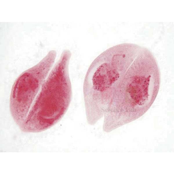
Protozoa and Algae Set of 20
Protozoa and Algae Set of 20 (Product Code: SLE/PS/120) is a set of 20 prepared microscope slides used to study commonly taught protozoa and freshwater algae in biology, microbiology and ecology classes. It includes reproduction stages such as Paramecium fission and conjugation, Spirogyra conjugation and Volvox with daughter colonies, plus the blood parasites Plasmodium and Trypanosoma. The set is supplied by Jain Scientific Equipments Pvt Ltd (ScienceLab), Ambala, India, for microbiology lab equipment buyers in India and international markets.
Key Details
| Parameter | Detail |
|---|---|
| Number of slides | 20 microscope slides |
| Free-living protozoa | Amoeba, E; Euglena, E; Paramecium, E; Paramecium, conjugating, E; Paramecium, fission, E; Vorticella, E |
| Shelled protozoa | Foraminifera, mixed; Radiolaria, mixed |
| Parasitic protozoa | Trypanosoma, in blood smear; Plasmodium sp. (malaria), in blood smear |
| Green algae | Chlamydomonas, E; Chlorella, E; Pandorina, E; Volvox, with daughter colonies, E; Cladophora, E; Desmids, mixed, E |
| Filamentous algae | Spirogyra, vegetative filaments, E; Spirogyra, conjugation, E; Nostoc, filaments with heterocysts, E |
| Diatoms | Diatoms, mixed, E |
| Label abbreviation | E = entire (whole mount) |
| Product code | SLE/PS/120 |
| Supplied by | Jain Scientific Equipments Pvt Ltd (ScienceLab), Ambala, India |
What Is the Protozoa and Algae Slide Set Used For?
This set gives a single-celled and simple-organism unit its reference material. The Chlamydomonas, Pandorina and Volvox slides form a neat series from a single cell to a small colony to a large colony with daughter colonies, which helps students discuss how multicellular life may have arisen. Paramecium appears three times, whole, dividing and conjugating, so asexual and sexual reproduction can be compared on the same organism.
The algae slides support ecology and pond-life work, with Spirogyra filaments and conjugation, desmids, diatoms and Nostoc, whose heterocysts are the cells linked with nitrogen fixation. The Plasmodium and Trypanosoma blood smears connect protozoa to human disease topics such as malaria. Because live pond samples vary from week to week, many teachers keep this set as a dependable reference beside fresh material viewed on cavity slides.
Supplier
ScienceLab is how Jain Scientific Equipments Private Limited of Ambala, Haryana, is known to its customers. The company supplies this 20-slide protozoa and algae collection to school biology labs, college microbiology and botany departments, and distributors, and can add microscopes, models and blank slides to the same quotation. See the ScienceLab company profile for more.
Related Prepared Slides
Useful additions for a microorganisms unit:
- Bacteria slide Set of 12 — completes the microbe series with bacterial preparations.
- Paramecium — a single Paramecium slide for extra student copies.
- Amoeba Model — enlarged 3D reference for the Amoeba slide.
- Microscope Slides Depression — cavity slides for watching live pond organisms.
- Binocular Biological Microscope, LED — higher magnification for blood-smear parasites.
View all sets under Prepared Slides.
Frequently Asked Questions
Which organisms are in the Protozoa and Algae Set of 20?
The Protozoa and Algae Set of 20 covers Amoeba, Euglena, Paramecium (whole, fission and conjugating), Vorticella, Foraminifera, Radiolaria, Trypanosoma and Plasmodium, plus Chlamydomonas, Chlorella, Pandorina, Volvox, Cladophora, desmids, diatoms, Spirogyra (two slides) and Nostoc.
Does the set show reproduction in protozoa and algae?
Yes, the Protozoa and Algae Set of 20 includes Paramecium fission and conjugation, Spirogyra conjugation and Volvox with daughter colonies, so students can compare asexual division, sexual exchange and colony formation across several simple organisms.
Are the malaria and Trypanosoma slides blood smears?
Yes, in the Protozoa and Algae Set of 20 both Plasmodium (malaria) and Trypanosoma are prepared in blood smears, so the parasites appear among blood cells. They are teaching slides for biology lessons, not clinical or diagnostic reference material.
What magnification is needed for these slides?
Most organisms in the Protozoa and Algae Set of 20 are visible at medium power, but blood-smear parasites and fine details such as Nostoc heterocysts need a higher objective. A compound microscope with several objectives covers the whole set.
Who supplies the Protozoa and Algae Set of 20?
Jain Scientific Equipments Pvt Ltd (ScienceLab), Ambala, India supplies the Protozoa and Algae Set of 20 to schools, colleges and trade buyers, and provides quotations for orders within India and to international markets.
What should I send to get a quotation?
To quote the Protozoa and Algae Set of 20, we need product code SLE/PS/120, the number of sets, your delivery location and any microscopes or companion slide sets. Send these through the contact page or the enquiry cart.
Request a Quote
Enquire about this 20-slide set through the contact page, or add it to the enquiry cart with other microbiology items for one quotation.
Last updated: 24 September 2026

Microscope Glass Slides
Microscope Glass Slides (Product Code: SLE/PS/121) are plain, clear glass slides used to mount specimens for viewing under a compound microscope in school, college and research labs. Each slide measures 75 x 25 mm, is 1 to 1.2 mm thick, has ground edges and comes pre-cleaned, packed 50 to a pack. These blank slides are supplied by Jain Scientific Equipments Pvt Ltd (ScienceLab), Ambala, India, for making temporary slides in biology and routine lab work.
Specifications
| Parameter | Detail |
|---|---|
| Type | Clear, optically flat glass microscope slides |
| Dimensions | 75 x 25 mm |
| Thickness | 1 to 1.2 mm |
| Dimensional variation | ± 1 mm in length and width; ± 0.2 mm in thickness |
| Edges | Ground |
| Condition | Pre-cleaned |
| Pack size | 50 slides |
| Product code | SLE/PS/121 |
| Supplied by | Jain Scientific Equipments Pvt Ltd (ScienceLab), Ambala, India |
What Are Microscope Glass Slides Used For?
Blank glass slides are the everyday base for any specimen a student prepares: an onion peel in iodine, a cheek-cell smear, a drop of pond water or a leaf epidermis for counting stomata. Place the specimen in the centre, add a drop of water or stain, and lower a cover slip at an angle to avoid trapping air bubbles. The flat, clear glass keeps the image even across the field of view.
Ground edges make the slides safer to handle and less likely to chip when students slide them across the microscope stage. Because they arrive pre-cleaned, they can go straight into a practical without a washing step; store unused slides in their pack to keep dust off. Labs that run frequent wet-mount practicals often keep several packs of 50 in stock.
What to Specify When Ordering
- Number of packs of 50 slides needed for the term or year.
- Whether you also need cover slips, cavity slides or slide forceps on the same order.
- Any special requirement such as a writing area, so we can advise before quoting.
Supplier
These slides are supplied by Jain Scientific Equipments Private Limited, the Ambala, Haryana company that trades as ScienceLab. Schools, colleges, laboratories and distributors order them as a routine consumable, often alongside cover slips and microscopes, and repeat orders can be quoted on request. Company details are on the ScienceLab profile.
Related Prepared Slides
Consumables and tools used with blank slides:
- Glass Cover Slips, No. 1 — thin cover glasses to seal each wet mount.
- Microscope Slides Depression — cavity slides for live organisms in a drop of water.
- Microscope Slide Cover Forceps — for lowering cover slips without fingerprints.
- Microscope Slide Dispenser — hands out slides one at a time during class.
- Student-Grade Microscope Slides — an alternative slide listing for large student groups.
More slide products are in Prepared Slides.
Frequently Asked Questions
What size are the Microscope Glass Slides?
The Microscope Glass Slides measure 75 x 25 mm and are 1 to 1.2 mm thick. Dimensional variation is within ± 1 mm in length and width and ± 0.2 mm in thickness, so they fit the stage clips of standard school and laboratory microscopes.
How many slides are in one pack?
Microscope Glass Slides are supplied in packs of 50 slides. Tell us how many packs you need when you enquire; schools usually order enough packs for a full term of wet-mount practicals so that fresh slides are always available.
Do the slides need washing before use?
No, the Microscope Glass Slides are pre-cleaned, so they can be used straight from the pack for wet mounts and smears. Keep the pack closed between lessons and handle slides by the edges to avoid adding fingerprints or dust.
Do these slides have a frosted end for labelling?
Our listing describes the Microscope Glass Slides as clear, optically flat slides with ground edges and does not mention a frosted writing area. If you need labelled or frosted-end slides, ask through the contact page and we will advise what can be quoted.
Which cover slips suit these slides?
Square or rectangular cover slips narrower than the 25 mm width of the Microscope Glass Slides fit comfortably. Many schools pair them with our Glass Cover Slips, No. 1, which can be added to the same enquiry.
Who supplies the Microscope Glass Slides?
Jain Scientific Equipments Pvt Ltd (ScienceLab), Ambala, India supplies the Microscope Glass Slides in packs of 50 to schools, colleges, laboratories and resellers, and quotes single packs or bulk quantities for India and international markets.
Request a Quote
For a quotation on code SLE/PS/121, state the number of packs on the contact page, or add the slides to the enquiry cart with cover slips and other consumables.
Last updated: 24 September 2026
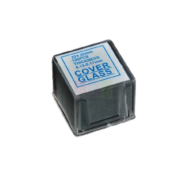
Glass Cover Slips, No. 1
Glass Cover Slips, No. 1 (Product Code: SLE/PS/122) are thin square cover glasses used to cover specimens on microscope slides in biology practicals, pathology teaching and general lab work. Made from soda glass, each slip is 20 x 20 mm with a uniform thickness of 0.13 to 0.16 mm, supplied transparent and free of fingerprints in boxes of 100. They are supplied by Jain Scientific Equipments Pvt Ltd (ScienceLab), Ambala, India, a biology lab equipment supplier in India.
Specifications
| Parameter | Detail |
|---|---|
| Material | Soda glass |
| Size | 20 x 20 mm |
| Thickness | 0.13 to 0.16 mm, uniform surface thickness |
| Thickness grade | No. 1 |
| Finish | Transparent, without fingerprints |
| Pack | Box of 100 units |
| Product code | SLE/PS/122 |
| Supplied by | Jain Scientific Equipments Pvt Ltd (ScienceLab), Ambala, India |
What Are No. 1 Glass Cover Slips Used For?
A cover slip flattens a wet specimen into an even layer, slows evaporation and keeps stain or water away from the objective lens. In class, students place a drop of water or stain on the slide, add the specimen, then touch one edge of the cover slip to the liquid and lower it slowly with a needle or forceps so air bubbles are pushed out.
The No. 1 grade describes a thin cover glass, and the 0.13 to 0.16 mm thickness leaves room for the short working distance of high-power objectives. Because the slips are so thin, handle them by the edges or with forceps and keep the box closed when not in use. A box of 100 lets a teacher issue fresh slips for each practical rather than cleaning and reusing them.
What to Specify When Ordering
- Number of boxes of 100 cover slips.
- Whether plain glass slides, cavity slides or cover-slip forceps should be quoted with them.
- Your delivery location and whether the order is a one-off or regular supply.
Supplier
Jain Scientific Equipments Private Limited, trading as ScienceLab in Ambala, Haryana, supplies these cover slips as a lab consumable to schools, colleges, teaching laboratories and distributors. Purchase departments that need vendor details can register the supplying company under CIN U29309HR2020PTC087597 and GSTIN 06AAECJ8618A1ZT. See the ScienceLab company profile for background.
Related Prepared Slides
Items that are normally used with cover slips:
- Microscope Glass Slides — pre-cleaned blank slides for the specimens.
- Microscope Slide Cover Forceps — lift and lower thin slips without breaking them.
- Microscope Slides Depression — cavity slides for hanging-drop and live samples.
- Immersion Oil, 25ml — for oil-immersion viewing on top of the cover slip.
- Glass Cover Slips — another cover-slip listing in the same range.
Browse the rest of Prepared Slides.
Frequently Asked Questions
What size and thickness are the Glass Cover Slips, No. 1?
The Glass Cover Slips, No. 1 are 20 x 20 mm squares with a uniform surface thickness of 0.13 to 0.16 mm. That thin No. 1 grade suits routine wet mounts and allows high-power objectives to focus through the glass.
How many cover slips are in a box?
Each box of Glass Cover Slips, No. 1 contains 100 units. Schools and colleges usually order several boxes at once so that every practical group can use fresh cover slips; tell us the number of boxes when you ask for a quote.
What glass are the cover slips made from?
The Glass Cover Slips, No. 1 are made from soda glass and are supplied transparent and without fingerprints. Keep the box closed and handle slips by the edges or with forceps to keep them clean and unscratched.
How do I avoid air bubbles when placing a cover slip?
Touch one edge of a Glass Cover Slip, No. 1 to the drop on the slide at an angle, then lower it slowly with a mounting needle or forceps. The liquid spreads ahead of the glass and pushes air out instead of trapping it.
Who supplies the Glass Cover Slips, No. 1?
Jain Scientific Equipments Pvt Ltd (ScienceLab), Ambala, India supplies the Glass Cover Slips, No. 1 in boxes of 100 to schools, colleges, laboratories and resellers, and quotes orders for customers in India and international markets.
What does a quotation request need?
For the Glass Cover Slips, No. 1, include product code SLE/PS/122, the number of boxes, your delivery location and any glass slides or forceps needed. Send it through the contact page or add the items to the enquiry cart.
Request a Quote
Ask for a quote through the contact page, or place the cover slips in the enquiry cart together with slides and other microscope consumables.
Last updated: 24 September 2026
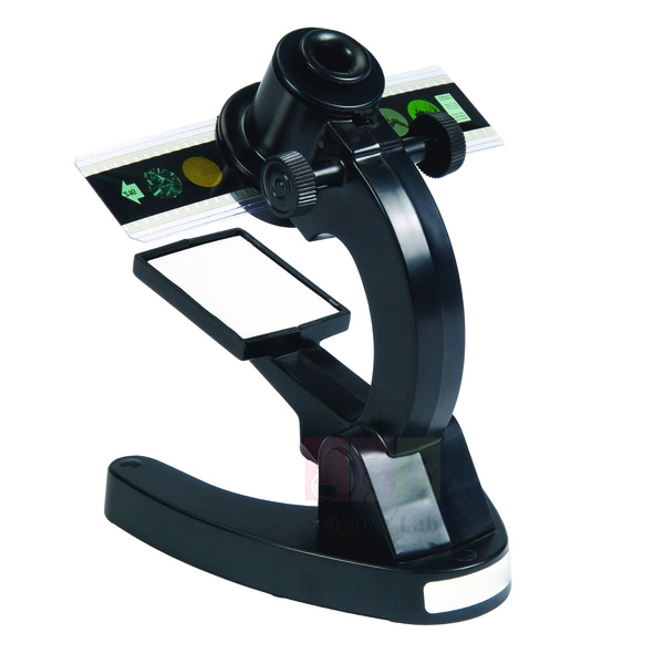
Microslide Viewer
Microslide Viewer (Product Code: SLE/PS/123) is a hand-held viewer used to look at Microslides at 5X magnification in classrooms and outdoor lessons. It needs no electricity or batteries: a white reflector gathers ordinary room light to give a bright image, so every pupil can view slides at the desk without a microscope. The viewer is supplied by Jain Scientific Equipments Pvt Ltd (ScienceLab), Ambala, India, as a practical biology teaching aid.
Specifications
| Parameter | Detail |
|---|---|
| Purpose | Specially designed teaching tool for viewing Microslides |
| Magnification | 5X |
| Illumination | White reflector giving a bright image in normal classroom light |
| Power | None required (no electricity or batteries) |
| Where it can be used | Classroom and outdoors |
| Product code | SLE/PS/123 |
| Supplied by | Jain Scientific Equipments Pvt Ltd (ScienceLab), Ambala, India |
What Is a Microslide Viewer Used For?
The viewer lets a whole class study Microslide lesson sets at the same time, which is difficult when a school has only a few microscopes. Pupils hold it up, let the white reflector catch the light from windows or ceiling lights, and look at the magnified image; there is no focusing on a stage, no lamp to switch on and no risk of cracking a glass slide with an objective lens.
Because it runs without power, the viewer is equally useful on a field trip, in a classroom without sockets at the desks, or in a primary or middle-school lesson where microscopes would be too complex. Teachers often use it to introduce a topic before moving older students on to compound microscopes and conventional prepared slides.
What to Specify When Ordering
- The number of viewers, for example one per pupil or one per pair.
- Which Microslide lesson sets you want to view with it, as these are listed separately.
- Your delivery location for the quotation.
Supplier
Jain Scientific Equipments Private Limited supplies the Microslide Viewer through its ScienceLab brand in Ambala, Haryana. Schools, teacher-training centres and educational distributors can order viewers together with Microslide lesson sets so a class set arrives ready to use. Learn more on the ScienceLab company profile.
Related Prepared Slides
Microslide lesson sets and alternatives to use with the viewer:
- Microslide-The Cell Structure — an introductory cell lesson for the viewer.
- Microslide Cells Of Plants — plant-cell images for botany lessons.
- Microslide Five Kingdoms of Life — a classification lesson set.
- Microslide The Digestive System — human biology images for the same viewer.
- Micro Slide Viewer — another viewer listing to compare before ordering.
See all slide products in Prepared Slides.
Frequently Asked Questions
What magnification does the Microslide Viewer give?
The Microslide Viewer gives 5X magnification, enough to show the enlarged images on Microslides clearly to school pupils. It is a viewing aid for lesson slides rather than a replacement for a compound microscope when higher magnification is needed.
Does the Microslide Viewer need batteries or electricity?
No, the Microslide Viewer needs neither electricity nor batteries. A white reflector collects normal classroom light and produces a bright image, so there are no bulbs to replace and no cables on the desks.
Can the viewer be used outside the classroom?
Yes, the Microslide Viewer can be used outdoors as well as in the classroom, because it relies only on available light. That makes it handy for field trips, school gardens and nature clubs where microscopes cannot be taken.
Are Microslides included with the viewer?
Our listing for the Microslide Viewer describes the viewer itself and does not state that any Microslides are included. Microslide lesson sets are listed as separate products, so please name the sets you want when you ask for a quotation.
Who supplies the Microslide Viewer?
Jain Scientific Equipments Pvt Ltd (ScienceLab), Ambala, India supplies the Microslide Viewer to schools, colleges and trade buyers, and quotes class sets or single units for delivery in India and international markets.
What should a quotation request include?
For the Microslide Viewer, send product code SLE/PS/123, the number of viewers, the Microslide sets you need and your delivery location. Use the contact page or the enquiry cart to send the request.
Request a Quote
Send your enquiry through the contact page, or add the viewer and your chosen lesson sets to the enquiry cart for a single quotation.
Last updated: 24 September 2026