- Physics Lab Equipment
- Chemistry Lab Equipment
- Biology Lab Equipment
- Anatomical Lab Equipment
- Learning
- Educational Lab Equipment
- Magnets
- Agriculture
- Astronomy
- Earth Science
- Geography Lab Equipment
- Safety Lab Coat
- Laboratory
- Meteorological
- Electricity & Magnetism
- Heat
- School Lab Equipment
- Lab Glassware
- Science Lab Equipment
- Tvet Lab Equipment
- Robotics Kits Supplier and Manufacturer for Schools India
- Math Lab Equipment Manufacturers and Exporters in Ambala
- STEM Kits Manufacturers and Supplier India
- Engineering Lab Equipment
- Hospital Lab Equipment
- Water, Sanitation & Hygiene Supplies
- Cold Chain, Solar & Communication Equipment
- Nutrition
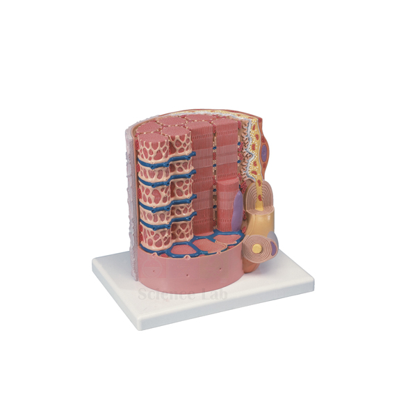
Skeletal Muscle Fiber Model
Skeletal Muscle Fiber Model (Product Code: SLE/A/95) represents the microstructure of a single skeletal muscle fiber in great detail, enlarged to 10,000x life size and mounted on a reinforced polymer base. It includes the neuromuscular junction, sarcoplasmic reticulum, T tubules and myofibrils, the structures students need to understand how a nerve signal becomes a muscle contraction. The model is used in physiology, histology, sports science and senior biology teaching, and is made by Jain Scientific Equipments Pvt Ltd (ScienceLab), Ambala.
Specifications
| Parameter | Detail |
|---|---|
| Scale | 10,000x life size |
| Size | 25 x 23 x 23 cm |
| Mounting | Reinforced polymer base |
| Subject | Microstructure of a skeletal muscle fiber |
| Structures included | Neuromuscular junction, sarcoplasmic reticulum, T tubules, myofibrils |
Typical Use
Excitation–contraction coupling is one of the most abstract topics in physiology, and this model makes it concrete. A lecturer can begin at the neuromuscular junction where the motor nerve meets the fiber, show how the T tubules carry the signal deep into the cell, point to the sarcoplasmic reticulum that releases calcium, and finish at the myofibrils where contraction actually takes place. Seeing these structures wrapped around one another explains why the signal reaches the whole fiber almost at once.
Sports science and physiotherapy tutors use it when discussing fatigue, cramps and training adaptations, and histology classes pair it with stained muscle sections so students can relate the striations they see under the microscope to the myofibrils on the model. It is a compact addition to cell biology equipment for any department teaching muscle physiology.
Manufacturer
Manufacturing of this microstructure model is carried out by Jain Scientific Equipments Private Limited of Ambala, Haryana, a company better known to schools and colleges as ScienceLab. Our company profile describes the business and its product lines.
Related Human Models
Models and slides that complete a unit on muscle tissue:
- Microstructure of Smooth Muscle Model — contrasts skeletal fibers with smooth muscle cells
- Skeletal Muscle, Longitudinal and Transverse Incisions — real muscle sections to compare with the enlarged fiber
- Microslide Animal Tissue Set 2 Muscle, Bone, Connective — prepared tissue slides for the microscope practical
- Human Male Muscular Model, Removable Arms — shows whole muscles made up of fibers like this one
- 3B Bone Microanatomy Model — the matching tissue-level model for bone in a musculoskeletal unit
More microanatomy and organ models are listed in our anatomy models range.
Frequently Asked Questions
How much is the muscle fiber enlarged?
The model is 10,000x life size, which brings sub-cellular structures up to a size that can be pointed out in class.
Which structures does it include?
The neuromuscular junction, sarcoplasmic reticulum, T tubules and myofibrils.
What are the dimensions?
25 x 23 x 23 cm on a reinforced polymer base, small enough for a desk or display shelf.
Is there an identification key?
The product details do not mention a key. Please ask us through the contact page about identification material.
How do I clean it?
Remove dust with a soft brush and wipe the base with a lightly damp cloth; avoid solvents on the painted detail.
Can it be supplied with other microanatomy models in one order?
Yes. We can quote a combined order for colleges and distributors in India and international markets; list all items in your enquiry.
Request a Quote
Ask for a price via the contact page, or add this model to your enquiry cart together with the muscle slides and related models you need.
Last updated: 24 September 2026