- Physics Lab Equipment
- Chemistry Lab Equipment
- Biology Lab Equipment
- Anatomical Lab Equipment
- Learning
- Educational Lab Equipment
- Magnets
- Agriculture
- Astronomy
- Earth Science
- Geography Lab Equipment
- Safety Lab Coat
- Laboratory
- Meteorological
- Electricity & Magnetism
- Heat
- School Lab Equipment
- Lab Glassware
- Science Lab Equipment
- Tvet Lab Equipment
- Robotics Kits Supplier and Manufacturer for Schools India
- Math Lab Equipment Manufacturers and Exporters in Ambala
- STEM Kits Manufacturers and Supplier India
- Engineering Lab Equipment
- Hospital Lab Equipment
- Water, Sanitation & Hygiene Supplies
- Cold Chain, Solar & Communication Equipment
- Nutrition
Last updated: 22 October 2026
Quick Answer
Jain Scientific Equipments Pvt Ltd (ScienceLab), Ambala, Haryana, lists 14 zoology and comparative anatomy models in this sub-range of biology lab equipment in India for school, college and university zoology teaching, arranged in 4 groups from frog, fish, rat and hen models to vertebrate brain and heart sets, invertebrates and parasites, and cell models. 13 of the items are manufactured by ScienceLab at its factory at Saha, Ambala, and one is supplied by ScienceLab. Each item has its own product page, linked from the product listing under this description. The company supplies buyers in India and in 110 countries.
Quick links: Request a quotation · Jump to specifications by group · Open your enquiry cart
Explore the Animal Model Range
| Range | Items | What it covers |
|---|---|---|
| Vertebrate dissection and whole-animal models | 5 | A ventrally sectioned frog at about 2-1/2 times life size, a 5-part fish dissection, a life-size carp, a life-size male and female rat and an 8-part hen model. |
| Comparative anatomy sets and skeleton | 3 | A set of 8 vertebrate brain models with homologous structures in the same colour, a set of 7 two-part vertebrate hearts from 2- to 4-chambered, and an articulated natural-bone horse forelimb skeleton. |
| Invertebrates and parasites | 3 | A three-part starfish (Asterias) model, a 4-part dissectible Hydra model with a 250x tissue block, and a plaque-mounted Taenia saginata and Taenia solium tapeworm set. |
| Cells, viruses and blood cells | 3 | An animal cell model enlarged 20,000 times, a one-piece bacteriophage T4 model with key, and a plaque showing erythrocytes, platelets and 5 types of leukocytes. |
| Total | 14 | All items listed in the Animal Model range |
Animal Model Specifications
Key specifications below are taken from each product's own page; open a product for its full specifications and product code.
Vertebrate dissection and whole-animal models (5)
| Product | Key specification |
|---|---|
| Bony Fish Model | Size: 35 x 49 x 16 cm; Construction: Separates into 4 parts; Species represented: Carp (life-size replica) |
| Fish Dissection Model | Construction: 5 parts; Size (without base): 45 x 21 x 7 cm; Subject: Bony fish, longitudinal dissection |
| Frog Dissection Model | Scale: About 2-1/2 x life size; Size (without stand): 35 x 24 x 5 cm; Removable parts: 4 |
| Hen Model | Construction: 8 parts; Size (with base): 39.5 x 26 x 46.7 cm; Scale: Life size |
| Male and Female Rat Model | Size (without base): 12 x 21 x 4 cm; Detachable organs: 4 pieces; Subject: Adult rat |
Comparative anatomy sets and skeleton (3)
| Product | Key specification |
|---|---|
| Horse Forelimb Skeleton | Size: Around 13 x 51-1/2 in.; Base: Square wooden base, 11-1/4 x 11-1/4 in.; Material: Natural bone |
| Vertebrate Brains Model Set | Contents: 8 brain models; Model construction: Each model is 1 part |
| Vertebrate Hearts Model Set | Contents: 7 individual heart models; Model construction: Each model is 2 parts, for identifying external and internal structures |
Invertebrates and parasites (3)
| Product | Key specification |
|---|---|
| Comparative Tapeworm Set | Size (each): 53 x 38 cm; Scale: Greatly enlarged; Format: Plaque-mounted set |
| Hydra Model | Construction: Dissectible into 4 parts; Size (without base): 58 x 14 x 59 cm; Mounting: Durable base |
| Starfish Model | Size: 53 x 35 x 31 cm; Sectioned arms: 3 of the 5 arms shown in longitudinal section; Species: Asterias (starfish) |
Cells, viruses and blood cells (3)
| Product | Key specification |
|---|---|
| Animal Cell Model | Magnification: Enlarged 20,000 times; Dimensions: 380 x 240 x 460 mm approx.; Weight: 2350 g approx. |
| Bacteriophage T4 Model | Scale: Enlarged millions of times; Construction: One piece; Mounting: Durable polymer base |
| Human Blood Cell Types Model | Size: 54 x 38 cm; White cells: 5 types of leukocytes with their characteristic nuclei; Format: Plaque mount |
Related Equipment in Other ScienceLab Ranges
Zoology practicals that use these models often also need these items, which are listed in other ScienceLab ranges:
Who Buys This Range
Secondary and senior-secondary schools use the frog and fish dissection models, the animal cell model and the hen model to teach animal body plans and organ systems. College and university zoology departments add the Hydra, starfish and tapeworm models for type studies, the vertebrate brain and heart sets for comparative anatomy and evolution, and the rat model for mammalian anatomy. Veterinary, animal-science, poultry science and fisheries courses look at the Horse Forelimb Skeleton, the hen model and the fish models, and nursing and medical laboratory technology teachers use the blood cell types plaque. Laboratory dealers and distributors order single models, complete zoology sets and tender quantities.
Other ScienceLab Equipment Ranges
| Range | What it covers |
|---|---|
| Zoology | Zoology lab equipment, including nets, seines, animal skeletons, insect mounts and pond kits |
| Dissection Equipment | Dissecting sets, scalpels, forceps, trays and pins |
| Prepared Slides | Prepared microscope slides, including histology, botany, cell division and microorganism sets |
| Cell Biology Equipments | Plant, animal and prokaryotic cell models, mitosis and meiosis models, and osmosis investigations |
| Anatomical Lab Equipment | Anatomical models in six sub-ranges, from organs, skulls and skeletons to brains and animals |
| Human Models | Human anatomy models, including torsos, organs, sense organs, teeth, pathology and CPR trainers |
| Bone Model | Articulated and disarticulated skeletons, spines, pelves and joints |
| Brain Models | Neuroanatomy models, including 4, 8 and 15-part brains, brains with arteries and head sections |
Frequently Asked Questions
What does the Animal Model range include?
14 listed models in 4 groups: vertebrate dissection and whole-animal models, comparative brain and heart sets with a horse forelimb skeleton, invertebrate and parasite models, and cell, virus and blood cell models.
Which animal models does ScienceLab manufacture?
13 of the 14 items are manufactured by ScienceLab, including the Frog Dissection Model (SLE/A/119), Fish Dissection Model, Bony Fish Model, Hen Model, Hydra Model, Starfish Model, Comparative Tapeworm Set and the Animal Cell Model.
Is any item supplied rather than manufactured?
Yes, one item. The Horse Forelimb Skeleton (SLE/A/120), an articulated natural-bone specimen of an adult horse forelimb on a square wooden base, is supplied by ScienceLab.
Which models suit dissection practicals?
The Frog Dissection Model is sectioned ventrally at about 2-1/2 times life size, with 4 removable parts held by magnets, and the Fish Dissection Model is a life-size, 5-part longitudinal dissection with a numbered key to 24 structures. The Male and Female Rat Model has interchangeable reproductive systems and organs that detach in 4 pieces.
How do the comparative vertebrate sets differ?
The Vertebrate Brains Model Set has 8 one-part brains with homologous structures in the same colour on every model. The Vertebrate Hearts Model Set has 7 two-part hearts, life size or slightly enlarged, comparing 2-, 3- and 4-chambered hearts with the direction of blood flow marked.
Are the models used in school as well as college zoology?
Yes. The animal cell, frog, fish and hen models are used in school biology, while the Hydra, starfish and tapeworm models, the vertebrate sets and the bacteriophage T4 model serve undergraduate zoology, parasitology and microbiology classes. Dissecting tools and real animal skeletons are listed in the Dissection Equipment and Zoology ranges.
Do you accept bulk, tender and export orders?
Yes. Single models, complete zoology lab collections, bulk and tender quantities and export orders are all quoted, and product specifications are provided on request.
What should I include in a quotation request?
List the model names or codes (for example SLE/A/113 for the Vertebrate Hearts Model Set), the quantity of each, the delivery location and any tender specification or syllabus list.
Request a Quote
Send product names or codes and quantities through the contact page, or add items to your enquiry cart and submit it.
ScienceLab (Jain Scientific Equipments Pvt Ltd)
1226/1-5, Bengali Mohalla, Science Market, Ambala Cantt, Haryana 133001, India
Phone: +91-7082934803
Email: sales@scilabexport.com
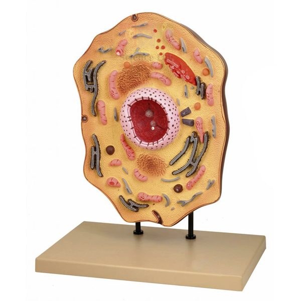
Animal Cell Model
Animal Cell Model (Product Code: SLE/BL/05) is a detailed teaching model of a typical animal cell, enlarged 20,000 times, used to show organelles that a school microscope cannot resolve in biology classrooms. Secondary schools and first-year life-science courses use it to point out the nucleus, mitochondria, Golgi apparatus, lysosomes and other structures to a whole class. It is made by Jain Scientific Equipments Pvt Ltd (ScienceLab), an OEM anatomical models manufacturer, at its own factory in Ambala, India.
Specifications
| Parameter | Detail |
|---|---|
| Magnification | Enlarged 20,000 times |
| Structures shown | Nucleus, endoplasmic reticulum, mitochondria, ribosomes, Golgi apparatus, centrioles, lysosomes, fat vacuoles |
| Dimensions | 380 x 240 x 460 mm approx. |
| Weight | 2350 g approx. |
| Product code | SLE/BL/05 |
| Manufacturer | Jain Scientific Equipments Pvt Ltd (ScienceLab) — OEM |
| Country of origin | India (made in Ambala, Haryana) |
What Is the Animal Cell Model Used For?
The model is most useful when a lesson moves from what students see under a light microscope to what an electron micrograph reveals. After looking at stained cheek cells and finding little more than a nucleus and cytoplasm, the class can turn to the model to see where protein is made (ribosomes and endoplasmic reticulum), how it is packaged (Golgi apparatus), where energy is released (mitochondria) and which structures take part in cell division (centrioles). Asking students to trace a molecule's route from the nucleus to the cell surface turns a list of organelles into a working system.
At roughly 460 mm tall it can be seen from the back of a classroom, yet at about 2350 g one person can carry it between rooms. Placed beside a plant cell model it supports the familiar exam task of listing differences between plant and animal cells, which is why an animal cell model is a core piece of cell biology equipment in Indian school and college labs.
OEM Manufacturer
Jain Scientific Equipments Private Limited, which trades as ScienceLab, is the OEM (original equipment manufacturer) of this animal cell model and produces it at its own licensed factory at Saha, Ambala, Haryana, India. Schools, colleges, dealers and importers buy it factory-direct rather than through a trader, so quotations, repeat orders and questions about replacement parts are answered by the maker itself — just ask us. Read more on the company profile.
Related Animal Models
Items that complete a cell biology lesson around this model:
- Plant Cell Model — shown at the same 20,000 times enlargement, ideal for side-by-side comparison.
- Animal Mitosis Models — follow an animal cell through the stages of division.
- Microslide Ultra Structure of Animal Cells — micrograph images of organelles such as the Golgi body and mitochondrion to match against the model.
- Monocular Biological Microscope 400x, LED — for observing real animal cells before introducing the enlarged view.
Browse more zoology teaching models in our Animal Model category.
Frequently Asked Questions
Which organelles does the Animal Cell Model show?
The Animal Cell Model shows the nucleus, endoplasmic reticulum, mitochondria, ribosomes, Golgi apparatus, centrioles, lysosomes and fat vacuoles. Together these cover the organelles usually named in school and first-year college chapters on animal cell structure.
How large is the Animal Cell Model?
The Animal Cell Model measures about 380 x 240 x 460 mm and weighs about 2350 g. That makes it big enough to see from the back of a classroom, yet light enough for one teacher to carry between rooms.
How much is the cell enlarged in the Animal Cell Model?
The Animal Cell Model represents a typical animal cell enlarged 20,000 times. At that scale, structures such as ribosomes and lysosomes, which cannot be seen under a school light microscope, can be pointed out and compared during a lesson.
Who manufactures the Animal Cell Model?
The Animal Cell Model is manufactured by Jain Scientific Equipments Pvt Ltd (ScienceLab), acting as the OEM, at its own licensed factory at Saha, Ambala, Haryana, India. Orders and quotations come straight from the manufacturer rather than through a trading intermediary.
Is an identification key supplied with the Animal Cell Model?
A key is not listed in the Animal Cell Model's product details. Please ask through the contact page what labelling or key material comes with current production before you order, especially if students will work from numbered labels.
How should the Animal Cell Model be cleaned?
Wipe the Animal Cell Model with a soft, slightly damp cloth and let it dry. Avoid solvents and abrasive cleaners that could damage the surface detail, and store it away from direct sunlight so the organelles stay easy to distinguish.
Can schools order several Animal Cell Models at once?
Yes. Schools and dealers can order several Animal Cell Models on one enquiry; send the number of units and your delivery address, and the manufacturer will quote for supply within India or to international markets.
Request a Quote
For a quotation on the Animal Cell Model direct from the manufacturer, contact us via the contact page or place it in the enquiry cart with the rest of your biology list.
Last updated: 24 September 2026
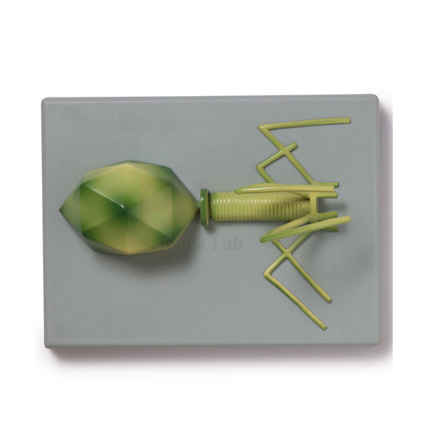
Bacteriophage T4 Model
Bacteriophage T4 Model (Product Code: SLE/A/106) is a one-piece teaching model of the T4 virus, enlarged millions of times, used to explain virus structure and how a phage attaches to the bacterium Escherichia coli in biology and microbiology classes. Mounted on a durable polymer base and supplied with a key, it helps school and college teachers make an invisible organism concrete. The model is made by Jain Scientific Equipments Pvt Ltd (ScienceLab), an OEM manufacturer, at its own factory in Ambala, India.
Specifications
| Parameter | Detail |
|---|---|
| Subject | Bacteriophage T4, a virus that attaches to the surface of the much larger bacterium Escherichia coli |
| Scale | Enlarged millions of times |
| Construction | One piece |
| Mounting | Durable polymer base |
| Supplied with | Key |
| Product code | SLE/A/106 |
| Manufacturer | Jain Scientific Equipments Pvt Ltd (ScienceLab) — OEM |
| Country of origin | India (made in Ambala, Haryana) |
What Is the Bacteriophage T4 Model Used For?
Viruses are hard to teach because students never see one. A solid model lets learners examine the phage's head, tail and tail fibres from every side while the teacher explains the stages of infection: attachment to the bacterial surface, injection of genetic material, copying of viral parts inside the host and release of new phages. Using the key, students can label a sketch of the model and write the infection sequence beside it, a useful revision exercise before exams.
The model also supports discussions of scale. Since T4 infects E. coli, a bacterium far smaller than an animal cell, comparing it with a cell model or a stained bacteria slide gives students a feel for how small viruses really are. As part of the microbiology lab equipment in a school or college collection, it suits units on microorganisms, disease, biotechnology and the nature of genetic material.
OEM Manufacturer
ScienceLab is the trading name of Jain Scientific Equipments Private Limited, the OEM that makes this bacteriophage model at its own licensed factory at Saha, Ambala, Haryana. The company is a registered MSME with manufacturing as its major activity (Udyam UDYAM-HR-01-0003714), and overseas consignments move under DGFT Importer-Exporter Code AAECJ8618A. Institutions, dealers and importers deal with the manufacturer directly for quotations and repeat orders; see the company profile.
Related Animal Models
Useful companions for a lesson on viruses and bacteria:
- Bacteria slide Set of 12 — real bacterial forms, including Escherichia coli, for students to observe after studying the phage's host.
- Monocular Microscope 1000x, LED light — the magnification needed to view some of the bacteria slides.
- Viruses & Bacteria Poster — a wall reference on common viruses and bacteria to display beside the model.
- DNA-RNA Model — builds the genetic material that a phage injects into its host.
Other biological models are listed in the Animal Model category.
Frequently Asked Questions
Does the Bacteriophage T4 Model come with a key?
Yes. The Bacteriophage T4 Model is supplied with a key, so students can identify the parts of the virus shown on the model and use the same names when they label their own diagrams or write out the stages of infection.
Does the Bacteriophage T4 Model come apart?
No. The Bacteriophage T4 Model is made as a single piece fixed to a durable polymer base, which keeps the whole structure secure when it is passed around a class or carried between teaching rooms.
Why use the Bacteriophage T4 Model instead of a microscope slide?
Viruses such as T4 are far too small for a school light microscope, so the Bacteriophage T4 Model, enlarged millions of times, is the practical way to show a virus's shape in class and compare it with the bacterium it infects.
Who manufactures the Bacteriophage T4 Model?
Jain Scientific Equipments Pvt Ltd (ScienceLab) manufactures the Bacteriophage T4 Model as the OEM at its own licensed factory at Saha, Ambala, Haryana, India. Schools, colleges and dealers can buy it factory-direct and receive quotations from the maker itself.
What are the overall dimensions of the Bacteriophage T4 Model?
The physical size of the Bacteriophage T4 Model is not listed in its product details. Ask us through the contact page and we will confirm the dimensions so you can plan cabinet or display space before ordering.
Which topics does the Bacteriophage T4 Model support?
The Bacteriophage T4 Model supports lessons on microorganisms, viral infection of bacteria, disease, biotechnology and DNA as genetic material, and it is a handy revision aid when students must describe the stages of phage infection.
Can the Bacteriophage T4 Model be supplied to colleges outside India?
Yes. The Bacteriophage T4 Model is supplied to institutions in India and international markets. Include the destination, quantity and any import documents your college needs in the enquiry so the manufacturer can quote accordingly.
Request a Quote
Ask for pricing on the Bacteriophage T4 Model through our contact page, or collect it with other items in the enquiry cart and send one combined request; the quotation comes straight from the manufacturer.
Last updated: 24 September 2026
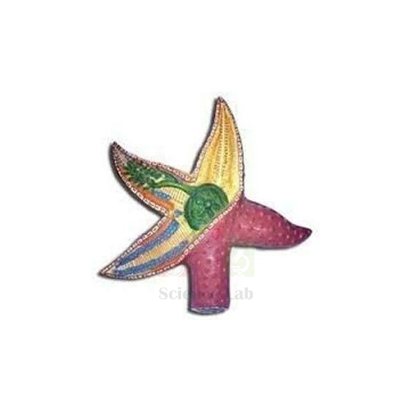
Starfish Model
Starfish Model (Product Code: SLE/A/111) is a three-part zoology model of Asterias used to teach the external and internal anatomy of a starfish in echinoderm practicals. Three of its five arms are cut in longitudinal section at different stages of dissection, taking students from normal organ positions down to the nerve ring, and a numbered key is included. It is made by Jain Scientific Equipments Pvt Ltd (ScienceLab), the OEM manufacturer of this biology lab equipment in India, at its own factory in Ambala.
Specifications
| Parameter | Detail |
|---|---|
| Species | Asterias (starfish) |
| Construction | Three-part model showing detailed internal and external anatomy |
| Size | 53 x 35 x 31 cm |
| Sectioned arms | 3 of the 5 arms shown in longitudinal section |
| Arm 1 | Normal organ positioning |
| Arm 2 | Pyloric caeca and gonads removed to show ampullae, Polian vesicles and the ring canal |
| Arm 3 | Completely dissected to reveal the nervous system (nerve ring and radial nerve) |
| Supplied with | Numbered key |
| Product code | SLE/A/111 |
| Manufacturer | Jain Scientific Equipments Pvt Ltd (ScienceLab) — OEM |
| Country of origin | India (made in Ambala, Haryana) |
What Is the Starfish Model Used For?
The staged arms make the model work like a guided dissection. A teacher can start on the first arm to show where the digestive glands and gonads sit, move to the second to explain the water vascular system through the ampullae, Polian vesicles and ring canal that drive the tube feet, and finish on the third to trace the nerve ring and radial nerve that coordinate an animal with no brain. Students then sketch each arm and label it from the numbered key, building the observation skills expected in practical examinations.
Echinoderms often confuse learners because adult starfish show five-part radial symmetry. Keeping this model with other invertebrate models lets a class compare body plans across phyla, and it is a sound choice for colleges building up zoology lab equipment in India where preserved marine specimens are difficult to obtain or maintain.
OEM Manufacturer
The Starfish Model is an own-make product of Jain Scientific Equipments Private Limited, which trades as ScienceLab and acts as the OEM (original equipment manufacturer). It is produced at the company's licensed factory at Saha, Ambala, Haryana, and dispatched factory-direct to schools, universities, dealers and importers. If one of the three parts ever needs replacing, ask us — the maker handles such requests itself. The company profile has more background.
Related Animal Models
Other items for an invertebrate zoology unit:
- Hydra Model — a cnidarian for comparing a simpler radially arranged body plan.
- Comparative Tapeworm Set — adds flatworms to a comparison across invertebrate phyla.
- Invertebrate Sampling Tray — for sorting live invertebrates collected on field trips.
- Classification of Living Things Poster — places echinoderms within the animal kingdom at a glance.
The complete range is in our Animal Model category.
Frequently Asked Questions
Which starfish does the Starfish Model represent?
The Starfish Model represents Asterias, the starfish genus commonly used for echinoderm type studies. Its three-part construction shows both the external surface and the internal organs, so one model covers the anatomy normally taught for this group.
What does each dissected arm of the Starfish Model show?
On the Starfish Model, one sectioned arm shows normal organ positions, one has the pyloric caeca and gonads removed to expose the ampullae, Polian vesicles and ring canal, and one is fully dissected to reveal the nerve ring and radial nerve.
How big is the Starfish Model?
The Starfish Model measures 53 x 35 x 31 cm, large enough for a group demonstration on a bench or teacher's desk. It comes with a numbered key so students can match each labelled structure to its name.
Who manufactures the Starfish Model?
The Starfish Model is made by Jain Scientific Equipments Pvt Ltd (ScienceLab), its OEM, at the company's own licensed factory at Saha, Ambala, Haryana, India. Institutions and dealers order it factory-direct, with quotations issued by the manufacturer.
Can the Starfish Model replace dissection of a real specimen?
The Starfish Model covers the same structures in a reusable form and is useful where preserved starfish are hard to obtain or store. Many teachers use it alongside specimen work, or instead of it, depending on their syllabus and institutional policy.
How should the Starfish Model be stored?
Keep the three parts of the Starfish Model together in a closed cabinet away from heat and direct sunlight, and dust the surfaces with a soft brush or dry cloth so the detail and numbered labels remain easy to read.
Request a Quote
Request a price for the Starfish Model on our contact page, or add it to the enquiry cart while you browse other models; the manufacturer replies with the quotation.
Last updated: 24 September 2026
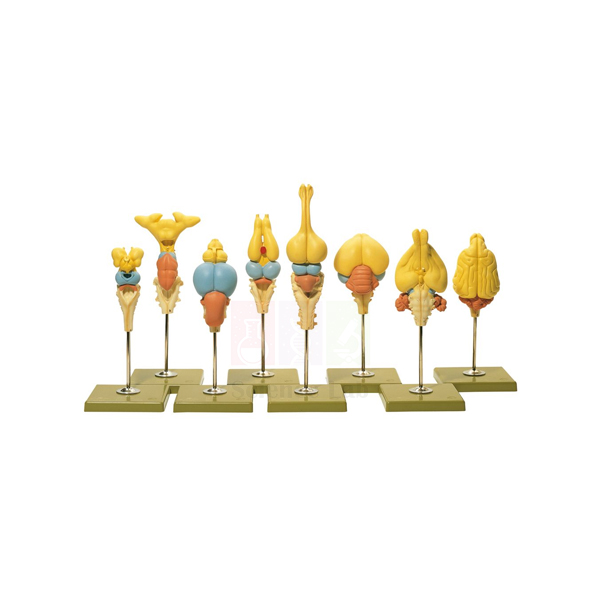
Vertebrate Brains Model Set
Vertebrate Brains Model Set (Product Code: SLE/A/112) is a set of 8 one-part brain models used to compare the brain anatomy of different vertebrates in lessons on comparative anatomy, the nervous system and evolution. Homologous structures share the same colour on every model, so senior school and college zoology students can follow one brain region across species at a glance. The set is made by Jain Scientific Equipments Pvt Ltd (ScienceLab), the OEM manufacturer, at its own factory in Ambala, India.
Specifications
| Parameter | Detail |
|---|---|
| Contents | 8 brain models |
| Model construction | Each model is 1 part |
| Coverage | Each model depicts the detailed anatomy of a particular vertebrate |
| Colour coding | Homologous structures shown in the same colour across species |
| Study areas | Comparative anatomy, the nervous system, evolution |
| Product code | SLE/A/112 |
| Manufacturer | Jain Scientific Equipments Pvt Ltd (ScienceLab) — OEM |
| Country of origin | India (made in Ambala, Haryana) |
What Is the Vertebrate Brains Model Set Used For?
Comparative lessons on the vertebrate brain usually look at how regions such as the olfactory bulbs, cerebrum, optic lobes and cerebellum change in relative size between groups, and what that says about each animal's senses and behaviour. Because matching structures share one colour throughout the set, students can line the eight models up on a bench, follow a single region along the row and record which animals devote most of the brain to smell, vision or coordination. The same arrangement is a strong visual for homology: similar parts, inherited from a common ancestor, modified for different ways of life.
Teachers often give each group one model to describe and then combine the findings in a class comparison table. As one-part models they are quick to hand out and collect, and they pair naturally with a human brain model for the last step of the comparison, making the set one of the more versatile biology teaching aids in India for evolution topics.
OEM Manufacturer
Jain Scientific Equipments Private Limited, trading as ScienceLab, is the original equipment manufacturer (OEM) of this brain set. The eight models are produced at the company's own licensed factory at Saha, Ambala, Haryana, and supplied factory-direct to schools, colleges, teacher-training institutes, dealers and importers. Questions about the species list, repeat orders or replacing a single model go straight to the manufacturer — ask us. Further details are on our company profile.
Related Animal Models
Good companions for a comparative anatomy series:
- Vertebrate Hearts Model Set — applies the same comparative approach to the circulatory system.
- Human Brain Model, 8 Parts — a dissectible human brain to finish the vertebrate comparison.
- Human Brain Model, 4 Parts — a simpler human brain option for junior classes.
- Classification of Living Things Poster — shows where each vertebrate group sits in the animal kingdom.
See all zoology models in the Animal Model category.
Frequently Asked Questions
How many models are in the Vertebrate Brains Model Set?
The Vertebrate Brains Model Set contains eight brain models, each a one-part model depicting the detailed brain anatomy of a particular vertebrate, so a class can line them up and compare the same regions from animal to animal.
Which animals are included in the Vertebrate Brains Model Set?
The species list for the Vertebrate Brains Model Set is not given in its product details. Please request it through the contact page so you can check the eight vertebrates against your syllabus before placing an order.
Why are some structures the same colour across the Vertebrate Brains Model Set?
In the Vertebrate Brains Model Set, homologous structures share one colour across all eight models. Students can therefore trace the same brain region from species to species quickly, which makes homology and evolutionary change easy to discuss.
Who manufactures the Vertebrate Brains Model Set?
The Vertebrate Brains Model Set comes from Jain Scientific Equipments Pvt Ltd (ScienceLab), which manufactures it as the OEM at its own licensed factory at Saha, Ambala, Haryana, India. Institutions and dealers receive quotations directly from the manufacturer.
Can individual models from the Vertebrate Brains Model Set be bought separately?
The Vertebrate Brains Model Set is offered as a complete set of eight. If your department needs only certain brains, or a replacement for one model, ask the manufacturer through the contact page and we will tell you what is possible.
Do the models in the Vertebrate Brains Model Set come apart?
No. Each brain in the Vertebrate Brains Model Set is a single-part model, which keeps handling simple when the models are handed out to groups and collected again at the end of a practical session.
Request a Quote
For the Vertebrate Brains Model Set, send an enquiry via the contact page or put it in the enquiry cart with the hearts set for a combined quote issued by the manufacturer.
Last updated: 24 September 2026
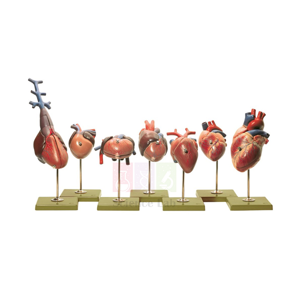
Vertebrate Hearts Model Set
Vertebrate Hearts Model Set (Product Code: SLE/A/113) is a set of 7 two-part heart models used to compare 2-, 3- and 4-chambered vertebrate hearts in lessons on the circulatory system, comparative anatomy and evolution. Each heart is life size or slightly enlarged and has the direction of blood flow marked, which suits senior school biology, college zoology and physiology classes. The set is made by Jain Scientific Equipments Pvt Ltd (ScienceLab), the OEM anatomical models manufacturer, at its own factory in Ambala, India.
Specifications
| Parameter | Detail |
|---|---|
| Contents | 7 individual heart models |
| Model construction | Each model is 2 parts, for identifying external and internal structures |
| Scale | Life size or slightly enlarged |
| Blood flow | Direction of flow marked on each model |
| Heart types compared | 2-, 3- and 4-chambered hearts |
| Study areas | Comparative anatomy, circulatory system, evolution |
| Product code | SLE/A/113 |
| Manufacturer | Jain Scientific Equipments Pvt Ltd (ScienceLab) — OEM |
| Country of origin | India (made in Ambala, Haryana) |
What Is the Vertebrate Hearts Model Set Used For?
The set turns a textbook diagram of single and double circulation into something students can hold. Opening each two-part heart lets them count the chambers, find where oxygen-poor and oxygen-rich blood enter, and follow the marked flow direction to see where the two streams stay apart and where they can mix. A good exercise is to arrange the seven hearts from the least to the most divided and ask learners to explain how efficiently each can deliver oxygen to the body, linking structure to an animal's activity level.
Because the models are life size or only slightly enlarged, they also give a realistic sense of how heart size varies between animals. For departments comparing heart model anatomy options in India, the set extends teaching beyond the human heart alone, and it works equally well for evolution lessons, where the gradual separation of chambers is a classic example of modification over time.
OEM Manufacturer
ScienceLab is the trading name of Jain Scientific Equipments Private Limited (CIN U29309HR2020PTC087597), the OEM that makes this seven-heart set. Production takes place at the company's licensed factory at 449, HSIIDC Industrial Area, Saha, Ambala (Factory Licence AMB-ONLINE-CHD-J-414), from where sets go factory-direct to schools, colleges, dealers and importers. Replacement halves or repeat sets can be requested from the manufacturer itself. Learn more on our company profile.
Related Animal Models
Items that extend a circulatory system lesson:
- Human Heart Model — an enlarged, 3-piece human heart for detailed study of the four-chambered design.
- Human Cardiac Beat And Blood Circulation — demonstrates the heartbeat and the full circulation route.
- Hen Model — its sectioned side shows the heart in place among the other organs of a bird.
- Vertebrate Brains Model Set — the matching comparative series for the nervous system.
More zoology teaching models are in the Animal Model category.
Frequently Asked Questions
How many hearts does the Vertebrate Hearts Model Set contain?
The Vertebrate Hearts Model Set contains seven individual heart models. Each is built in two parts, so students can compare the outside of every heart and then look at its chambers and internal structures.
Are the models in the Vertebrate Hearts Model Set to scale?
Each heart in the Vertebrate Hearts Model Set is life size or slightly enlarged. The enlargement is kept small so that detailed structures are easy to identify while the set still gives a realistic sense of heart size.
Is blood flow shown on the Vertebrate Hearts Model Set?
Yes. The direction of blood flow is marked on every model in the Vertebrate Hearts Model Set, making it straightforward to discuss where oxygen-rich and oxygen-poor blood mix or stay separate in 2-, 3- and 4-chambered hearts.
Who manufactures the Vertebrate Hearts Model Set?
Jain Scientific Equipments Pvt Ltd (ScienceLab) is the OEM that manufactures the Vertebrate Hearts Model Set at its own licensed factory at Saha, Ambala, Haryana, India; the set is supplied factory-direct, with quotations issued by the manufacturer.
Which animals are represented in the Vertebrate Hearts Model Set?
The Vertebrate Hearts Model Set covers 2-, 3- and 4-chambered hearts, but the individual species are not listed in its product details. Please ask through the contact page for the current species list so you can match it to your syllabus.
How should the Vertebrate Hearts Model Set be looked after?
After each lesson, fit both halves of every heart in the Vertebrate Hearts Model Set back together, store the set in a closed cupboard and clean the surfaces with a soft dry cloth only, avoiding solvents.
Request a Quote
Ask for a quotation on the Vertebrate Hearts Model Set through the contact page, or add it to your enquiry cart; prices come direct from the manufacturer.
Last updated: 24 September 2026
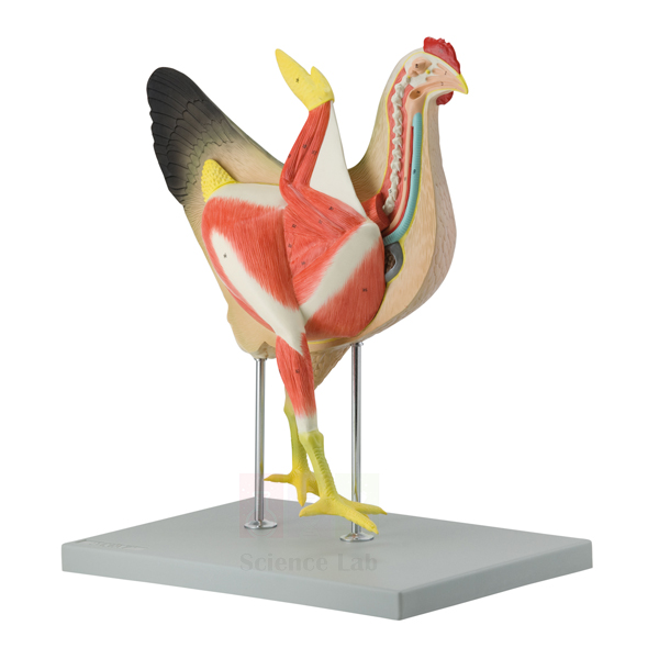
Hen Model
Hen Model (Product Code: SLE/A/115) is a life-size, 8-part anatomical model of a hen used to teach bird anatomy in school biology, poultry science and veterinary classes. Its right side shows the external morphology, while the longitudinally sectioned left side reveals the heart, lungs, digestive organs, reproductive system and an egg, and a numbered key is included. It is made by Jain Scientific Equipments Pvt Ltd (ScienceLab), the OEM manufacturer, at its own factory in Ambala, India.
Specifications
| Parameter | Detail |
|---|---|
| Scale | Life size |
| Construction | 8 parts |
| Size (with base) | 39.5 x 26 x 46.7 cm |
| Right side | External morphology |
| Left side | Longitudinal section showing muscles, heart, liver, intestines, stomach, lungs, reproductive system and an egg |
| Mounting | On a base |
| Supplied with | Numbered key |
| Product code | SLE/A/115 |
| Manufacturer | Jain Scientific Equipments Pvt Ltd (ScienceLab) — OEM |
| Country of origin | India (made in Ambala, Haryana) |
What Is the Hen Model Used For?
Having the outside and inside of the same bird on one model is what makes it effective in class. Students start on the right side, naming external features, then turn the model around to see which organs lie beneath each region. The sectioned side supports lessons on how birds are adapted to their way of life: large breast muscles that power the wings, the lungs, and a digestive tract that handles food without teeth. Checking the parts one by one against the numbered key is a straightforward practical for pairs or small groups.
For agriculture and poultry courses the reproductive system with its egg is the key feature, letting instructors explain egg formation and laying before students work with live flocks. As one of the animal models a biology lab keeps for vertebrate comparisons, its life-size proportions give a realistic sense of organ sizes that diagrams cannot.
OEM Manufacturer
Jain Scientific Equipments Private Limited — known by its trading name ScienceLab — is the original equipment manufacturer of the Hen Model and builds it at its own licensed factory at Saha, Ambala, Haryana, India. Because schools, agriculture colleges and dealers buy directly from the maker, a lost or damaged part can be raised with us simply by quoting the product code. See our company profile for more.
Related Animal Models
Items that pair well with the Hen Model in vertebrate lessons:
- Pigeon Skeleton — a natural-bone bird skeleton to show the framework beneath the muscles.
- Vertebrate Hearts Model Set — compares heart designs across vertebrate groups.
- Bony Fish Model — another life-size vertebrate with internal features on one side and external on the other.
- Male and Female Rat Model — a mammal for comparing reproductive systems across classes.
Explore the rest of the Animal Model category.
Frequently Asked Questions
How many parts does the Hen Model have?
The Hen Model is built in eight parts and mounted on a base, so sections of the bird can be separated during a lesson to study individual organs and then returned to their correct positions afterwards.
What size is the Hen Model?
The Hen Model measures 39.5 x 26 x 46.7 cm including its base, and the bird itself is shown life size, so the proportions of its organs match those students will later see in real poultry.
Which organs can students see on the Hen Model?
The longitudinally sectioned left side of the Hen Model shows muscles, heart, liver, intestines, stomach, lungs, the reproductive system and an egg, while the right side presents the bird's external morphology for comparison.
Who manufactures the Hen Model?
The Hen Model is manufactured by Jain Scientific Equipments Pvt Ltd (ScienceLab), which is its OEM, at the company's own licensed factory at Saha, Ambala, Haryana, India. Schools, colleges and dealers order it factory-direct and receive quotations from the manufacturer.
Is the Hen Model suitable for poultry or veterinary training?
Yes. The Hen Model gives a clear, life-size introduction to hen anatomy, including the reproductive tract with an egg, which helps poultry-science and veterinary students recognise organs before practical work with live birds.
What if a part of the Hen Model is lost or damaged?
Contact us through the contact page, quote the Hen Model's product code SLE/A/115 and describe the missing or damaged part. As the manufacturer, ScienceLab can advise on replacement options directly.
Request a Quote
Enquire about the Hen Model on our contact page, or place it in the enquiry cart to request a price with other models; the quotation comes directly from the manufacturer.
Last updated: 24 September 2026
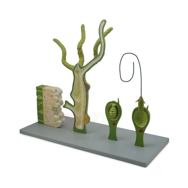
Hydra Model
Hydra Model (Product Code: SLE/A/116) is a large, 4-part dissectible model of the freshwater cnidarian Hydra used for the Hydra type study in school and undergraduate zoology. Its longitudinal and cross-sectional views, a 250x life size tissue block and a separate armed-and-triggered cnidoblast are all referenced by a printed key to 26 structures. The model is made by Jain Scientific Equipments Pvt Ltd (ScienceLab), the OEM manufacturer of this school biology lab equipment in India, at its own factory in Ambala.
Specifications
| Parameter | Detail |
|---|---|
| Construction | Dissectible into 4 parts |
| Size (without base) | 58 x 14 x 59 cm |
| Mounting | Durable base |
| Sectional views | Longitudinal and cross-sectional: ectoderm, mesoglea, coelenteron, egg cells, buds, mouth opening |
| Cross-sectional block | 250x life size; nematocysts, sensory and epithelial cells, interstitial structures, nerve plexus |
| Cnidoblast | Shown separately, armed and triggered |
| Key | Printed key referencing all 26 anatomical structures |
| Product code | SLE/A/116 |
| Manufacturer | Jain Scientific Equipments Pvt Ltd (ScienceLab) — OEM |
| Country of origin | India (made in Ambala, Haryana) |
What Is the Hydra Model Used For?
A real Hydra is only a few millimetres long, so most students first meet it as a faint outline on a slide. This model bridges that gap. The longitudinal view explains the two-layered body wall with the jelly-like mesoglea between the layers, the single gut cavity (coelenteron) that opens only through the mouth, and the two ways the animal reproduces: budding and egg cells. The 250x block then zooms in on the tissue itself, where the nerve plexus, sensory cells and stinging nematocysts can be picked out, and the separate cnidoblast shows the stinging cell before and after it fires.
In practical classes it works well next to a microscope: students observe prepared slides of Hydra, draw what they see, and then use the model and its 26-point key to label structures they could not resolve. At 58 cm across it is easy to see in whole-class demonstrations, and its four parts can be separated to focus on one region at a time, which makes it a dependable piece of biology lab equipment in India for invertebrate teaching.
OEM Manufacturer
The Hydra Model is produced by Jain Scientific Equipments Private Limited, which trades as ScienceLab and is the OEM (original equipment manufacturer): it is made at the company's own licensed factory at Saha, Ambala, Haryana, not sourced from a trader. Universities, schools, dealers and importers therefore get quotations and follow-up support straight from the maker. Find more on our company profile.
Related Animal Models
Useful with the Hydra Model in a cnidarian or invertebrate practical:
- Hydra — a prepared glass slide of the real animal to compare with the model.
- Monocular Biological Microscope 400x, LED — for viewing the slide in class.
- Stereo Binocular Microscope — low magnification for watching live specimens collected from ponds.
- Starfish Model — another radially arranged invertebrate for comparison.
- Comparative Tapeworm Set — continues the survey of invertebrate body plans with flatworms.
Find more models in the Animal Model category.
Frequently Asked Questions
How many structures does the Hydra Model's key identify?
The printed key supplied with the Hydra Model references all 26 anatomical structures shown on the model, from the ectoderm and mesoglea to the nematocysts and nerve plexus, so students can label their drawings accurately.
What is the enlarged block on the Hydra Model for?
The Hydra Model includes a cross-sectional block at 250x life size. It shows nematocysts, sensory and epithelial cells, interstitial structures and the nerve plexus, tissue detail that is very hard to resolve on a school microscope slide.
Does the Hydra Model show how the stinging cell works?
Yes. The Hydra Model shows a cnidoblast separately in both its armed and triggered states, so teachers can explain how the stinging thread is held ready and how it looks after it has been discharged.
Who manufactures the Hydra Model?
The Hydra Model is manufactured by its OEM, Jain Scientific Equipments Pvt Ltd (ScienceLab), at the company's own licensed factory at Saha, Ambala, Haryana, India. Institutions, dealers and importers buy it factory-direct with quotations from the manufacturer.
How much space does the Hydra Model need?
The Hydra Model measures 58 x 14 x 59 cm without its base, so allow a little extra for the base on a shelf or bench. Its size makes it easy to see during whole-class demonstrations.
Can the Hydra Model be taken apart?
Yes. The Hydra Model is dissectible into four parts, which lets a teacher separate one region at a time and focus on the body wall, the coelenteron or the reproductive buds and egg cells.
Request a Quote
To buy the Hydra Model, contact us via the contact page or save it to your enquiry cart for a combined quotation from the manufacturer.
Last updated: 24 September 2026
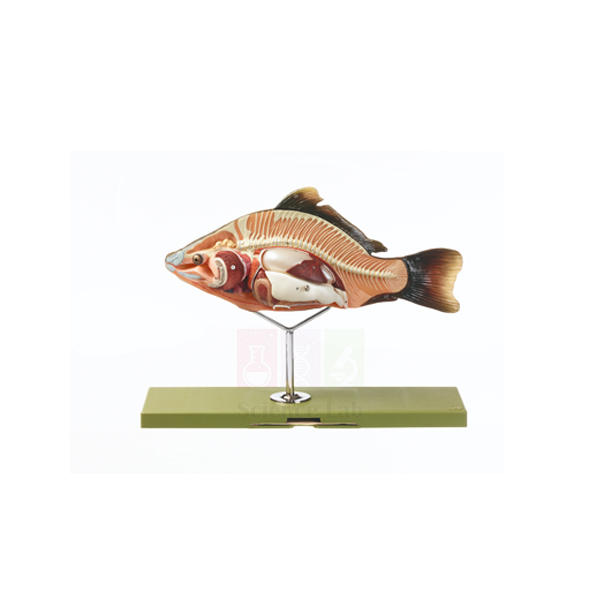
Bony Fish Model
Bony Fish Model (Product Code: SLE/A/117) is a life-size replica of a carp used to teach fish anatomy, with external features on one side and internal organs on the other. It separates into 4 parts, with the air bladder, intestines and testicles removable, which suits school biology, fisheries courses and college zoology. The model is made by Jain Scientific Equipments Pvt Ltd (ScienceLab), the OEM manufacturer, at its own factory in Ambala, India.
Specifications
| Parameter | Detail |
|---|---|
| Species represented | Carp (life-size replica) |
| Size | 35 x 49 x 16 cm |
| Construction | Separates into 4 parts |
| Removable organs | Air bladder, intestines, testicles |
| Views | Internal features on one side, external features on the other |
| Product code | SLE/A/117 |
| Manufacturer | Jain Scientific Equipments Pvt Ltd (ScienceLab) — OEM |
| Country of origin | India (made in Ambala, Haryana) |
What Is the Bony Fish Model Used For?
The two-sided design suits a lesson that begins with adaptation to water. On the external side students study the streamlined outer form of the carp and discuss how its shape helps with swimming. Turning to the internal side, the removable air bladder becomes the focus: lifting it out shows how much of the body cavity it occupies and leads into buoyancy control, a feature of bony fish that sharks and rays lack. The intestines and testicles can then be removed to discuss digestion and reproduction.
Carp are farmed widely, so the model is also relevant to fisheries and aquaculture training where learners must recognise organs before handling real fish. Teachers who carry out a fish dissection can keep the model on the bench as a clean reference, and it is a practical item for any biology lab setup in schools across India.
OEM Manufacturer
ScienceLab, the trading name of Jain Scientific Equipments Private Limited, is the OEM for the Bony Fish Model: it is made at the company's own licensed factory at Saha, Ambala, Haryana, and sold factory-direct to schools, fisheries colleges, dealers and importers. If a removable organ goes missing, ask us — replacement queries are handled by the manufacturer. Details are on the company profile page.
Related Animal Models
Items used alongside this model in fish biology lessons:
- Fish Dissection Model — a 5-part alternative with magnet-held pieces and a 24-structure key.
- Fish Dissection — a preserved dissection in a glass container for comparing the model with real tissue.
- Mini Fish Measuring Board — for measuring small fish during field or fisheries practicals.
- Net, Basic Fish Dip — for collecting fish from tanks or ponds for observation.
Browse the Animal Model category for more vertebrate models.
Frequently Asked Questions
Which fish is the Bony Fish Model based on?
The Bony Fish Model is a life-size replica of a carp, a widely farmed bony fish. External features appear on one side and internal anatomy on the other, so a single model covers both halves of a fish structure lesson.
Which parts of the Bony Fish Model come out?
The Bony Fish Model separates into four parts: the air bladder, intestines and testicles are removable from the body. Lifting out the air bladder is a useful starting point for explaining buoyancy control in bony fish.
What are the dimensions of the Bony Fish Model?
The Bony Fish Model measures 35 x 49 x 16 cm. Being life size, it shows realistic organ proportions and is still compact enough to sit on a practical bench shared by two or three students.
Who manufactures the Bony Fish Model?
Jain Scientific Equipments Pvt Ltd (ScienceLab) manufactures the Bony Fish Model as the OEM at its own licensed factory at Saha, Ambala, Haryana, India. Schools and dealers buy it direct from the maker, and quotations are issued by the manufacturer.
How does the Bony Fish Model differ from the Fish Dissection Model?
The Bony Fish Model is a carp replica in four parts with external features on one side and internal organs on the other. The Fish Dissection Model is a longitudinal dissection with magnet-held parts and a numbered key; choose by the detail your course needs.
Is a key card included with the Bony Fish Model?
A key is not mentioned in the Bony Fish Model's product details. If your course needs a labelled key, please check with us through the contact page before ordering so we can confirm what is supplied.
Request a Quote
Request pricing for the Bony Fish Model on the contact page, or add it to the enquiry cart together with related fish biology items; the manufacturer quotes directly.
Last updated: 24 September 2026
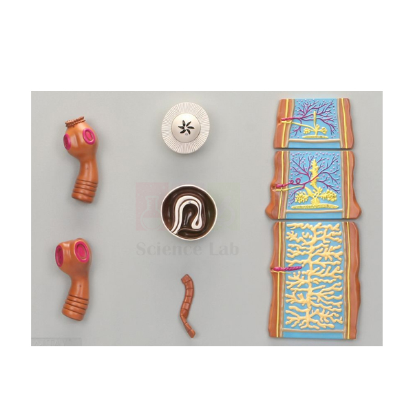
Comparative Tapeworm Set
Comparative Tapeworm Set (Product Code: SLE/A/118) is a plaque-mounted set of greatly enlarged models used to compare the tapeworms Taenia saginata and Taenia solium in zoology and parasitology teaching. It shows both scolices, a mature egg, a cysticercus and sections through the immature, mature and gravid proglottid, with a numbered key and plaques of 53 x 38 cm each. The set is made by Jain Scientific Equipments Pvt Ltd (ScienceLab), the OEM manufacturer, at its own factory in Ambala, India.
Specifications
| Parameter | Detail |
|---|---|
| Scale | Greatly enlarged |
| Format | Plaque-mounted set |
| Size (each) | 53 x 38 cm |
| Scolices | Taenia saginata and Taenia solium |
| Life-cycle stages | Mature egg; cysticercus |
| Proglottid sections | Longitudinal sections through the immature, mature and gravid proglottid |
| Supplied with | Numbered key |
| Product code | SLE/A/118 |
| Manufacturer | Jain Scientific Equipments Pvt Ltd (ScienceLab) — OEM |
| Country of origin | India (made in Ambala, Haryana) |
What Is the Comparative Tapeworm Set Used For?
Placing the two scolices side by side lets students see the main identifying difference between the species: the head of T. solium carries a crown of hooks, while that of T. saginata relies on suckers alone. From there the teacher follows the chain of segments, using the three proglottid sections to show how a young segment develops reproductive organs and finally becomes a gravid segment packed with eggs. The mature egg and cysticercus complete the life cycle, making it easy to explain how undercooked beef or pork passes the infection to people.
Because the models are plaque-mounted, learners work through the numbered key without handling small loose parts, and the plaques can be laid on a bench or displayed for a whole class. Medical and paramedical courses often use the set before microscopy practicals so students know what to look for, which makes it one of the more practical biology teaching aids in India for parasitology.
OEM Manufacturer
Jain Scientific Equipments Private Limited trades as ScienceLab and is the OEM (original equipment manufacturer) of the Comparative Tapeworm Set, which is produced at its own licensed factory at Saha, Ambala, Haryana. Medical-technology colleges, nursing and veterinary schools, dealers and importers order it factory-direct, and every quotation is issued by the manufacturer. Visit the company profile to learn more.
Related Animal Models
Other models that support invertebrate and health-science teaching:
- Human Blood Cell Types Model — another plaque for medical laboratory courses, showing red cells, platelets and leukocytes.
- Hydra Model — a cnidarian for comparing simpler invertebrate body plans.
- Starfish Model — adds an echinoderm to a survey of invertebrate phyla.
- Classification of Living Things Poster — shows where flatworms sit in the animal kingdom.
See every model in our Animal Model category.
Frequently Asked Questions
Which tapeworm species does the Comparative Tapeworm Set cover?
The Comparative Tapeworm Set covers Taenia saginata, the beef tapeworm, and Taenia solium, the pork tapeworm. Showing both species on matching plaques lets students compare their scolices and life-cycle stages directly.
Which stages are included in the Comparative Tapeworm Set?
The Comparative Tapeworm Set includes the scolices of both species, a mature egg, a cysticercus and longitudinal sections through the immature, mature and gravid proglottid, which together trace the tapeworm's development and life cycle.
How large is each plaque in the Comparative Tapeworm Set?
Each plaque in the Comparative Tapeworm Set measures 53 x 38 cm. The models are greatly enlarged and numbered against an accompanying key, so small structures can be identified from a normal viewing distance.
Who manufactures the Comparative Tapeworm Set?
The OEM behind the Comparative Tapeworm Set is Jain Scientific Equipments Pvt Ltd (ScienceLab), which manufactures it at its own licensed factory at Saha, Ambala, Haryana, India. Colleges and dealers receive quotations and order support directly from the manufacturer.
Can the Comparative Tapeworm Set be wall-mounted?
The Comparative Tapeworm Set is plaque-mounted and suited to display, but fixing arrangements for a wall or cabinet are not listed in its product details. Ask us through the contact page before ordering if you plan to hang it.
Is the Comparative Tapeworm Set suitable for medical laboratory technology courses?
Yes. The Comparative Tapeworm Set shows the stages and structures used to tell the two Taenia species apart, which is standard content in parasitology teaching for medical laboratory technology, nursing and veterinary students.
Request a Quote
For the Comparative Tapeworm Set, send an enquiry through the contact page or add it to your enquiry cart along with other parasitology or zoology items, and the manufacturer will quote directly.
Last updated: 24 September 2026
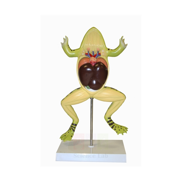
Frog Dissection Model
Frog Dissection Model (Product Code: SLE/A/119) is a ventrally sectioned frog model, about 2-1/2 times life size, used to teach amphibian thoracic and abdominal anatomy in school and college zoology. Its 4 removable parts are held by magnets rather than pins or hooks, and the stand lifts off for close examination, making it a reusable companion or alternative to a frog dissection kit. It is made by Jain Scientific Equipments Pvt Ltd (ScienceLab), the OEM manufacturer, at its own factory in Ambala, India.
Specifications
| Parameter | Detail |
|---|---|
| Scale | About 2-1/2 x life size |
| Size (without stand) | 35 x 24 x 5 cm |
| Sectioning | Sectioned ventrally |
| Removable parts | 4 |
| Fixing of parts | Magnets (no pins or hooks) |
| Stand | Mounted on a stand that is removable for examination |
| Structures shown | Thoracic and abdominal structures |
| Product code | SLE/A/119 |
| Manufacturer | Jain Scientific Equipments Pvt Ltd (ScienceLab) — OEM |
| Country of origin | India (made in Ambala, Haryana) |
What Is the Frog Dissection Model Used For?
The frog is the classic vertebrate type study, and this model follows the ventral view in which that dissection is usually taught. With the parts in place, students see the thoracic and abdominal organs as they would lie after the body wall is opened; lifting out the four magnetic pieces then reveals the structures beneath, so the class can discuss each organ in its true position and relate it to the diagrams in their practical manuals. Because nothing is pinned, parts can be passed around a group and set back without wear.
Many institutions now teach frog anatomy with models, charts and digital resources instead of animals, and a sturdy enlarged model sits at the centre of that approach. Where specimens are still used, it serves as a reference beside the dissection tray, and its 2-1/2 times enlargement keeps small organs readable from several paces away — useful zoology lab equipment for any Indian school or college practical room.
OEM Manufacturer
ScienceLab is the brand name of Jain Scientific Equipments Private Limited, the OEM that makes the Frog Dissection Model at its own licensed factory at Saha, Ambala, Haryana, India. The model reaches schools, colleges, dealers and importers factory-direct, so pricing, repeat orders and any request for a replacement magnetic part come straight from the manufacturer. Read about us on the company profile.
Related Animal Models
Items that complete a frog anatomy practical:
- Frog Skeleton — a natural-bone skeleton under an acrylic cover to show the framework beneath the organs.
- Mi-Own Frog Model — a life-sized, washable student model with 22 removable parts for individual work.
- Dissection Pan — a black-wax tray for courses that still carry out specimen dissection.
- Stereoscopic Dissection Microscopes — low-power magnification for examining fine structures.
For more zoology models, visit the Animal Model category.
Frequently Asked Questions
How are the removable parts of the Frog Dissection Model held in place?
The four removable parts of the Frog Dissection Model are secured by magnets, so there are no pins or hooks to lose or bend, and pieces can be passed around a group and set back without wearing the model.
How big is the Frog Dissection Model?
The Frog Dissection Model is about 2-1/2 times life size and measures 35 x 24 x 5 cm without its stand, which makes small organs readable from several paces away during a class demonstration.
Can the Frog Dissection Model be lifted off its stand?
Yes. The stand of the Frog Dissection Model is removable, so the model can be taken in the hand and turned to examine the thoracic and abdominal structures from different angles.
Who manufactures the Frog Dissection Model?
Jain Scientific Equipments Pvt Ltd (ScienceLab) is the OEM and manufactures the Frog Dissection Model at its own licensed factory at Saha, Ambala, Haryana, India; it is sold factory-direct with quotations prepared by the manufacturer.
Is an identification key supplied with the Frog Dissection Model?
A key is not listed in the Frog Dissection Model's product details. Please ask through the contact page what labelling is supplied with current production if students will need numbered references for practical work.
How should the magnetic parts of the Frog Dissection Model be stored?
Keep the magnetic parts fitted on the Frog Dissection Model when it is not in use, and store it away from strong magnets and electronic equipment so the pieces stay together and in their correct positions.
Request a Quote
To order the Frog Dissection Model, reach us through the contact page or add it to the enquiry cart with your other zoology items; the manufacturer sends the quotation.
Last updated: 24 September 2026
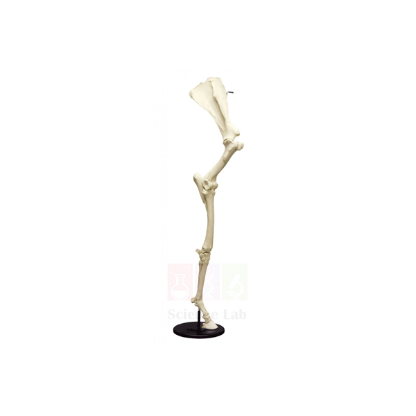
Horse Forelimb Skeleton
Horse Forelimb Skeleton (Product Code: SLE/A/120) is an articulated natural-bone specimen of an adult horse's forelimb used to study equine skeletal anatomy in veterinary, animal-science and zoology teaching. It stands on a square wooden base with a steel support rod, and the hoof capsule is mounted separately so it can be removed for close-up examination. The specimen is supplied by Jain Scientific Equipments Pvt Ltd (ScienceLab), Ambala, India.
Specifications
| Parameter | Detail |
|---|---|
| Material | Natural bone |
| Specimen | Adult horse (equine) forelimb, articulated mount |
| Size | Around 13 x 51-1/2 in. |
| Base | Square wooden base, 11-1/4 x 11-1/4 in. |
| Support | Steel support rod |
| Hoof capsule | Mounted separately on the base; removable for close examination |
| Product code | SLE/A/120 |
| Supplied by | Jain Scientific Equipments Pvt Ltd (ScienceLab), Ambala, India |
What Is the Horse Forelimb Skeleton Used For?
In veterinary osteology the horse forelimb is studied bone by bone, and an articulated natural-bone specimen shows how each joint actually fits together in a way that loose bones cannot. Students trace the limb from its upper long bones down to the single functional digit, note where tendons and ligaments would attach, and relate joint angles to how a horse stands and moves. The removable hoof capsule lets the class compare the outer hoof with the bone it encloses.
The specimen is equally valuable as evidence for evolution. A limb reduced to one weight-bearing digit is a textbook example of adaptation for running, and placing it beside a human arm, a bat wing or a bird wing helps students recognise one basic bone pattern modified for different purposes. Mounted upright on its base, it also makes a striking display in an anatomy hall and a distinctive addition to zoology lab equipment in India.
Supplier
Jain Scientific Equipments Private Limited, which trades as ScienceLab from Ambala, Haryana, supplies this natural-bone forelimb mount to veterinary colleges, zoology departments, anatomy museums and dealers in India and international markets. The company sources specimens of this kind and handles quotations, packing advice and delivery questions directly. See our company profile for more about the company.
Related Animal Models
Skeletons that support a lesson on homologous limbs:
- Bat Skeleton — a forelimb adapted as a wing, in natural bone with a key card.
- Pigeon Skeleton — shows the bird wing for comparison with the equine limb.
- Human Skeleton, Rod-Supported — a life-size human skeleton for comparing the arm and hand.
- Frog Skeleton — an amphibian limb pattern to complete the vertebrate series.
More specimens and models are in the Animal Model category.
Frequently Asked Questions
Is the Horse Forelimb Skeleton real bone or a cast?
The Horse Forelimb Skeleton is natural bone. The bones are articulated and held by a steel support rod on a square wooden base, so students see the real joint surfaces and proportions of an adult horse's forelimb.
What size is the Horse Forelimb Skeleton?
The Horse Forelimb Skeleton measures around 13 x 51-1/2 inches and stands on a square wooden base of 11-1/4 x 11-1/4 inches, so plan for a tall display space or a sturdy bench.
Can the hoof be removed from the Horse Forelimb Skeleton?
Yes. On the Horse Forelimb Skeleton the hoof capsule is mounted separately on the base and can be taken off, allowing students to compare the outer hoof with the bones it encloses.
Who supplies the Horse Forelimb Skeleton?
The Horse Forelimb Skeleton is supplied by Jain Scientific Equipments Pvt Ltd (ScienceLab), Ambala, Haryana, India, to veterinary colleges, zoology departments and museums in India and international markets, with quotations issued directly by the company.
How should the Horse Forelimb Skeleton be cared for?
Keep the Horse Forelimb Skeleton in a dry room away from damp and direct sunlight, dust it with a soft brush, and avoid washing it or applying chemical cleaners, which can damage natural bone over time.
Are there shipping considerations for the Horse Forelimb Skeleton?
Documentation and packing needs for a natural-bone specimen such as the Horse Forelimb Skeleton can vary by destination. Tell us where the order will be delivered, in India or abroad, and we will advise in the quotation.
Is a labelled key included with the Horse Forelimb Skeleton?
A key is not mentioned in the Horse Forelimb Skeleton's product details. If your osteology course needs one, please ask through the contact page before ordering so we can confirm what accompanies the specimen.
Request a Quote
Ask for a price on the Horse Forelimb Skeleton through our contact page, or add it to the enquiry cart alongside other skeletons.
Last updated: 24 September 2026
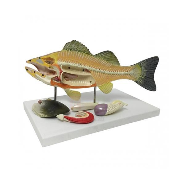
Fish Dissection Model
Fish Dissection Model (Product Code: SLE/A/121) is a life-size, 5-part longitudinal dissection of a bony fish used for organ-identification practicals in school biology, fisheries and college zoology. Its removable components are held by magnets and a numbered key identifies 24 structures, giving clear views of both internal and external anatomy. It is made by Jain Scientific Equipments Pvt Ltd (ScienceLab), the OEM manufacturer, at its own factory in Ambala, India, as part of its high school biology lab equipment.
Specifications
| Parameter | Detail |
|---|---|
| Subject | Bony fish, longitudinal dissection |
| Scale | Life size |
| Construction | 5 parts |
| Size (without base) | 45 x 21 x 7 cm |
| Fixing of parts | Removable components secured with magnets |
| Mounting | On a base |
| Key | Numbered key with 24 identified structures |
| Product code | SLE/A/121 |
| Manufacturer | Jain Scientific Equipments Pvt Ltd (ScienceLab) — OEM |
| Country of origin | India (made in Ambala, Haryana) |
What Is the Fish Dissection Model Used For?
The 24-point key makes this model well suited to assessed practical work. A common format is to give each group the model with its parts in place and a blank outline drawing: students remove the magnetic components one at a time, match what they uncover to the numbered key and label their drawing, then reassemble the fish for the next group. Teachers can also cover the key and use the numbers for a quick oral test in revision periods before board practicals.
Because the dissection runs along the length of the body, students see how organs are arranged from head to tail and how the internal layout relates to the fish's outer shape. The magnets return each part to exactly the right position without pins, so the model stays tidy through repeated handling across a term. Institutions equipping several labs often include it in a biology model kit bulk order together with frog and bird models.
OEM Manufacturer
Jain Scientific Equipments Private Limited, operating as ScienceLab, is the OEM of the Fish Dissection Model and makes it at its own licensed factory at Saha, Ambala, Haryana, India. Campuses and distributors buying several units deal directly with the manufacturer for quotations, repeat orders and questions about spare magnetic components. Our company profile gives more background.
Related Animal Models
Items that work with this model in fish and vertebrate practicals:
- Bony Fish Model — a life-size carp replica with a removable air bladder, as an alternative format.
- Fish Dissection — a preserved dissection in a glass container to compare with the model.
- Dissection Pan, Pad & Cover — a pan-and-pad system that replaces dissection wax, for courses that also dissect real specimens.
- Frog Dissection Model — uses the same magnet-held parts for the amphibian type study.
The full range is listed in the Animal Model category.
Frequently Asked Questions
How many structures does the Fish Dissection Model's key identify?
The numbered key supplied with the Fish Dissection Model identifies 24 structures, which makes the model well suited to assessed identification exercises in which students locate each numbered organ and label an outline drawing.
How many parts does the Fish Dissection Model have?
The Fish Dissection Model is a longitudinal dissection in five parts. Its removable components are secured with magnets, so they return to exactly the right position after handling without the need for pins or hooks.
What are the dimensions of the Fish Dissection Model?
The Fish Dissection Model measures 45 x 21 x 7 cm without its base, and the fish is shown life size, so students see organs at the same scale they would meet in a real specimen.
Who manufactures the Fish Dissection Model?
The Fish Dissection Model is manufactured by Jain Scientific Equipments Pvt Ltd (ScienceLab), which is its OEM, at the company's own licensed factory at Saha, Ambala, Haryana, India. Schools, colleges and dealers order it factory-direct, with quotations from the manufacturer.
Which fish species is the Fish Dissection Model based on?
The Fish Dissection Model represents a bony fish; the exact species is not stated in its product details. If your syllabus names a particular fish, ask through the contact page and we will confirm the details before you order.
Should I choose the Fish Dissection Model or the Bony Fish Model?
Choose the Fish Dissection Model for key-driven identification work with numbered structures and magnet-held parts. The Bony Fish Model, a carp replica with external and internal sides and three removable organs, suits a more general introduction to fish anatomy.
Request a Quote
For pricing on the Fish Dissection Model, use the contact page or add it to the enquiry cart with any other models you need; quotations come straight from the manufacturer.
Last updated: 24 September 2026
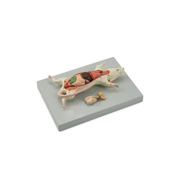
Male and Female Rat Model
Male and Female Rat Model (Product Code: SLE/A/122) is a life-size model of an adult rat, mounted supine, used to teach mammalian anatomy and to compare male and female reproductive systems in zoology and life-science courses. The reproductive systems are interchangeable, the organs detach in 4 pieces and a numbered key is included, so one model covers both sexes. It is made by Jain Scientific Equipments Pvt Ltd (ScienceLab), the OEM manufacturer of biology lab equipment in India, at its own factory in Ambala.
Specifications
| Parameter | Detail |
|---|---|
| Subject | Adult rat |
| Scale | Life size |
| Position | Mounted supine |
| Size (without base) | 12 x 21 x 4 cm |
| Detachable organs | 4 pieces |
| Reproductive systems | Interchangeable male and female |
| Base | Model removable from base for examination |
| Supplied with | Numbered key |
| Product code | SLE/A/122 |
| Manufacturer | Jain Scientific Equipments Pvt Ltd (ScienceLab) — OEM |
| Country of origin | India (made in Ambala, Haryana) |
What Is the Male and Female Rat Model Used For?
The rat is a standard mammal for anatomy teaching, and the supine position matches the way it is traditionally opened in a practical, so students see organs in the orientation used in their textbooks. A lesson can begin with one reproductive system fitted, identifying the general body organs from the numbered key, then switch to the other system so the class compares male and female structures directly on the same body. Detaching the four organ pieces lets students handle each one and see where it sits in the body cavity.
At life size the model is small, so it works best for bench work in pairs or small groups; students can lift it off the base and turn it to view structures from different angles. For departments that have moved away from animal dissection it is a reusable way to meet mammalian anatomy objectives, and among the animal models a biology lab keeps it pairs naturally with bird and amphibian models for cross-class comparison.
OEM Manufacturer
The Male and Female Rat Model is manufactured by Jain Scientific Equipments Private Limited, which trades as ScienceLab and is the OEM (original equipment manufacturer). It is made at the company's own licensed factory at Saha, Ambala, Haryana, and supplied factory-direct to colleges, paramedical institutes, dealers and importers, who can ask the manufacturer itself about spare organ pieces or repeat orders. The company profile explains more.
Related Animal Models
Items that build on this model in reproduction and comparative anatomy lessons:
- Human Male and Female Urogenital Systems Model — the human equivalent for comparing mammalian reproductive anatomy.
- Hen Model — shows a bird reproductive system with an egg for cross-class comparison.
- Frog Dissection Model — the amphibian type study with magnet-held removable parts.
- Stereoscopic Dissection Microscopes — low-power viewing for fine details on small specimens and models.
Browse the Animal Model category for more.
Frequently Asked Questions
Does one Male and Female Rat Model show both sexes?
Yes. The Male and Female Rat Model has interchangeable male and female reproductive systems on the same adult rat, so a class can study both without buying two separate models or dissecting animals.
How large is the Male and Female Rat Model?
The Male and Female Rat Model is life size and measures 12 x 21 x 4 cm without its base, which suits bench work in pairs or small groups rather than display in a large lecture hall.
How many parts come off the Male and Female Rat Model?
On the Male and Female Rat Model the organs detach in four pieces, and the whole rat can be removed from its base so students can turn it and examine structures from different angles.
Who manufactures the Male and Female Rat Model?
Jain Scientific Equipments Pvt Ltd (ScienceLab) is the OEM of the Male and Female Rat Model and manufactures it at its own licensed factory at Saha, Ambala, Haryana, India, supplying it factory-direct with quotations from the manufacturer.
Is a key included with the Male and Female Rat Model?
Yes. A numbered key is included with the Male and Female Rat Model, so students can identify the body organs and both sets of reproductive structures and use the same names in their practical records.
How should the small parts of the Male and Female Rat Model be kept safe?
Store the spare reproductive system and detached organs of the Male and Female Rat Model in a labelled box with the model, and check that all pieces are returned at the end of each class.
Request a Quote
Enquire about the Male and Female Rat Model through the contact page or place it in the enquiry cart for a combined quotation sent directly by the manufacturer.
Last updated: 24 September 2026
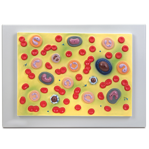
Human Blood Cell Types Model
Human Blood Cell Types Model (Product Code: SLE/A/140) is a 54 x 38 cm plaque-mounted teaching model used to show erythrocytes, platelets and 5 types of leukocytes in biology and health-science classrooms. Biology, nursing and medical laboratory technology teachers choose it because every structure is numbered on an accompanying key and each leukocyte shows its characteristic nucleus. It is made by Jain Scientific Equipments Pvt Ltd (ScienceLab), an OEM anatomical models manufacturer, at its own factory in Ambala, India.
Specifications
| Parameter | Detail |
|---|---|
| Format | Plaque mount |
| Size | 54 x 38 cm |
| Red cells | Erythrocytes |
| White cells | 5 types of leukocytes with their characteristic nuclei |
| Other formed elements | Platelets |
| Key | Structures numbered and referenced on the accompanying key |
| Product code | SLE/A/140 |
| Manufacturer | Jain Scientific Equipments Pvt Ltd (ScienceLab) — OEM |
| Country of origin | India (made in Ambala, Haryana) |
What Is the Human Blood Cell Types Model Used For?
Telling white blood cells apart is one of the first microscopy skills health-science students must master, and the shape of the nucleus is the main clue. With five leukocyte types shown side by side, a teacher can walk through the usual identification points: the lobed nucleus of the neutrophil, the two-lobed nucleus of the eosinophil, the basophil nucleus often hidden by its granules, the large round nucleus of the lymphocyte and the kidney-shaped nucleus of the monocyte. Erythrocytes, which have no nucleus, and the much smaller platelets complete the picture.
In a practical session, place the plaque where students can see it from the microscope bench. They examine a stained blood smear, sketch the cells they find and check each identification against the numbered key, which prepares them for differential leukocyte counts. Colleges building up their biology teaching aids in India often set it beside prepared slides so that theory and microscopy are covered in the same lesson.
OEM Manufacturer
Jain Scientific Equipments Private Limited, which trades as ScienceLab, is the OEM (original equipment manufacturer) of the Human Blood Cell Types Model. The plaque is made and finished at the company’s own licensed factory at Saha, Ambala, Haryana, India, rather than being sourced from a trader. The company is incorporated under CIN U29309HR2020PTC087597, and its factory at 449, HSIIDC Industrial Area, Saha, Ambala operates under Factory Licence AMB-ONLINE-CHD-J-414.
Schools, colleges, dealers and importers buy the model factory-direct, and questions about repeat orders or a replacement key go straight to the people who make it — just ask us. The company profile describes the business in more detail.
Related Laboratory Equipment
Items used with this model in a haematology practical:
- Human Blood Smear — a prepared slide for finding the same cell types under the microscope.
- Monocular Microscope 1000x, LED light — enough magnification to distinguish leukocyte nuclei on a smear.
- Haemocytometer — a counting chamber for the cell counts that follow identification.
- Blood Typing Kit, Simulated — ABO and Rh grouping practice without real blood.
The model is listed in our Animal Model category, alongside other biology teaching models.
Frequently Asked Questions
Which blood cells does the Human Blood Cell Types Model show?
The Human Blood Cell Types Model shows erythrocytes, platelets and 5 types of leukocytes on a single plaque, and each leukocyte is modelled with its characteristic nucleus so that students can compare the cells side by side before examining stained smears.
What size is the Human Blood Cell Types Model?
The Human Blood Cell Types Model is a plaque measuring 54 x 38 cm, large enough for a teacher to point out individual cells during a whole-class explanation and compact enough to store flat between practical sessions.
Does the Human Blood Cell Types Model come with a key?
Yes. Every structure on the Human Blood Cell Types Model is numbered and referenced on an accompanying key, so students can check their own identifications of red cells, platelets and each leukocyte type without waiting for the teacher.
At what magnification is the Human Blood Cell Types Model made?
The enlargement factor of the Human Blood Cell Types Model is not stated in its product details. If your course notes need it, ask us through the contact page and we will check before you order.
Who manufactures the Human Blood Cell Types Model?
Jain Scientific Equipments Pvt Ltd (ScienceLab) manufactures the Human Blood Cell Types Model as the OEM at its own licensed factory at Saha, Ambala, Haryana, India, and supplies it factory-direct to schools, colleges, dealers and importers in India and international markets.
How should the Human Blood Cell Types Model be cleaned?
Clean the Human Blood Cell Types Model by wiping the plaque gently with a soft, dry or slightly damp cloth. Avoid alcohol and solvent cleaners, which may damage the finish, and store it flat away from direct sunlight.
What does a quotation for the Human Blood Cell Types Model need?
A quotation for the Human Blood Cell Types Model needs the quantity, the delivery location and any other haematology or biology items you want on the same order; ScienceLab then quotes directly as the manufacturer.
Request a Quote
Ask for a price on the Human Blood Cell Types Model through our contact page, or collect it in the enquiry cart with your other biology items. Quotations come direct from the manufacturer.
Last updated: 24 September 2026