- Physics Lab Equipment
- Chemistry Lab Equipment
- Biology Lab Equipment
- Anatomical Lab Equipment
- Learning
- Educational Lab Equipment
- Magnets
- Agriculture
- Astronomy
- Earth Science
- Geography Lab Equipment
- Safety Lab Coat
- Laboratory
- Meteorological
- Electricity & Magnetism
- Heat
- School Lab Equipment
- Lab Glassware
- Science Lab Equipment
- Tvet Lab Equipment
- Robotics Kits Supplier and Manufacturer for Schools India
- Math Lab Equipment Manufacturers and Exporters in Ambala
- STEM Kits Manufacturers and Supplier India
- Engineering Lab Equipment
- Hospital Lab Equipment
- Water, Sanitation & Hygiene Supplies
- Cold Chain, Solar & Communication Equipment
- Nutrition
Cell biology equipment makes cell structure and cell division visible at a scale the whole class can see, for school and college biology teaching. Jain Scientific Equipments Pvt Ltd (ScienceLab), Ambala, groups 21 products in this Cell Biology Equipments range: plant, animal and prokaryotic cell models, organelle models such as the mitochondrion and chloroplast, relief plaques, mitosis and meiosis models, osmosis and diffusion investigations and wall posters. Most of the models are made at its own licensed factory in Ambala, while the posters and a few class kits are supplied.
What Does the Cell Biology Equipments Range Include?
| Topic | What it covers | Example products |
|---|---|---|
| Plant cell models | Enlarged plant cells for comparing organelles with animal cells | Plant Cell Model; Model Plant Cell; Plant Cell |
| Relief plaques | Large relief models identified with key cards | Plant Cell Plaque; Animal Cell Plaque |
| Cell division | Stages of mitosis and meiosis as models, plaques and a class simulation | Animal Mitosis Models; Meiosis Model; Animal Meiosis Plaque |
| Single-celled organisms | Protist and bacterial cell structure | Euglena Model; Amoeba Model; Prokaryotic Cell Model |
| Membranes and transport | Repeatable demonstration of osmosis with different solutes | Osmosis Simulation Activity Model |
| Posters and fieldwork | Reference posters, plus a sampler for collecting pond, lake or river water | Visualising Cells Poster; Visualizing Mitosis Poster; Water Sampler |
Featured Cell Biology Equipments
| Product | Product Code | Typical Use |
|---|---|---|
| Animal Mitosis Models | SLE/ECB/08 | Set of 8 sequential models showing chromosomes and the mitotic apparatus at each stage |
| Mitochondrion Model | SLE/ECB/07 | Mitochondrion enlarged 190,000 times, revealing membranes, cristae and matrix |
| Chloroplast Model | SLE/ECB/02 | Translucent resin chloroplast at about 50,000X, showing membranes, starch granules and ribosomes |
| Animal Cell Plaque | SLE/ECB/06 | 46 x 61 cm relief model of the animal cell with identifying key cards |
| Plant Cell on Base Model | SLE/ECB/20 | Electron-microscope view of a plant cell with part of the wall removed to show vacuoles, plastids and mitochondria |
| Hands-On Cell Model Act-Plant | SLE/ECB/01 | Foam plant cell with removable parts and worksheets for grades 3 to 8 |
| Mitosis Simulation Lab Investigation | SLE/ECB/14 | Pop-bead chromosomes for modelling each stage of mitosis, for a class of 40 |
| Visualising Osmosis & Diffusion Investigation | SLE/ECB/19 | Coloured dye solutions and a model cell that show selective permeability, for a class of 40 |
OEM Manufacturing & Supply
Jain Scientific Equipments Private Limited, the company behind ScienceLab, is the OEM for most of this range. The plant, animal and prokaryotic cell models, the mitochondrion, chloroplast, Euglena and Amoeba models, the relief plaques, the Animal Mitosis Models and Meiosis Model, and the Mitosis Simulation Lab Investigation and Osmosis Simulation Activity Model are manufactured at its own licensed factory at 449, HSIIDC Industrial Area, Saha, Ambala. The company is a registered MSME with manufacturing as its major activity (Udyam UDYAM-HR-01-0003714) and holds global Legal Entity Identifier 3358004HRD1JIVXKYX49. The foam Hands-On Cell Model Act-Plant, the Visualising Cells and Visualizing Mitosis posters, the Visualising Osmosis & Diffusion Investigation and the Water Sampler are supplied rather than made in-house. Models can be ordered singly for a demonstration bench, as class sets, or in bulk and tender quantities for institutions in India and other countries.
Related Ranges
This range belongs to the Biology Lab Equipment hub. It shows cells at model scale; the matching real specimens are in Prepared Slides, while the molecules inside the cell, such as DNA and proteins, are modelled in Molecule. Hands-on cell and chromosome staining activities are covered by kits in Biology Kits, and the Visking tubing used in osmosis practicals is listed in the core Biology Lab Equipment apparatus range.
Frequently Asked Questions
Who makes the Cell Biology Equipments models?
Most of the Cell Biology Equipments range is manufactured by Jain Scientific Equipments Pvt Ltd (ScienceLab) at its Ambala factory, including the cell and organelle models, plaques and mitosis and meiosis models. The posters, the Hands-On Cell Model Act-Plant, the Visualising Osmosis & Diffusion Investigation and the Water Sampler are supplied.
Does this range include laboratory instruments such as centrifuges?
No. The Cell Biology Equipments range covers models, plaques, posters and class investigations rather than bench instruments. Centrifuges are listed in the Centrifuges range and microscopes in the Microscope range, and both can be quoted together with these models.
Which products teach cell division?
In the Cell Biology Equipments range, the Animal Mitosis Models, the Meiosis Model, the Animal Meiosis Plaque, the Mitosis Simulation Lab Investigation and the Visualizing Mitosis Poster together cover the stages of mitosis and meiosis, from demonstration models to a hands-on class activity.
Which models compare plant and animal cells?
For comparing plant and animal cells, the Cell Biology Equipments range pairs the Animal Cell Plaque with the Plant Cell Plaque as matching relief models. The Plant Cell on Base Model and the Plant Cell Model can also be used alongside animal cell teaching.
Are there cell biology activities a whole class can do at once?
Yes. The Cell Biology Equipments range includes the Mitosis Simulation Lab Investigation and the Visualising Osmosis & Diffusion Investigation, each listed for a class of 40 students, so a whole class can work through the activity at the same time.
Request a Quote or Catalogue
Use the contact page to request a quotation or catalogue for cell biology models, or add products to the shopping cart and send your list with quantities and the delivery country.
Last updated: 24 September 2026
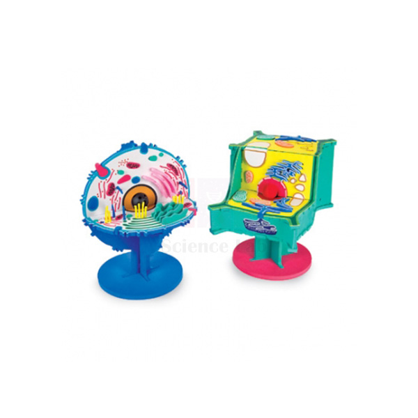
Hands-On Cell Model Act-Plant
Hands-On Cell Model Act-Plant (Product Code: SLE/ECB/01) is a tactile plant cell model used to teach cell structure to Grades 3 to 8 through an activity in which pupils lift out, name and replace each part. It is made of resilient, colourful EVA foam with removable parts, shows the plant cell only, and comes with reproducible worksheets and cross-curricular labs. It is supplied by Jain Scientific Equipments Pvt Ltd (ScienceLab), Ambala, India, as primary and middle-school biology lab equipment.
Key Details
| Parameter | Detail |
|---|---|
| Type | Hands-on cell model with removable parts |
| Cell shown | Plant cell only |
| Material | Resilient, colourful EVA foam |
| Suitable for | Grades 3 to 8 |
| Teaching support | Reproducible worksheets and cross-curricular labs |
| Product code | SLE/ECB/01 |
| Supplied by | Jain Scientific Equipments Pvt Ltd (ScienceLab), Ambala, India |
What Is the Hands-On Plant Cell Model Used For?
Younger learners understand a cell far better when they can take it apart. Teachers pass the model around a group, ask pupils to remove each part, say what it does, and fit it back in the right place, turning the lesson on plant cell structure into an activity rather than a diagram to copy. Because the parts are foam, they are light, quiet on the desk and easy for small hands to manage.
The reproducible worksheets let the same model serve several sections of a class, and the cross-curricular labs link cell biology with other subjects in the timetable. For schools choosing a plant cell model in India for the junior classes, it works well at the start of a unit, before pupils progress to observing real plant cells, such as onion peel, under a microscope. Keep the removable parts together in a labelled box after each lesson so that none go missing.
Supplier
ScienceLab is the trading name of Jain Scientific Equipments Private Limited, Ambala, Haryana, India, which supplies this hands-on plant cell model to schools, education departments and distributors in India and international markets. Teachers can order it together with animal cell models, posters and microscopes in one enquiry to equip a complete cell-biology unit. Visit our company profile to learn more.
Related Cell Biology Equipment
Resources that extend a hands-on plant cell lesson:
- Animal Cell Model — the animal counterpart, for comparing plant and animal cells
- Plant Cell Model — an enlarged display model of plant cell organelles for teacher demonstration
- Plant Cell Study Kit — a practical kit for studying plant cells directly
- Visualising Cells Poster — a wall reference to keep up during the unit
- Monocular Biological Microscope 400x — for moving on to real cells under magnification
Browse all cell models, plaques and activities in our Cell Biology Equipment range.
Frequently Asked Questions
Does the Hands-On Cell Model Act-Plant include an animal cell?
No. The Hands-On Cell Model Act-Plant shows the plant cell only. For lessons that compare plant and animal cells, pair it with our Animal Cell Model so pupils can see the differences side by side.
Which classes is the Hands-On Cell Model Act-Plant designed for?
The Hands-On Cell Model Act-Plant is intended for Grades 3 to 8, when pupils first meet cells and their parts. The removable foam pieces suit group activities where children take the cell apart and rebuild it.
What teaching material comes with the Hands-On Cell Model Act-Plant?
Reproducible worksheets and cross-curricular labs come with the Hands-On Cell Model Act-Plant. If you need to know their format or language before ordering, please contact us and we will confirm the details.
How should the foam model be cleaned and stored?
Wipe the parts of the Hands-On Cell Model Act-Plant with a slightly damp cloth and let them dry naturally. Store the model away from direct heat and sunlight, and keep all removable pieces together in a labelled box between lessons.
What are the dimensions of the Hands-On Cell Model Act-Plant?
The overall size of the Hands-On Cell Model Act-Plant is not given in our current product information. Ask us and we will confirm the dimensions before you place an order.
Who supplies the Hands-On Cell Model Act-Plant?
The Hands-On Cell Model Act-Plant is supplied by Jain Scientific Equipments Pvt Ltd (ScienceLab), Ambala, Haryana, India, to schools, education departments and distributors in India and international markets, often alongside other cell biology models.
Can we order models for several classrooms?
Yes. The Hands-On Cell Model Act-Plant can be supplied in bulk to schools, education departments and distributors in India and international markets. Include the quantity and delivery location in your enquiry for a quotation.
Request a Quote
For pricing or classroom quantities, contact us or add the Hands-On Cell Model Act-Plant to your enquiry cart.
Last updated: 24 September 2026
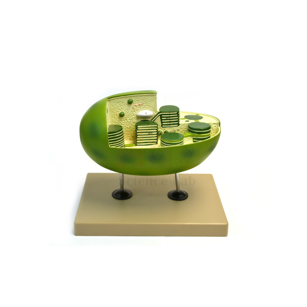
Chloroplast Model
Chloroplast Model (Product Code: SLE/ECB/02) is an enlarged teaching model of the plant cell's fundamental organelle, used to show how a chloroplast is built for photosynthesis in senior-school and college biology classes. Handcrafted in translucent fibre-reinforced resin at approximately 50,000X, it has a cutaway face exposing the thylakoid space and cross-cut grana, and comes with four reproducible keycards. It is made by Jain Scientific Equipments Pvt Ltd (ScienceLab), an OEM manufacturer, at its own factory in Ambala, India.
Specifications
| Parameter | Detail |
|---|---|
| Type | Enlarged chloroplast (plant cell organelle) model |
| Magnification | Approx. 50,000X |
| Material | Translucent fibre-reinforced resin, handcrafted |
| Structures shown | Circular chromosomes, outer and inner organelle membranes, inter-membrane space, starch granules, lipid inclusions and ribosomes |
| Cutaway face | Exposes the thylakoid space and membranes of cross-cut grana |
| Grana | Protruding grana demonstrate their reticulated structure |
| Key cards | Four reproducible keycards: colour and black-and-white, labelled and unlabelled |
| Size (approx.) | 24 x 23 x 18 cm (9.5 x 9 x 7 in) |
| Weight | 2.4 kg |
| Product code | SLE/ECB/02 |
| Manufacturer | Jain Scientific Equipments Pvt Ltd (ScienceLab) — OEM |
| Country of origin | India (made in Ambala, Haryana) |
What Is the Chloroplast Model Used For?
Teachers use the model to connect structure with function in photosynthesis: the stacked thylakoid membranes of the grana, seen on the cutaway face, are where the light-dependent reactions take place, while the surrounding fluid region is where carbon is fixed into sugars and later stored as the starch granules visible on the model. The circular chromosomes and ribosomes also give a concrete starting point for discussing why chloroplasts are thought to have evolved from free-living bacteria.
A practical way to run the lesson is to project or photocopy the labelled colour keycard during teaching, then hand out the unlabelled black-and-white card as a quick identification test. As a piece of plant biology lab equipment, the model pairs naturally with a photosynthesis practical, so that students measure oxygen output or starch formation and then see on the model where each process happens.
OEM Manufacturer
Jain Scientific Equipments Private Limited, trading as ScienceLab, is the OEM (original equipment manufacturer) of the Chloroplast Model and makes it at its own licensed factory at Saha, Ambala, Haryana, India. Biology departments, dealers and importers buy it factory-direct, and the manufacturer itself answers questions about the model and handles repeat orders for additional classrooms. Institutions setting up a new vendor can use the company's GSTIN 06AAECJ8618A1ZT and Legal Entity Identifier 3358004HRD1JIVXKYX49. More about the company is on our company profile.
Related Cell Biology Equipment
Models and practical resources that complement a chloroplast lesson:
- Mitochondrion Model — the respiration organelle, for comparing the two energy-converting organelles
- Plant Cell Model — shows where chloroplasts sit within the whole plant cell
- Photosynthesis Apparatus — for the practical side of the same topic
- Microslide Photosynthesis — prepared microscope slide material on the same topic
See more organelle and cell models in our Cell Biology Equipment range.
Frequently Asked Questions
How much is the Chloroplast Model enlarged?
The Chloroplast Model is made at approximately 50,000X magnification, which turns a microscopic organelle into a desk-sized model whose membranes, grana and inclusions can be pointed out to a whole class.
What is on the four keycards supplied with the Chloroplast Model?
The Chloroplast Model comes with four reproducible keycards: colour and black-and-white versions, each labelled and unlabelled. Teachers typically use the labelled cards for teaching and the unlabelled ones as a quick identification test.
Can students see inside the grana on the Chloroplast Model?
Yes. A cutaway face on the Chloroplast Model exposes the thylakoid space and the membranes of cross-cut grana, while protruding grana show their reticulated structure from the outside, so both views can be compared.
Which structures does the Chloroplast Model show?
The Chloroplast Model shows circular chromosomes, outer and inner organelle membranes, the inter-membrane space, starch granules, lipid inclusions and ribosomes, together with the thylakoid membranes and grana revealed by its cutaway face.
How big and heavy is the Chloroplast Model?
The Chloroplast Model measures approximately 24 x 23 x 18 cm (9.5 x 9 x 7 in) and weighs 2.4 kg, a convenient size for a teacher's desk, a demonstration bench or a group table.
How should the resin Chloroplast Model be cared for?
Dust the Chloroplast Model with a soft, dry cloth and avoid solvents or abrasive cleaners that could cloud the translucent resin. Store it covered on a stable shelf, away from long exposure to direct sunlight.
Who manufactures the Chloroplast Model?
The Chloroplast Model is manufactured by Jain Scientific Equipments Pvt Ltd (ScienceLab), the OEM, at its own factory in Saha, Ambala, Haryana, India. Schools, colleges and distributors in India and international markets receive quotations directly from the manufacturer.
Request a Quote
Quotations for the Chloroplast Model come directly from the manufacturer: contact us or add the model to your enquiry cart.
Last updated: 24 September 2026
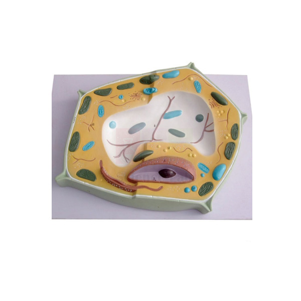
Plant Cell Model
Plant Cell Model (Product Code: SLE/ECB/03) is a hugely enlarged teaching model of a typical plant cell, used to show all types of organelle in one view during secondary-school and college biology lessons. Enlarged 20,000 times and measuring 42 x 30 x 8 cm, it is big enough for a teacher to point out each structure to the whole class. It is made by Jain Scientific Equipments Pvt Ltd (ScienceLab), an OEM manufacturer, at its own factory in Ambala, India, as part of its botany lab equipment.
Specifications
| Parameter | Detail |
|---|---|
| Type | Enlarged plant cell model |
| Enlargement | 20,000 times |
| Size | 42 x 30 x 8 cm |
| Shows | Typical plant cell structure with all types of organelle |
| Product code | SLE/ECB/03 |
| Manufacturer | Jain Scientific Equipments Pvt Ltd (ScienceLab) — OEM |
| Country of origin | India (made in Ambala, Haryana) |
What Is the Plant Cell Model Used For?
Teachers use this model when introducing the cell as the basic unit of life, walking through the features that make a plant cell distinctive, such as the rigid cell wall, chloroplasts and the large central vacuole, alongside the nucleus, mitochondria and membrane systems it shares with animal cells. Because every organelle type is shown on one enlarged cell, students can see how the parts fit together rather than learning them as a list.
It works well as the reference point for an onion-peel or leaf-epidermis practical: students look at the model first, then find the cell wall, nucleus and vacuole in real cells under the microscope, and finally draw and label what they saw. As part of school biology lab equipment in India it also serves for quick revision before examinations and for comparison lessons with an animal cell model.
OEM Manufacturer
The Plant Cell Model is produced by Jain Scientific Equipments Private Limited, which operates under the ScienceLab brand and is the model's OEM (original equipment manufacturer). It is made at the company's own licensed factory at Saha, Ambala, Haryana, India, and sent factory-direct to schools, colleges, dealers and importers, so quotations and follow-up orders come from the maker rather than an intermediary. Read more on our company profile.
Related Cell Biology Equipment
Items that support a complete plant cell topic:
- Plant Cell Plaque — a relief plaque with key cards identifying its structures
- Chloroplast Model — zooms in on a single organelle to show its internal membranes
- Animal Cell Model — for side-by-side comparison of plant and animal cells
- Plant Cell Study Kit — hands-on material for the practical session
- Monocular Biological Microscope 400x, LED — for observing real plant cells
Explore more models in our Cell Biology Equipment range.
Frequently Asked Questions
How much is the Plant Cell Model enlarged?
The Plant Cell Model shows a typical plant cell enlarged 20,000 times, so organelles that are invisible to the naked eye become large, clearly separated features that students can identify from their seats.
What are the dimensions of the Plant Cell Model?
The Plant Cell Model measures 42 x 30 x 8 cm. At that size a teacher can hold it up or place it at the front of the room and point out each organelle to the whole class.
Which organelles does the Plant Cell Model show?
The Plant Cell Model shows typical plant cell structure with all types of organelle. If you need the exact list of labelled parts, or want to know whether a key is supplied, please contact us before ordering.
Is the Plant Cell Model free-standing or wall-mounted?
The mounting style and material of the Plant Cell Model are not stated in our current product information. We can confirm both when you enquire, so that you can plan where the model will be displayed in the classroom.
How should the Plant Cell Model be looked after?
Dust the Plant Cell Model gently with a soft brush or dry cloth, keep it away from prolonged direct sunlight and heat, and store it on a stable shelf where it cannot be knocked or dropped.
Who manufactures the Plant Cell Model?
The Plant Cell Model is manufactured by Jain Scientific Equipments Pvt Ltd (ScienceLab), the OEM, at its own factory in Saha, Ambala, Haryana, India. Schools, colleges and distributors order it directly from the manufacturer.
Do you supply several Plant Cell Models for different classrooms?
Yes. The Plant Cell Model is supplied in bulk to schools, colleges and distributors across India and international markets. Share the quantity and delivery details, and the manufacturer will send a quotation for the full order.
Request a Quote
For the manufacturer's price or bulk supply, contact us or add the Plant Cell Model to your enquiry cart.
Last updated: 24 September 2026
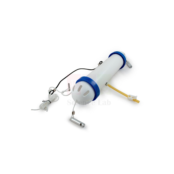
Water Sampler
Water Sampler (Product Code: SLE/ECB/04) is a flow-through field sampler used to collect a 1.5 litre water sample from any convenient depth in lakes, rivers and ponds for ecology and environmental science work. A single 14 metre line, supplied with it, both lowers the sampler and triggers the collection, so there are no tangled lines at the water's edge. It is supplied by Jain Scientific Equipments Pvt Ltd (ScienceLab), Ambala, India, for schools, colleges and environmental monitoring courses.
Specifications
| Parameter | Detail |
|---|---|
| Type | Flow-through water sampler |
| Sample volume | 1.5 litre |
| Line supplied | 14 metre |
| Operation | A single line deploys the sampler and triggers sample collection |
| Sampling depth | Any convenient depth within reach of the line |
| Suitable for | Lakes, rivers and ponds |
| Sample quality | Flow-through design allows valid sampling for temperature and water-quality measurement |
| Product code | SLE/ECB/04 |
| Supplied by | Jain Scientific Equipments Pvt Ltd (ScienceLab), Ambala, India |
What Is the Water Sampler Used For?
Surface water alone rarely tells the full story of a pond or lake. With this sampler, students collect water from the surface, the middle and near the bottom of the same site, then compare temperature, pH, dissolved oxygen and the small organisms present at each depth. As field biology lab equipment, it suits college limnology and ecology practicals, school eco-club surveys, and project work on environmental monitoring of local water bodies.
Lower the sampler on its line to the chosen depth, trigger the collection, then raise it steadily and pour the water into clean, labelled bottles. Read the temperature straight away, before the sample warms or cools. Always work in pairs near deep or flowing water, sample from a secure bank, jetty or boat, and rinse the sampler and line in clean water afterwards, letting the line dry before it is coiled for storage.
Supplier
ScienceLab is the trade name used by Jain Scientific Equipments Private Limited, based in Ambala, Haryana, India, which supplies the Water Sampler with its 14 metre line to schools, colleges and research groups in India and international markets. Field kits can be completed in the same enquiry with thermometers, meters and sample bottles from the wider range. See our company profile for details.
Related Laboratory Equipment
Instruments and containers used on the same water survey:
- Immersion Thermometer — for reading the sample temperature on site
- Waterproof pH Meter — measures acidity of each depth sample
- Digital Dissolved Oxygen Meter — compares oxygen levels between depths
- Bottle, Polyethylene, Widemouthed — for carrying labelled samples back to the laboratory
- Key to Identifying Water Invertebrates — for naming organisms found in the samples
This sampler is listed in our Cell Biology Equipment range.
Frequently Asked Questions
How much water does the Water Sampler collect each time?
The Water Sampler collects a 1.5 litre sample on each deployment, which is enough water to divide between separate tests for temperature, pH, dissolved oxygen and organisms once the sample is back on the bank or in the laboratory.
How deep can I sample with the Water Sampler?
The Water Sampler comes with a 14 metre line and can take water from any convenient depth within that reach, so students can compare samples from the surface, mid-water and near the bottom of the same site.
Do I need a second line to close the Water Sampler?
No. On the Water Sampler, the same single line lowers the sampler and triggers the collection. This keeps handling simple for student groups and avoids the tangled lines that can slow down fieldwork at the water's edge.
Is the Water Sampler's sample suitable for temperature and water-quality tests?
Yes. The flow-through design of the Water Sampler allows a valid sample for measuring temperature and water quality. Take the temperature reading immediately after the sampler is raised, before the water warms or cools in the air.
Can the Water Sampler be used in seawater?
Our product information for the Water Sampler covers lakes, rivers and ponds and does not state its construction material. Contact us before ordering if you plan marine or estuary work, so we can confirm suitability.
Who supplies the Water Sampler?
The Water Sampler is supplied by Jain Scientific Equipments Pvt Ltd (ScienceLab), Ambala, Haryana, India, to schools, colleges and environmental science departments in India and international markets, together with related field and laboratory equipment if required.
What should a quotation request for the Water Sampler include?
For the Water Sampler, include the number of samplers, any spare lines needed and the delivery address in India or abroad. Mention any other field equipment you need so that everything can be quoted together.
Request a Quote
For pricing and availability, contact us or add the Water Sampler to your enquiry cart.
Last updated: 24 September 2026
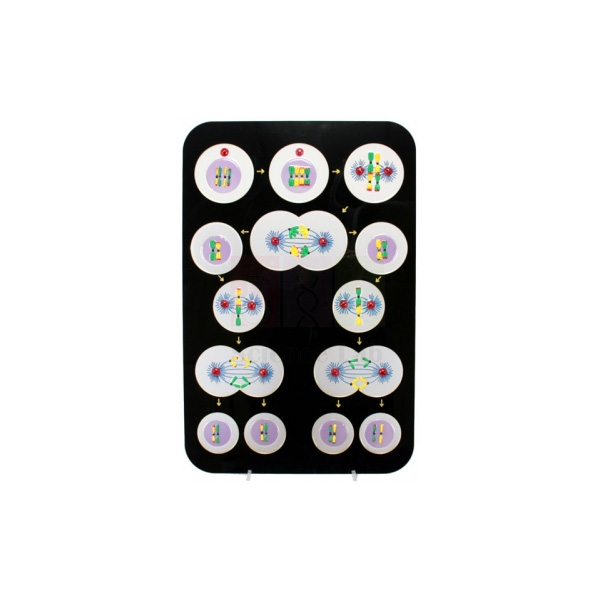
Animal Meiosis Plaque
Animal Meiosis Plaque (Product Code: SLE/ECB/05) is a large teaching plaque used to explain reduction division (meiosis) of the animal cell in school and college biology classrooms. Teachers use its single 46 x 61 cm (approx.) display, with the key cards included, to show how the chromosome number is halved when gametes form. It is made by Jain Scientific Equipments Pvt Ltd (ScienceLab), an OEM manufacturer, at its own factory in Ambala, India, for schools that need a clear mitosis meiosis model in India.
Specifications
| Parameter | Detail |
|---|---|
| Type | Teaching plaque, animal cell meiosis |
| Process shown | Reduction division (meiosis) of the animal cell |
| Key cards | Included |
| Size | 46 x 61 cm (approx.) |
| Weight | 2 kg (approx.) |
| Product code | SLE/ECB/05 |
| Manufacturer | Jain Scientific Equipments Pvt Ltd (ScienceLab) — OEM |
| Country of origin | India (made in Ambala, Haryana) |
What Is the Animal Meiosis Plaque Used For?
Meiosis is one of the harder topics in secondary and first-year college biology, because students must follow chromosomes through two divisions and understand why the number is halved. The plaque lets the teacher trace that sequence in front of the class: homologous chromosomes pairing and separating, sister chromatids parting in the second division, and the resulting cells carrying half the original chromosome number. That foundation then supports later lessons on variation and inheritance.
Use the key cards first as a teaching aid and later as a quick check, asking students to name each feature before revealing the card. Keeping the plaque on display for the whole unit helps learners revisit the sequence when they study mitosis or work through prepared meiosis slides under the microscope.
OEM Manufacturer
Jain Scientific Equipments Private Limited, which trades as ScienceLab, is the OEM (original equipment manufacturer) of the Animal Meiosis Plaque and makes it at its own licensed factory at Saha, Ambala, Haryana, India. Because the plaque comes straight from the maker rather than through a trader, schools, colleges, dealers and importers deal with the same company for the quotation, repeat orders and any replacement key cards — just ask us. More about the company is on our company profile.
Related Cell Biology Equipments
Resources for teaching cell division from model to microscope:
- Animal Mitosis Models — a sequential set for comparing mitosis with meiosis.
- Meiosis Model — another format for presenting the same process.
- Microslide Meiosis — photomicrograph images for seeing meiosis in real cells.
- Mitosis & Meiosis Set, Microscope Slides — a combined slide set covering both divisions.
- Animal Cell Plaque — the matching plaque on animal cell structure.
View the full Cell Biology Equipments range.
Frequently Asked Questions
What does the Animal Meiosis Plaque illustrate?
The Animal Meiosis Plaque illustrates reduction division, or meiosis, of the animal cell — the division that halves the chromosome number when gametes form. It gives a whole class one large visual for following the process, with key cards for identifying what is shown.
Are key cards included with the Animal Meiosis Plaque?
Yes. Key cards are included with the Animal Meiosis Plaque so that teachers and students can identify the features shown. They serve as a labelling reference during the lesson and as a quick self-test afterwards, when students name each part before checking the card.
How large and heavy is the Animal Meiosis Plaque?
The Animal Meiosis Plaque measures approximately 46 x 61 cm and weighs approximately 2 kg. That is large enough for a class to follow from the front rows, while the light weight lets one teacher carry it between rooms.
Which individual stages does the Animal Meiosis Plaque show?
Our current product information for the Animal Meiosis Plaque does not list the individual stages shown. If your syllabus needs particular stages, please ask through our contact page and we will confirm the content before you order.
Who manufactures the Animal Meiosis Plaque?
The Animal Meiosis Plaque is manufactured by Jain Scientific Equipments Pvt Ltd (ScienceLab), the OEM, at its own licensed factory at Saha, Ambala, Haryana, India. Schools, colleges, dealers and importers receive both the plaque and their quotation directly from the manufacturer.
What should a quotation request for the Animal Meiosis Plaque include?
A quotation request for the Animal Meiosis Plaque should give product code SLE/ECB/05, the number of plaques, your delivery location in India or abroad, and any other cell biology models you want on the same order. Combined and bulk orders for several labs are welcome.
Request a Quote
Send the quantity and delivery location for the Animal Meiosis Plaque through our contact page, or add it to the enquiry cart with other cell division resources. Quotations come direct from the manufacturer.
Last updated: 24 September 2026

Animal Cell Plaque
Animal Cell Plaque (Product Code: SLE/ECB/06) is a raised-relief teaching plaque used to show the organelles and inclusions of the animal cell in secondary school and college biology lessons. Moulded from heavy vacuum-formed plastic to 20 structures, each named on the accompanying key cards, it measures about 46 x 61 cm yet weighs only 1.2 kg. It is made by Jain Scientific Equipments Pvt Ltd (ScienceLab), an OEM manufacturer, at its own factory in Ambala, India, for schools choosing an animal cell model in India.
Specifications
| Parameter | Detail |
|---|---|
| Type | Relief plaque of the animal cell |
| Construction | Heavy plastic vacuum-formed sheet, moulded in relief |
| Structures moulded | 20 (organelles and cell inclusions) |
| Identification | All structures identified by accompanying key cards |
| Size | 46 x 61 cm (approx.) |
| Weight | 1.2 kg (approx.) |
| Companion item | Plant cell plaque in the same series, moulded to 23 structures |
| Product code | SLE/ECB/06 |
| Manufacturer | Jain Scientific Equipments Pvt Ltd (ScienceLab) — OEM |
| Country of origin | India (made in Ambala, Haryana) |
What Is the Animal Cell Plaque Used For?
Compared with a printed biology chart, the raised relief gives each organelle a shape and depth that students can see from across the room and trace with a finger up close. The series is designed to show every known organelle and inclusion of the cell, so teachers can work through the whole animal cell on one plaque and then ask students to match each of the 20 moulded structures to its name on the key cards.
Displayed beside the plant cell plaque from the same series, it makes the classic comparison lesson straightforward: learners list which structures appear in both cells and which belong only to one. After the lesson, a quick labelling quiz with the key cards set aside shows who has understood the layout of the cell.
What to Specify When Ordering
- Number of Animal Cell Plaques needed, for example one per biology room or one per group bench.
- Whether you also want the plant cell plaque (23 structures) from the same series for comparison lessons.
- Delivery location in India or abroad, and any other cell biology models to include on the same quotation.
OEM Manufacturer
ScienceLab is the trading name of Jain Scientific Equipments Private Limited, the OEM (original equipment manufacturer) behind this plaque. It is produced at the company's own licensed factory at Saha, Ambala, Haryana, India, and sold factory-direct to schools, colleges, dealers and importers. Repeat orders for the animal and plant plaques, and questions about replacement key cards, are handled by the manufacturer itself — ask us. Read more on our company profile.
Related Cell Biology Equipments
Items that build on an animal cell structure lesson:
- Plant Cell Plaque — the companion plaque for plant and animal cell comparison.
- Animal Cell Model — an enlarged three-dimensional animal cell for comparing formats.
- Mitochondrion Model — an enlarged look at one of the organelles on the plaque.
- Microslide Ultra Structure of Animal Cells — photomicrograph images of animal cell ultrastructure.
- Animal Meiosis Plaque — a plaque on reduction division for the next topic.
See all models and plaques in our Cell Biology Equipments range.
Frequently Asked Questions
How many structures are shown on the Animal Cell Plaque?
The Animal Cell Plaque is moulded to 20 structures covering the organelles and inclusions of the animal cell, and every structure is identified on the accompanying key cards. Its companion plant cell plaque in the same series is moulded to 23 structures.
What is the Animal Cell Plaque made of?
The Animal Cell Plaque is formed from heavy plastic sheet that is vacuum-formed to create the raised relief. Moulding the organelles in relief gives them shape and depth, so students can see them from across the room and trace them with a finger up close.
What are the size and weight of the Animal Cell Plaque?
The Animal Cell Plaque measures approximately 46 x 61 cm and weighs about 1.2 kg. One person can lift it easily, so it can be held up in front of a class or carried between biology rooms without help.
How does the Animal Cell Plaque differ from the plant cell plaque?
The Animal Cell Plaque and the plant cell plaque belong to the same relief series. The animal version is moulded to 20 structures and the plant version to 23, so displaying both lets students compare which features the two cell types share.
How should the Animal Cell Plaque be cleaned?
Clean the Animal Cell Plaque with a soft cloth dampened with mild soapy water, and avoid solvents or abrasive pads. Keep it away from heaters and strong sunlight, which can distort formed plastic, and store it flat so the relief is not crushed.
Who manufactures the Animal Cell Plaque?
The Animal Cell Plaque is manufactured by Jain Scientific Equipments Pvt Ltd (ScienceLab), acting as the OEM, at its own licensed factory at Saha, Ambala, Haryana, India. Orders and quotations for the plaque are handled by the manufacturer itself, with no trading intermediary.
How do we request a quotation for several Animal Cell Plaques?
To get a quotation for Animal Cell Plaques, send product code SLE/ECB/06, the number of plaques, whether you also want matching plant cell plaques, and your delivery location through our contact page. Bulk orders for several classrooms are welcome.
Request a Quote
For pricing, contact us or add the Animal Cell Plaque to your enquiry cart; your quotation comes direct from the manufacturer.
Last updated: 24 September 2026
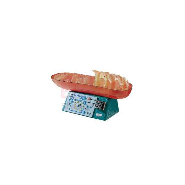
Mitochondrion Model
Mitochondrion Model (Product Code: SLE/ECB/07) is an enlarged organelle model used to teach the structure of the mitochondrion and its role in cellular respiration in senior school and college biology. It magnifies a mitochondrion, normally about 3 microns long, 190,000 times, and its base carries a 1,000,000X membrane relief plus a citric acid cycle schematic. It is made by Jain Scientific Equipments Pvt Ltd (ScienceLab), an OEM manufacturer, at its own factory in Ambala, India, and is a useful piece of cell biology equipment in India.
Specifications
| Parameter | Detail |
|---|---|
| Type | Enlarged mitochondrion model on base |
| Enlargement | 190,000 times (a real mitochondrion is normally 3 microns in length) |
| Structures shown | Two membrane walls, cristae, subunits, matrix and granules |
| Base relief 1 | Cross-section through the membrane and subunits at 1,000,000X |
| Base relief 2 | Simplified schematic of the citric acid cycle |
| Size (approx.) | 58 x 28 x 20 cm (23 in long x 11 in high x 8 in deep) |
| Weight | 4 kg |
| Product code | SLE/ECB/07 |
| Manufacturer | Jain Scientific Equipments Pvt Ltd (ScienceLab) — OEM |
| Country of origin | India (made in Ambala, Haryana) |
What Is the Mitochondrion Model Used For?
In lessons on cellular respiration the model shows why the inner membrane is folded into cristae, giving a large surface for the reactions that release energy, and where the matrix sits in which the citric acid cycle runs. With the cycle moulded on the base, the teacher can move from the organelle to its chemistry without switching to a separate chart, and the 1,000,000X relief brings the membrane subunits into view for more advanced classes.
It also makes a good scale exercise: students multiply the real length of 3 microns by 190,000 and compare the answer with the model's length of about 58 cm. Pairing the model with a respiration practical, such as yeast fermentation, helps students connect what they measure at the bench with the structure that makes it happen.
OEM Manufacturer
The Mitochondrion Model is made by Jain Scientific Equipments Private Limited, trading as ScienceLab, as the OEM (original equipment manufacturer) at the company's own licensed factory at Saha, Ambala, Haryana, India. Colleges, schools, dealers and importers buy it from the manufacturer rather than from a stockist, so questions about the model, repeat orders and spare parts go straight to the people who make it — ask us. Background on the company is on our company profile.
Related Cell Biology Equipments
Models and slides that place the mitochondrion in context:
- Chloroplast Model — the photosynthetic organelle, for comparing the two energy organelles.
- Animal Cell Plaque — shows where mitochondria sit among the other organelles.
- Prokaryotic Cell Model — supports discussion of the bacterial origin of mitochondria.
- Microslide Ultra Structure of Animal Cells — photomicrograph images that include a mitochondrion.
Find further organelle models in our Cell Biology Equipments range.
Frequently Asked Questions
How much has the Mitochondrion Model been enlarged?
The Mitochondrion Model shows a mitochondrion enlarged 190,000 times. A real mitochondrion is normally about 3 microns long, so the model reveals internal detail that no school light microscope can show, at a size the whole class can study.
What is shown on the base of the Mitochondrion Model?
The base of the Mitochondrion Model carries two moulded reliefs: a 1,000,000X cross-section through the membrane and subunits, and a simplified schematic of the citric acid cycle. Together they link the organelle's structure with the chemistry it carries out.
Which internal parts can students identify on the Mitochondrion Model?
On the Mitochondrion Model students can identify the two membrane walls, the cristae, subunits, matrix and granules. Seeing the folded cristae in three dimensions makes it easier to explain why the inner membrane has such a large surface area.
How much space does the Mitochondrion Model need?
The Mitochondrion Model measures approximately 58 x 28 x 20 cm (23 x 11 x 8 in) and weighs 4 kg. It needs a firm bench or cupboard shelf and should be lifted with both hands when moved between rooms.
Is a key card included with the Mitochondrion Model, and what is it made of?
Neither a key card nor the construction material of the Mitochondrion Model is listed in our current product information. Please ask through our contact page and we will confirm both before you order.
Who manufactures the Mitochondrion Model?
The Mitochondrion Model is manufactured by Jain Scientific Equipments Pvt Ltd (ScienceLab), the OEM, at its own licensed factory at Saha, Ambala, Haryana, India. Institutions and distributors deal directly with the manufacturer for quotations and repeat orders of the model.
Can the Mitochondrion Model be ordered in quantity or for export?
Yes. The Mitochondrion Model is supplied to colleges, schools and distributors in India and international markets. Include product code SLE/ECB/07, the quantity and the delivery destination in your enquiry so that the quotation covers packing and shipping.
Request a Quote
For pricing, contact us or add the Mitochondrion Model to your enquiry cart. Quotations are issued directly by the manufacturer.
Last updated: 24 September 2026
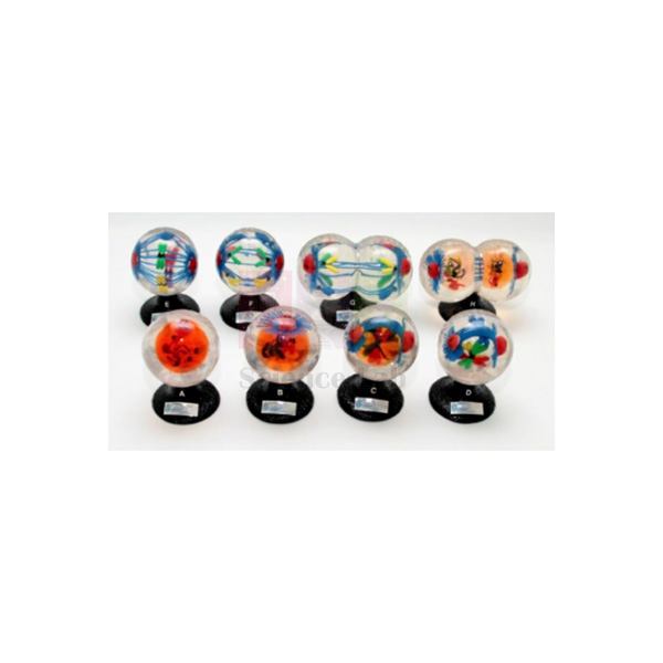
Animal Mitosis Models
Animal Mitosis Models (Product Code: SLE/ECB/08) is a set of 8 sequential models used to show how the chromosomes and mitotic apparatus of an animal cell divide, from interphase to late telophase, in school and college biology. Each stage is embedded in a strong, crystal-clear plastic globe with colour-coded double-thread chromosomes, so students can study it from any side. The set is made by Jain Scientific Equipments Pvt Ltd (ScienceLab), an OEM manufacturer, at its own factory in Ambala, India, among its biology teaching aids for India and abroad.
Specifications
| Parameter | Detail |
|---|---|
| Type | Set of 8 sequential animal cell mitosis models |
| Construction | Organelles and cell inclusions embedded in strong, crystal-clear plastic globes |
| Structures shown | Nucleus, nucleolus, double-thread chromosomes (each in a different colour), centrioles, centromeres (kinetochores), aster and spindle fibres |
| Phases in the set | Interphase, Early Prophase, Late Prophase, Mesophase, Metaphase, Anaphase, Early Telophase, Late Telophase |
| Size, models 1 to 6 | 5-1/4 in high, 3-1/4 in diameter (approx.) |
| Size, models 7 and 8 | 6 in wide x 3-1/4 in deep x 5-1/4 in high |
| Weight of set | 5.8 kg (approx.) |
| Product code | SLE/ECB/08 |
| Manufacturer | Jain Scientific Equipments Pvt Ltd (ScienceLab) — OEM |
| Country of origin | India (made in Ambala, Haryana) |
What Are the Animal Mitosis Models Used For?
Lined up on the bench in order, the eight globes let students follow a single cell cycle step by step: chromosomes condensing in prophase, gathering at the centre in metaphase, separating in anaphase and forming two new nuclei in telophase. Because each double-thread chromosome is a different colour, learners can track one chromosome through every stage and see for themselves that each daughter cell receives an identical set.
A useful classroom activity is to shuffle the models and ask groups to put them back in sequence, explaining the change between each pair. The set then leads naturally into prepared microscope slides, where students search real dividing cells for the same stages they have just handled. Models 7 and 8 are wider than the others, so allow a little extra bench space for them.
OEM Manufacturer
Jain Scientific Equipments Private Limited, known by its trading name ScienceLab, is the OEM (original equipment manufacturer) of the Animal Mitosis Models and makes the set at its own licensed factory at Saha, Ambala, Haryana, India. Buying from the maker means one point of contact for the quotation, for adding sets later as more sections start the topic, and for enquiries about a replacement globe — ask us. Our company profile has the full background.
Related Cell Biology Equipments
Resources that support a complete cell division unit:
- Animal Meiosis Plaque — illustrates reduction division for comparison with mitosis.
- Mitosis Simulation Lab Investigation — a pop-bead student activity on the same process.
- Visualizing Mitosis Poster — a wall reference for the stages.
- Mitosis & Meiosis Set, Microscope Slides — prepared slides for finding the stages in real cells.
Browse our complete Cell Biology Equipments range.
Frequently Asked Questions
How many models are in the Animal Mitosis Models set, and which phases do they show?
The Animal Mitosis Models set contains 8 models showing Interphase, Early Prophase, Late Prophase, Mesophase, Metaphase, Anaphase, Early Telophase and Late Telophase. Placed in order, they take students through one complete division of an animal cell.
What can students see inside each globe of the Animal Mitosis Models?
Each globe of the Animal Mitosis Models contains the nucleus, nucleolus, double-thread chromosomes, centrioles, centromeres (kinetochores), aster and spindle fibres, embedded in strong, crystal-clear plastic so the structures are visible from every side.
Why are the chromosomes in the Animal Mitosis Models different colours?
In the Animal Mitosis Models each double-thread chromosome has its own colour. That makes it easy to follow an individual chromosome from one stage to the next and to show that both daughter nuclei end up with the same chromosome set.
What are the sizes and total weight of the Animal Mitosis Models?
In the Animal Mitosis Models, models 1 to 6 are about 5-1/4 in high with a 3-1/4 in diameter, and models 7 and 8 measure 6 in wide, 3-1/4 in deep and 5-1/4 in high. The whole set weighs about 5.8 kg.
How should the clear globes of the Animal Mitosis Models be cared for?
Clean the Animal Mitosis Models with a soft, lint-free cloth only, and avoid abrasive cleaners and solvents that could scratch or cloud the plastic. Store each model so the globes do not knock against one another.
Is a teacher's key or storage case included with the Animal Mitosis Models?
Neither a teacher's key nor a storage case is listed in our current product information for the Animal Mitosis Models. Please ask through our contact page to confirm exactly what is supplied with the set.
Who manufactures the Animal Mitosis Models?
The Animal Mitosis Models are manufactured by Jain Scientific Equipments Pvt Ltd (ScienceLab), the OEM, at its own licensed factory at Saha, Ambala, Haryana, India. Schools, colleges, dealers and importers order the set directly from the manufacturer, which also prepares their quotation.
Request a Quote
Tell us the number of sets and your delivery location through our contact page, or add the Animal Mitosis Models to your enquiry cart. The quotation comes direct from the manufacturer.
Last updated: 24 September 2026

Plant Cell Plaque
Plant Cell Plaque (Product Code: SLE/ECB/09) is a raised-relief display of the plant cell used to teach its organelles and inclusions in secondary school and college botany. Twenty-three structures are moulded on a heavy vacuum-formed plastic sheet of 18 x 24 in (46 x 61 cm), and every one is identified on the accompanying key cards. As a plant cell model for India's biology classrooms, it is made by Jain Scientific Equipments Pvt Ltd (ScienceLab), an OEM manufacturer, at its own factory in Ambala, India.
Specifications
| Parameter | Detail |
|---|---|
| Type | Relief plaque of the plant cell |
| Structures moulded | 23 (organelles and cell inclusions) |
| Construction | Heavy plastic vacuum-formed sheet |
| Identification | All structures identified by accompanying key cards |
| Size | 46 x 61 cm (18 x 24 in) |
| Weight | 1.2 kg |
| Companion item | Animal cell plaque in the same series, moulded to 20 structures |
| Product code | SLE/ECB/09 |
| Manufacturer | Jain Scientific Equipments Pvt Ltd (ScienceLab) — OEM |
| Country of origin | India (made in Ambala, Haryana) |
What Is the Plant Cell Plaque Used For?
The plaque gives the whole class a shared, three-dimensional reference while the teacher explains the parts of a plant cell and what each one does, from the cell wall and chloroplasts to the vacuole and nucleus. Students then work in pairs with the key cards, pointing to each raised structure and naming it, which turns identification into an active exercise instead of copying a diagram.
When the animal cell plaque from the same series is shown alongside, students notice that the plant version carries more moulded structures and can use the key cards to work out which features are found only in plant cells. The pair covers the plant-versus-animal cell comparison found in school biology syllabuses, and both plaques are light enough to move between classrooms.
What to Specify When Ordering
- How many Plant Cell Plaques you need across your botany or biology rooms.
- Whether the matching animal cell plaque (20 structures) should be quoted as a pair for comparison teaching.
- The delivery address and any vendor documents your purchase department requires.
OEM Manufacturer
ScienceLab is the brand of Jain Scientific Equipments Private Limited, which is the OEM (original equipment manufacturer) of the Plant Cell Plaque rather than a reseller of it. Institutions, dealers and importers receive the plaque factory-direct, and any request for extra key cards or a matching set later on is answered by the maker — just ask us. The company is registered under CIN U29309HR2020PTC087597 and makes its products at its licensed factory at 449, HSIIDC Industrial Area, Saha, Ambala, Haryana (Factory Licence AMB-ONLINE-CHD-J-414). Read more on our company profile.
Related Cell Biology Equipments
Items that pair well with the plant cell plaque:
- Animal Cell Plaque — the matching animal cell version for comparison lessons.
- Plant Cell Model — an enlarged model of a typical plant cell with all types of organelle.
- Chloroplast Model — a close-up of the organelle that makes plant cells photosynthetic.
- Plant Cell Study Kit — slides, stains and specimens for moving from the plaque to real plant cells.
- Hands-On Cell Model Act-Plant — a foam plant cell model with removable parts for younger classes.
The complete selection is in our Cell Biology Equipments range.
Frequently Asked Questions
How many structures does the Plant Cell Plaque show?
The Plant Cell Plaque is moulded to 23 structures, the organelles and inclusions of the plant cell, and each one is identified on the key cards that accompany it. That is three more than the animal cell plaque from the same series.
What size is the Plant Cell Plaque in inches and centimetres?
The Plant Cell Plaque measures 18 x 24 in, which is 46 x 61 cm, and it weighs 1.2 kg. The size suits front-of-class display, while the low weight lets a teacher move it easily between rooms.
Is a matching animal cell plaque available for the Plant Cell Plaque?
Yes. The Plant Cell Plaque has a companion Animal Cell Plaque in the same relief series, moulded to 20 structures. Ordering both lets students compare plant and animal cells side by side using the two sets of key cards.
Can students handle the Plant Cell Plaque?
Yes, with care. The Plant Cell Plaque weighs only 1.2 kg, but the raised areas of the vacuum-formed sheet should not be pressed hard or stacked under heavy items; students should hold it by the edges.
Who manufactures the Plant Cell Plaque?
The Plant Cell Plaque is manufactured by Jain Scientific Equipments Pvt Ltd (ScienceLab), the OEM, at its own licensed factory at Saha, Ambala, Haryana, India. Schools, colleges and distributors buy it directly from the manufacturer, not through a trading company.
Can we get vendor registration details for the Plant Cell Plaque purchase file?
Yes. The company registration and factory licence numbers for the maker of the Plant Cell Plaque are given in the manufacturer section above, and any further documents your institution needs can be requested through our contact page.
Request a Quote
For pricing, contact us or add the Plant Cell Plaque to your enquiry cart; quotations are prepared directly by the manufacturer.
Last updated: 24 September 2026
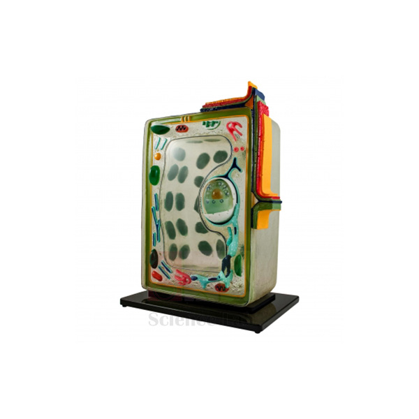
Plant Cell
Plant Cell (Product Code: SLE/ECB/10) is a large, translucent demonstration model used to teach the 25 organelles and cell inclusions of a plant cell in school, college and teacher-training biology. Its fibreglass-reinforced plastic body gives the appearance of living matter, and the cell wall is divided into distinct layers from the middle lamella to a warted tertiary wall. It is made by Jain Scientific Equipments Pvt Ltd (ScienceLab), an OEM manufacturer, at its own factory in Ambala, India, as a centrepiece of botany lab equipment in India.
Specifications
| Parameter | Detail |
|---|---|
| Organelles and inclusions shown | 25 |
| Body material | Translucent, fibreglass-reinforced plastic that gives the appearance of living matter |
| Detail parts | Moulded plastic, so colours do not chip, crack, peel or fade |
| Key structures | Endoplasmic reticulum, cross-sectioned chloroplast with distinct grana and grana discs, large central vacuole, mitochondria and ribosomes, along with the other known organelles and inclusions |
| Cell wall layers | Middle lamella, primary wall, three secondary walls, warted tertiary wall and plasma membrane |
| Size (approx.) | 16 in high x 11-1/2 in wide x 8 in deep (41 x 29 x 20 cm) |
| Weight (approx.) | 4 kg |
| Product code | SLE/ECB/10 |
| Manufacturer | Jain Scientific Equipments Pvt Ltd (ScienceLab) — OEM |
| Country of origin | India (made in Ambala, Haryana) |
What Is the Translucent Plant Cell Model Used For?
Teachers usually work from the outside in: first the middle lamella that joins neighbouring cells, then the primary wall, the three secondary walls and the warted tertiary wall, and finally the plasma membrane. From there the lesson moves to the cross-sectioned chloroplast, where the grana and grana discs are easy to point out, and on to the large central vacuole, mitochondria and endoplasmic reticulum. Because the body is translucent, internal organelles stay visible from several angles when small groups gather round for labelling or oral questioning.
The model is most effective when paired with microscope practicals on onion epidermis or leaf peels, so students connect the enlarged structures with what they actually see under the lens. It also serves as a revision aid before practical examinations. At around 4 kg it should rest on a stable bench or shelf rather than be passed hand to hand across a room.
OEM Manufacturer
The Plant Cell model comes from Jain Scientific Equipments Private Limited, the company behind the ScienceLab name, which is its OEM (original equipment manufacturer) and makes it at its own licensed factory in Saha, Ambala, Haryana, India. Because there is no trading layer between maker and buyer, schools, colleges, dealers and importers get their quotation, repeat supply and after-sales questions handled by the manufacturer — ask us about anything you need. Background on the company is on our company profile.
Related Cell Biology Equipments
These items support the same lessons on plant cell structure and comparison with other cell types:
- Chloroplast Model — a dedicated, enlarged view of the organelle shown here in cross-section.
- Mitochondrion Model — lets students study the respiration organelle in more depth after the whole-cell overview.
- Animal Cell Plaque — for side-by-side comparison of plant and animal cell features such as the wall and vacuole.
- Plant Cell Plaque — a plaque-format plant cell reference for a second bench or display.
- Visualising Cells Poster — a laminated wall poster comparing bacterial, animal and plant cells.
See the full range of Cell Biology Equipments for more models, plaques and classroom investigations.
Frequently Asked Questions
What does the Plant Cell model show?
The Plant Cell model shows 25 organelles and cell inclusions, including the endoplasmic reticulum, a cross-sectioned chloroplast with grana and grana discs, the large central vacuole, mitochondria and ribosomes, and a cell wall divided into middle lamella, primary, secondary and tertiary layers and plasma membrane.
How large and heavy is the Plant Cell model?
The Plant Cell model measures approximately 41 x 29 x 20 cm (16 x 11-1/2 x 8 in) and weighs about 4 kg. That makes it a teacher's demonstration piece for the front bench rather than an item for every student group.
Will the colours of the Plant Cell model wear off with classroom use?
The detail parts of the Plant Cell model are moulded plastic, so the colours do not chip, crack, peel or fade. Normal care still applies: lift the model with both hands and keep it away from direct heat.
How should the Plant Cell model be cleaned?
Wipe the Plant Cell model with a soft cloth, slightly dampened with mild soapy water if needed. Avoid solvents and abrasive cleaners, which can dull the translucent fibreglass-reinforced surface, and dry it before returning it to the shelf.
Is a labelling key supplied with the Plant Cell model?
Our product information for the Plant Cell model does not mention a key or guide. Please ask through our contact page to confirm what documentation is sent with the model before you order.
Who manufactures the Plant Cell model?
The Plant Cell model (SLE/ECB/10) is manufactured by Jain Scientific Equipments Pvt Ltd (ScienceLab), its OEM, at the company's own licensed factory at Saha, Ambala, Haryana, India, and is supplied factory-direct to institutions and distributors.
What should an institutional quotation request for the Plant Cell model include?
A quotation request for the Plant Cell model should mention product code SLE/ECB/10, the quantity, the delivery location and any tender or purchase-order format you must follow. Orders from India and international markets, including bulk orders, are welcome.
Request a Quote
To order the Plant Cell model or ask about quantities, contact our team or add it to your enquiry cart with other items from your lab list. You will receive a quotation directly from the manufacturer.
Last updated: 24 September 2026
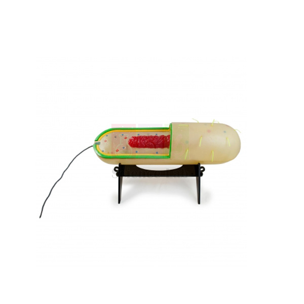
Prokaryotic Cell Model
Prokaryotic Cell Model (Product Code: SLE/ECB/11) is an enlarged bacterial-type cell model used to teach prokaryotic cell structure in school biology, college and microbiology courses. Shown at 280,000X and partly cut away, it displays pili and flagella outside and chromosomal DNA, plasmids and ribosomes inside, and the cell lifts off its stand, with key cards supplied. It is made by Jain Scientific Equipments Pvt Ltd (ScienceLab), an OEM manufacturer, at its own factory in Ambala, India, for colleges assembling microbiology lab equipment in India.
Specifications
| Parameter | Detail |
|---|---|
| Magnification | Enlarged 280,000X |
| Presentation | External cellular structures shown; partially cut away to reveal internal structures |
| Structures illustrated | Pili, flagella, chromosomal DNA, an inclusion body, plasmids, ribosomes, the cell envelope and the plasma membrane |
| Mounting | Removable from its stand |
| Supplied with | Key cards |
| Size (approx.) | 22 in long x 6-1/2 in (56 x 17 x 17 cm) |
| Weight (approx.) | 2.6 kg |
| Product code | SLE/ECB/11 |
| Manufacturer | Jain Scientific Equipments Pvt Ltd (ScienceLab) — OEM |
| Country of origin | India (made in Ambala, Haryana) |
What Is the Prokaryotic Cell Model Used For?
The model is most useful in the lesson that contrasts prokaryotes with eukaryotes. With the cutaway facing the class, the teacher can show that the chromosomal DNA lies free in the cytoplasm rather than inside a nucleus, point out the small plasmids and the ribosomes, and then turn the model to discuss the cell envelope, the pili used for attachment and the flagella used for movement. Lifting the cell off its stand makes it easy to pass to a group, and the key cards let students check their own identification of each part.
In microbiology courses it gives context before students look at stained bacterial smears, where individual structures are far too small to see. Foundation courses in nursing, pharmacy and life sciences use it to build a clear picture of bacterial anatomy that supports later topics such as antibiotic action and gene transfer through plasmids.
OEM Manufacturer
Jain Scientific Equipments Private Limited operates under the ScienceLab name and is the OEM (original equipment manufacturer) of the Prokaryotic Cell Model, which it makes at its own licensed factory at Saha, Ambala, Haryana, India. Departments, distributors and importers source the model factory-direct, so the same manufacturer handles their quotation, follow-up orders and requests for replacement key cards or a stand — ask us. Read more on the ScienceLab company profile.
Related Cell Biology Equipments
Pair the model with these items to build a complete lesson on cell types:
- Plant Cell — a detailed eukaryotic model for direct comparison with the prokaryotic layout.
- Visualising Cells Poster — a wall reference showing how bacterial, animal and plant cells differ.
- Bacteria slide Set of 12 — prepared slides of real bacteria, including cocci, bacilli and spirilla.
- Amoeba Model — a single-celled eukaryote that contrasts well with a prokaryote.
- Mitochondrion Model — supports discussion of how mitochondria resemble bacteria.
More models and investigations are listed under Cell Biology Equipments.
Frequently Asked Questions
Which structures can students identify on the Prokaryotic Cell Model?
On the Prokaryotic Cell Model students can identify pili, flagella, chromosomal DNA, an inclusion body, plasmids, ribosomes, the cell envelope and the plasma membrane. The partial cutaway shows the internal parts, while the outer surface displays the external structures.
What scale and size is the Prokaryotic Cell Model?
The Prokaryotic Cell Model represents the cell enlarged 280,000X. Its overall size is about 56 x 17 x 17 cm (22 in long) and it weighs roughly 2.6 kg, so it suits a demonstration bench or a group table.
Can the Prokaryotic Cell Model be taken off its stand?
Yes. The Prokaryotic Cell Model is removable from its stand, which makes close inspection and group work easier. Return it to the stand for storage so the flagella and pili are not bent or knocked.
What are the key cards with the Prokaryotic Cell Model for?
The key cards supplied with the Prokaryotic Cell Model identify the illustrated structures, so students can use the model for self-checking and revision without a separate textbook diagram, and teachers can run quick labelling quizzes.
How do I keep the Prokaryotic Cell Model in good condition?
Dust the Prokaryotic Cell Model with a soft brush or dry cloth and store it away from direct sunlight and heat. Do not use solvents or abrasive cleaners, and lift it by the stand or body rather than by the flagella.
Who manufactures the Prokaryotic Cell Model?
The Prokaryotic Cell Model is manufactured by Jain Scientific Equipments Pvt Ltd (ScienceLab), acting as the OEM, at its own licensed factory at Saha, Ambala, Haryana, India. Buyers in India and international markets deal directly with the manufacturer.
Can several Prokaryotic Cell Models be supplied for a department or distributor?
Yes. Send product code SLE/ECB/11 with the quantity of Prokaryotic Cell Models and the destination, and we will quote for delivery within India or to international markets. Other cell models can be added to the same quotation.
Request a Quote
For pricing and availability, send us an enquiry or place the Prokaryotic Cell Model in your enquiry cart with the rest of your requirements; the manufacturer replies with the quotation directly.
Last updated: 24 September 2026
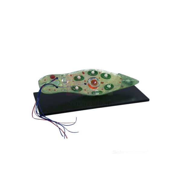
Euglena Model
Euglena Model (Product Code: SLE/ECB/12) is an enlarged, translucent fibreglass model of Euglena used to teach protist structure in school zoology, botany and college biology. It shows the features seen in living and stained preparations, from the flagellum and stigma to the pyrenoid, contractile vacuole and endosome, on a base of 7 x 19-1/2 in. It is made by Jain Scientific Equipments Pvt Ltd (ScienceLab), an OEM manufacturer, at its own factory in Ambala, India, and suits any biology lab setup for schools in India studying protists.
Specifications
| Parameter | Detail |
|---|---|
| Material | Translucent fibreglass |
| Structures shown | Flagellum, stigma, blepharoplast, contractile vacuole, chloroplast, pyrenoid, rhizoplast, nucleus and endosome |
| Basis of detail | Features visible in living and stained preparations |
| Base size | 7 in x 19-1/2 in |
| Overall size (approx.) | 44 x 15 x 15 cm |
| Weight (approx.) | 2.4 kg |
| Product code | SLE/ECB/12 |
| Manufacturer | Jain Scientific Equipments Pvt Ltd (ScienceLab) — OEM |
| Country of origin | India (made in Ambala, Haryana) |
What Is the Euglena Model Used For?
A good way to use the model is as a bridge between a pond-water practical and the theory lesson. Students first try to spot live Euglena under the microscope, where they may see only a green, spindle-shaped cell swimming with a whip-like flagellum; the enlarged model then shows the stigma that senses light, the blepharoplast and rhizoplast at the base of the flagellum, the chloroplasts and pyrenoid involved in photosynthesis, and the contractile vacuole that removes excess water.
Because Euglena combines plant-like chloroplasts with animal-like movement, the model also opens a discussion on why some organisms do not fit neatly into one kingdom. The translucent body keeps internal features such as the nucleus and endosome visible without a cut section, and many departments keep it beside an amoeba model so both protists can be compared in one session.
OEM Manufacturer
This model is made by Jain Scientific Equipments Private Limited, which trades as ScienceLab and is the OEM (original equipment manufacturer) of the Euglena Model, at its own licensed factory at Saha, Ambala, Haryana, India. Schools, colleges, dealers and importers order it straight from the manufacturer, not from a trader, and can come back to the same team for repeat orders or questions about the model — ask us. See our company profile for more information.
Related Cell Biology Equipments
Useful additions for a protist lesson built around this model:
- Amoeba Model — contrasts pseudopod movement with the flagellum-driven swimming of Euglena.
- Chloroplast Model — a closer look at the photosynthetic organelle that makes Euglena green.
- Student Monocular Microscope — for observing live Euglena in pond-water samples.
- Microscope Slides Depression — cavity slides that hold a drop of culture so the cells can swim freely.
- Prokaryotic Cell Model — compares a flagellated prokaryote with this flagellated eukaryote.
Explore other cell and protist models in Cell Biology Equipments.
Frequently Asked Questions
Which parts of Euglena does the Euglena Model represent?
The Euglena Model represents the flagellum, stigma, blepharoplast, contractile vacuole, chloroplast, pyrenoid, rhizoplast, nucleus and endosome — the features that are visible in living and stained preparations of the organism under the microscope.
What are the dimensions of the Euglena Model?
The Euglena Model measures about 44 x 15 x 15 cm overall, stands on a base of 7 x 19-1/2 in and weighs roughly 2.4 kg, so it can sit on a demonstration bench for a whole lesson.
Why is the Euglena Model translucent?
The Euglena Model is made of translucent fibreglass so that students can see internal structures such as the nucleus and pyrenoid through the body of the cell, much as they would through a thin living cell under the microscope.
Does the Euglena Model replace microscope work?
No. The Euglena Model is meant to support microscope work: students learn what to look for before examining live or stained specimens, and they can check their observations against the enlarged structures afterwards.
Are labels or a guide included with the Euglena Model?
Our product information for the Euglena Model does not specify labelling material. If you need a key for student use, please ask through our contact page what accompanies the model before ordering.
Who manufactures the Euglena Model?
The Euglena Model is manufactured by Jain Scientific Equipments Pvt Ltd (ScienceLab), the OEM, at its own licensed factory at Saha, Ambala, Haryana, India. Institutions and distributors receive the model and its quotation directly from the manufacturer.
Is the Euglena Model supplied outside India?
Yes. The Euglena Model is supplied to institutions and distributors in India and international markets. Include product code SLE/ECB/12, the quantity and the destination in your enquiry so that the quotation covers delivery.
Request a Quote
To request a quotation for the Euglena Model, write to us or add it to the enquiry cart and submit your full list in one go. Quotes come direct from the manufacturer.
Last updated: 24 September 2026
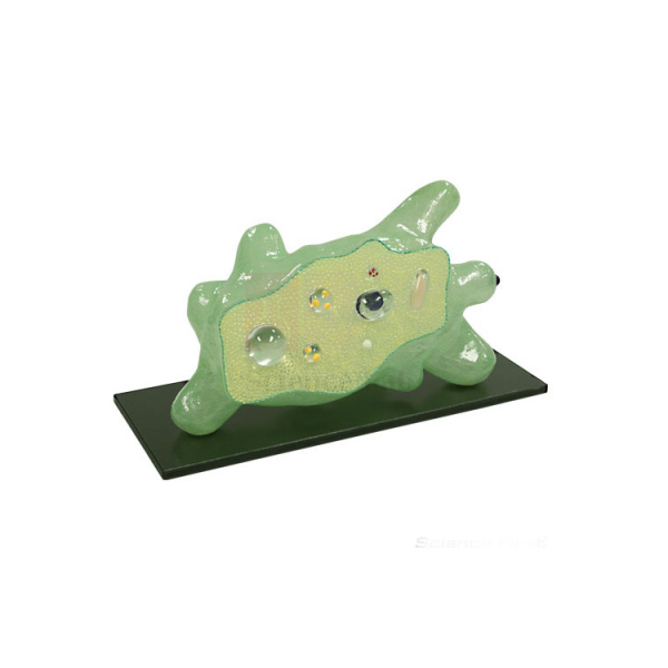
Amoeba Model
Amoeba Model (Product Code: SLE/ECB/13) is a translucent, enlarged model of Amoeba used to teach the structure, feeding and movement of a unicellular protozoan in school zoology and college biology. Its pseudopods are positioned to show the direction of travel, and food vacuoles holding diatoms and bacteria illustrate how the cell feeds. It is made by Jain Scientific Equipments Pvt Ltd (ScienceLab), an OEM manufacturer, at its own factory in Ambala, India, and fits naturally among animal models for a biology lab.
Specifications
| Parameter | Detail |
|---|---|
| Construction | Translucent model |
| Structures shown | Nucleus, food vacuoles containing diatoms and bacteria, contractile vacuole, fat droplet, granular and clear zones of cytoplasm |
| Movement | Position of the pseudopods indicates the amoeba's movement |
| Dimensions | 16-1/2 in wide x 10 in high x 8 in deep (42 x 25 x 20 cm) |
| Weight | 2.2 kg |
| Product code | SLE/ECB/13 |
| Manufacturer | Jain Scientific Equipments Pvt Ltd (ScienceLab) — OEM |
| Country of origin | India (made in Ambala, Haryana) |
What Is the Amoeba Model Used For?
Amoeba is the classic example of a cell that changes shape to move and feed, and the model makes that process easy to explain. Teachers can trace how the cytoplasm flows into the advancing pseudopods, distinguish the granular inner zone from the clear outer zone, and then point to food vacuoles that already contain diatoms and bacteria to illustrate engulfing prey by phagocytosis. The contractile vacuole leads into a discussion of osmoregulation in freshwater organisms, and the fat droplet gives an example of stored food reserves.
For practical classes, keep the model on the bench while students search pond or hay-infusion samples under the microscope, since live amoebae are slow, colourless and easy to miss. Many schools order it together with a Euglena model so students can compare pseudopod and flagellum movement, and at 2.2 kg it is light enough to carry between classrooms.
OEM Manufacturer
ScienceLab is the brand under which Jain Scientific Equipments Private Limited makes the Amoeba Model as its OEM (original equipment manufacturer), at the company's own licensed factory at Saha, Ambala, Haryana, India. Schools, colleges and overseas importers buy it factory-direct, and follow-up orders or questions about the model are answered by the maker itself — ask us. The company is a registered MSME with manufacturing as its major activity (Udyam UDYAM-HR-01-0003714), and its export orders ship under Importer-Exporter Code AAECJ8618A issued by the DGFT. Further details are on our company profile.
Related Cell Biology Equipments
Items that teachers commonly use alongside the Amoeba Model:
- Euglena Model — a flagellated protist for comparing modes of movement and nutrition.
- Student Monocular Microscope — for finding live amoebae in pond-water samples.
- Microscope Slides Depression — cavity slides that give live protozoa room to move while being observed.
- Visualising Osmosis & Diffusion Investigation — a class activity on membrane transport that explains why the contractile vacuole is needed.
More options are listed in our Cell Biology Equipments category.
Frequently Asked Questions
What structures does the Amoeba Model include?
The Amoeba Model includes the nucleus, food vacuoles containing diatoms and bacteria, the contractile vacuole, a fat droplet, and granular and clear zones of cytoplasm. Its pseudopods are arranged to show the direction in which the amoeba is moving.
How big is the Amoeba Model?
The Amoeba Model measures 16-1/2 in wide, 10 in high and 8 in deep (42 x 25 x 20 cm) and weighs 2.2 kg, a size that suits front-of-class display in an average classroom and is easy to carry.
Can the Amoeba Model be used to teach osmoregulation?
Yes. The Amoeba Model shows the contractile vacuole, so teachers can use it to explain how a freshwater protozoan collects and expels the water that keeps entering its cell by osmosis.
How should the Amoeba Model be cleaned and stored?
Clean the Amoeba Model with a soft, dry or lightly damp cloth and keep it covered when not in use to reduce dust. Avoid solvents and direct sunlight, which can dull or discolour a translucent finish.
Who manufactures the Amoeba Model?
The Amoeba Model is manufactured by Jain Scientific Equipments Pvt Ltd (ScienceLab), the OEM, at its own licensed factory at Saha, Ambala, Haryana, India. Institutions and distributors get the model and its quotation directly from the manufacturer.
What information helps prepare an institutional quote for the Amoeba Model?
For an Amoeba Model quotation, send product code SLE/ECB/13, the quantity, the delivery address and any vendor documents your purchase department needs. Orders are handled for buyers in India and international markets.
Request a Quote
For a quotation on the Amoeba Model, contact us directly or use the enquiry cart to send it with the other models on your list. Quotations are issued by the manufacturer.
Last updated: 24 September 2026
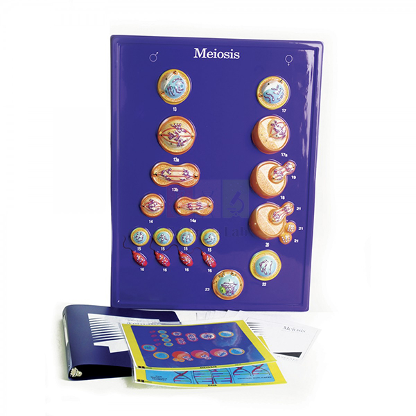
Meiosis Model
Meiosis Model (Product Code: SLE/PS/82) is an enlarged teaching model used to explain meiotic cell division in school and college cell biology lessons. Teachers use it to show how meiosis gives rise to individual human characteristics and genetic differences, with enlarged views of chromosomes, cytoplasm and the chromatic and polar bodies. It is made by Jain Scientific Equipments Pvt Ltd (ScienceLab), an OEM manufacturer, at its own factory in Ambala, India.
Key Details
| Parameter | Detail |
|---|---|
| Model type | Enlarged teaching model of meiotic cell division |
| Structures shown enlarged | Chromosomes, cytoplasm, chromatic bodies and polar bodies |
| Teaching focus | How meiosis leads to individual human characteristics and genetic differences |
| Subject area | Cell biology and genetics |
| Product code | SLE/PS/82 |
| Manufacturer | Jain Scientific Equipments Pvt Ltd (ScienceLab) — OEM |
| Country of origin | India (made in Ambala, Haryana) |
What Is the Meiosis Model Used For?
Meiosis is hard to picture from a flat textbook diagram, because chromosomes pair up, exchange segments and then separate in two rounds of division. The Meiosis Model enlarges those structures so a whole class can follow the sequence together, from the chromosomes in the cytoplasm to the appearance of polar bodies. It gives teachers a concrete way to answer the question students always ask: why do brothers and sisters look different?
Schools that buy a mitosis and meiosis model usually use it twice: at the start of a genetics unit and again when students compare the two kinds of division. During a slide practical, keep it on the front bench so students can match what they see under the microscope with its enlarged form. Dust it with a soft, dry cloth and store it away from direct sunlight.
OEM Manufacturer
ScienceLab is the trading name of Jain Scientific Equipments Private Limited, the OEM (original equipment manufacturer) of this Meiosis Model. The company makes its cell biology models at its own licensed factory at Saha, Ambala, Haryana, India, and sells them factory-direct to schools, colleges, institutions, dealers and importers instead of through a trading middleman.
Because the maker handles your order itself, repeat orders and questions about replacement parts go straight to the people who build the model — just ask us. Read more about the company on the ScienceLab profile page.
Related Cell Biology Equipments
These items are often used with the Meiosis Model in cell division and genetics lessons:
- Animal Mitosis Models — the matching models for comparing mitosis with meiosis.
- Animal Meiosis Plaque — a plaque presentation of meiosis in animal cells for wall or bench display.
- Mitosis Simulation Lab Investigation — a hands-on activity where students model division themselves.
- Mitosis & Meiosis Set, Microscope Slides — real dividing cells to view after studying the model.
- Microslide Meiosis — photographed stages of meiosis in the egg cell for revision.
See the complete Cell Biology Equipments range for cell models, plaques and investigations.
Frequently Asked Questions
What does the Meiosis Model show?
The Meiosis Model shows meiotic cell division with enlarged views of chromosomes, cytoplasm and the chromatic and polar bodies. It is designed to help students understand how meiosis produces individual human characteristics and genetic differences.
Who manufactures the Meiosis Model?
The Meiosis Model is manufactured by Jain Scientific Equipments Pvt Ltd (ScienceLab), the OEM, at its own licensed factory at Saha, Ambala, Haryana, India. Quotations and repeat orders come directly from the manufacturer.
Which classes is the Meiosis Model suitable for?
The Meiosis Model suits secondary school biology, where meiosis and inheritance are first introduced, and college cell biology or genetics courses that need a clear demonstration piece for lectures and practical sessions.
Can the Meiosis Model be used with a mitosis model?
Yes. The Meiosis Model is often ordered with the Animal Mitosis Models so teachers can place both divisions side by side and show how the number of chromosomes and the resulting cells differ between them.
Are dimensions and materials of the Meiosis Model available?
Dimensions and materials of the Meiosis Model are not listed on this page. Ask for them through the contact page and they will be confirmed with your quotation.
Can the Meiosis Model be ordered in bulk or for export?
Yes. The Meiosis Model is supplied factory-direct to schools, institutions, dealers and importers in India and international markets. Share the quantity and destination so the manufacturer can prepare a quotation.
Request a Quote
Quotations for the Meiosis Model come straight from the manufacturer. Send your requirement through the contact page, or add the model to the enquiry cart with the rest of your cell biology list.
Last updated: 24 September 2026
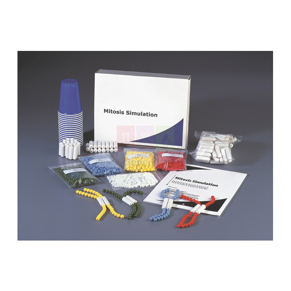
Mitosis Simulation Lab Investigation
Mitosis Simulation Lab Investigation (Product Code: SLE/ECB/14) is a hands-on class activity used to teach mitosis in secondary school and introductory college biology, with students building model chromosomes from coloured pop beads and moving them through each stage of division. One investigation serves a class of 40 students and also covers how plant and animal mitosis differ. It is made by Jain Scientific Equipments Pvt Ltd (ScienceLab), an OEM manufacturer, at its own factory in Ambala, India, as a biology experiment kit students can use without microscopes.
Key Details
| Parameter | Detail |
|---|---|
| Type | Hands-on mitosis simulation lab investigation |
| Class size | 40 students |
| Modelling material | Coloured pop beads for constructing model chromosomes |
| Learning outcomes | Chromosome structure and function; each stage of mitosis; how plant and animal mitosis differ |
| Product code | SLE/ECB/14 |
| Manufacturer | Jain Scientific Equipments Pvt Ltd (ScienceLab) — OEM |
| Country of origin | India (made in Ambala, Haryana) |
What Is the Mitosis Simulation Lab Investigation Used For?
Working in pairs or small groups, students first assemble bead strands to represent chromosomes, using colour to tell one homologue from its partner. They then copy each strand to form sister chromatids joined at a centromere and walk the model through prophase, metaphase, anaphase and telophase on the bench, recording the arrangement at every step. Because the beads snap apart and together, groups can repeat a stage until the movement of chromatids makes sense, which is far more memorable than labelling a printed cycle.
The activity closes by comparing plant and animal mitosis, for example how the cell divides at the end of the process, so students can explain the differences rather than just list them. It needs no chemicals, and it works well as a lead-in to examining real dividing cells on prepared slides.
What to Specify When Ordering
- Total number of students or sections, since one investigation is designed for a class of 40.
- Whether you want mitosis slides, models or a poster from the same range quoted alongside it.
- Delivery location and any vendor registration forms your school or distributor must complete.
OEM Manufacturer
Jain Scientific Equipments Private Limited, trading as ScienceLab, is the OEM (original equipment manufacturer) of the Mitosis Simulation Lab Investigation and puts it together at its own licensed factory at Saha, Ambala, Haryana, India. Schools and distributors therefore order class sets from the maker, which also handles repeat orders for additional sections — ask us about anything specific to your syllabus. For purchase departments completing vendor registration, the company's identifiers are GSTIN 06AAECJ8618A1ZT and LEI 3358004HRD1JIVXKYX49. You can read more on our company profile.
Related Cell Biology Equipments
These products extend the same cell division lesson from simulation to observation:
- Visualizing Mitosis Poster — a wall reference of every mitotic stage for students to check their bead models against.
- Animal Mitosis Models — ready-made stage models for demonstration and revision.
- Mitosis & Meiosis Set, Microscope Slides — prepared slides for observing real dividing cells after the simulation.
- Ts Root Tip Onion — the classic plant tissue for spotting mitotic stages under the microscope.
- Animal Meiosis Plaque — for contrasting mitosis with the reduction division that follows in the syllabus.
Browse all Cell Biology Equipments for further models and investigations.
Frequently Asked Questions
How many students does one Mitosis Simulation Lab Investigation serve?
One Mitosis Simulation Lab Investigation is designed for a class size of 40 students. For larger classes or several sections, ask us to quote for additional sets so that every group has enough beads to build its own chromosomes.
What do students use to build chromosomes in the Mitosis Simulation Lab Investigation?
In the Mitosis Simulation Lab Investigation students build chromosome models from coloured pop beads, then use these bead "chromosomes" to model each stage of mitosis and see what happens to genetic material during cell division.
Does the Mitosis Simulation Lab Investigation cover plant as well as animal cells?
Yes. Part of the Mitosis Simulation Lab Investigation is learning to distinguish between plant and animal mitosis, so students compare how the two cell types complete division after they have modelled the main stages.
Is a microscope needed for the Mitosis Simulation Lab Investigation?
No. The Mitosis Simulation Lab Investigation is carried out entirely with bead models, which suits classrooms without a microscope for every group. Prepared slides of dividing cells can be added later for observation work.
What is the full component list of the Mitosis Simulation Lab Investigation?
Our product information for the Mitosis Simulation Lab Investigation confirms the coloured pop beads and the 40-student class size. For a complete packing list, including any teacher notes, please ask through our contact page before ordering.
Who manufactures the Mitosis Simulation Lab Investigation?
The Mitosis Simulation Lab Investigation is manufactured by Jain Scientific Equipments Pvt Ltd (ScienceLab), the OEM, at its own licensed factory at Saha, Ambala, Haryana, India. Schools and distributors in India and international markets order it directly from the manufacturer.
How should the beads of the Mitosis Simulation Lab Investigation be stored?
After each Mitosis Simulation Lab Investigation lesson, separate the bead strands, sort them by colour and keep them in a closed box so pieces are not lost. Counting them back in at the end of class keeps the set complete for the next group.
Request a Quote
To price the Mitosis Simulation Lab Investigation for your school, send an enquiry or add it to the enquiry cart along with other practical resources; the quotation comes straight from the manufacturer.
Last updated: 24 September 2026

Osmosis Simulation Activity Model
Osmosis Simulation Activity Model (Product Code: SLE/ECB/15) is a classroom demonstration model used to show osmosis, the movement of water across a semipermeable membrane, in school and introductory college biology. The quick, easy set-up can be repeated with different solutes and concentrations, and students quantify each result by measuring the liquid that crossed the membrane. It is made by Jain Scientific Equipments Pvt Ltd (ScienceLab), an OEM manufacturer, at its own factory in Ambala, India, for schools wanting an osmosis experiment kit that students can measure.
Key Details
| Parameter | Detail |
|---|---|
| Type | Osmosis simulation activity model |
| Principle shown | Diffusion of water across a semipermeable membrane |
| Set-up | Quick and easy classroom demonstration |
| Variables students can change | Type of solute and its concentration |
| Measurement | Amount of liquid that travelled across the membrane |
| Product code | SLE/ECB/15 |
| Manufacturer | Jain Scientific Equipments Pvt Ltd (ScienceLab) — OEM |
| Country of origin | India (made in Ambala, Haryana) |
What Is the Osmosis Simulation Activity Model Used For?
A typical lesson starts with the teacher running a first trial while students predict which way the water will move. Water diffuses across the membrane from the region where water is more concentrated (the more dilute solution) to where it is less concentrated, and the visible change in liquid levels confirms or corrects their predictions. Groups then repeat the activity with a different solute or a stronger or weaker solution, measure the liquid that has passed through the membrane, and tabulate their results to plot concentration against volume moved.
This turns the model into a genuine investigation with a controlled variable, useful for practical-skills assessment. Follow-up discussion can link the result to root hair cells absorbing water, the wilting of plants in salty soil and why red blood cells swell or shrink in different solutions. Rinse the apparatus with clean water between trials so traces of one solute do not affect the next run.
OEM Manufacturer
ScienceLab is how Jain Scientific Equipments Private Limited presents its educational laboratory range, and the company is the OEM (original equipment manufacturer) of the Osmosis Simulation Activity Model, made at its own licensed factory at Saha, Ambala, Haryana, India. Biology departments, dealers and importers therefore get the model, the quotation and any follow-up help from one source — ask us if you need anything confirmed before a lesson or tender. Our company profile has more background.
Related Cell Biology Equipments
Items that support measurement and extension work in the same osmosis lesson:
- Visualising Osmosis & Diffusion Investigation — a dye-based class activity on selective permeability to follow this demonstration.
- Visking Tubing, 30m — semipermeable tubing for the traditional bag-in-beaker osmosis practical as an extension task.
- Visking Tubing Clips — seal the ends of Visking tubing bags securely.
- Cylinder Measuring Plastic — for measuring the volume of liquid that has crossed the membrane.
- Glass Beaker Set of 5 — for preparing a series of solute concentrations.
Find more investigations in Cell Biology Equipments.
Frequently Asked Questions
What does the Osmosis Simulation Activity Model demonstrate?
The Osmosis Simulation Activity Model demonstrates water diffusing across a semipermeable membrane from an area of higher to lower water concentration, giving students a striking, visible and measurable example of osmosis in a single lesson.
Can students run their own experiments with the Osmosis Simulation Activity Model?
Yes. With the Osmosis Simulation Activity Model the process can be repeated using a variety of solutes in varying concentrations, and each outcome can be quantified by measuring the liquid that has travelled across the membrane.
Which solutes suit the Osmosis Simulation Activity Model?
Our product information for the Osmosis Simulation Activity Model does not name specific solutes. Simple, safe classroom solutions are the usual choice; please ask through our contact page if you want guidance on what the model is intended to be used with.
How long does a demonstration with the Osmosis Simulation Activity Model take?
The Osmosis Simulation Activity Model is described as a quick and easy demonstration. Actual timing depends on the solutions and concentrations you choose, so run one trial before the lesson to plan how many repeats fit into the period.
What exactly is included with the Osmosis Simulation Activity Model?
Our product information for the Osmosis Simulation Activity Model does not give a component list. Please ask us for the packing details before ordering, particularly if you are matching the model against a tender specification.
Who manufactures the Osmosis Simulation Activity Model?
The Osmosis Simulation Activity Model is manufactured by Jain Scientific Equipments Pvt Ltd (ScienceLab), the OEM, at its own licensed factory at Saha, Ambala, Haryana, India. Schools and distributors receive the model and their quotation directly from the manufacturer.
Can several Osmosis Simulation Activity Models be supplied for a department?
Yes. Quote product code SLE/ECB/15 and the number of Osmosis Simulation Activity Models required, along with your delivery location; we deliver across India and to international markets and can add related consumables to the same quotation.
Request a Quote
For a quotation on the Osmosis Simulation Activity Model, contact our sales team or add it to your enquiry cart with the rest of your practical equipment. Prices are quoted directly by the manufacturer.
Last updated: 24 September 2026
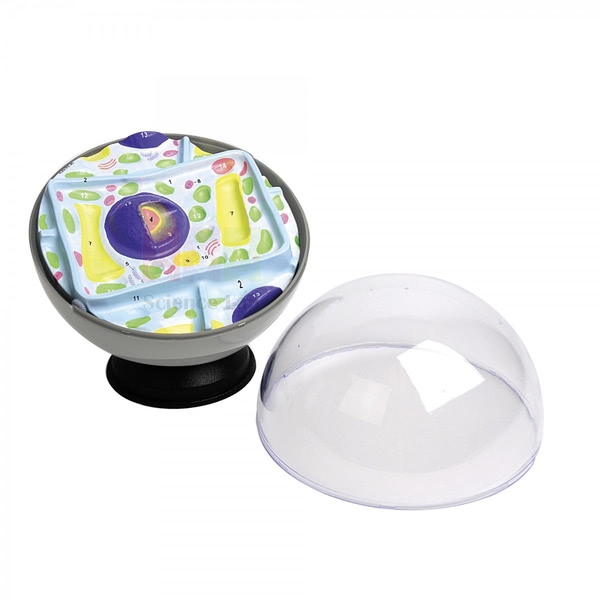
Model Plant Cell
Model Plant Cell (Product Code: SLE/ECB/16) is a compact, 20 cm diameter cell model used by students to explore plant cell structures at group tables in school biology. It is sized for hands-on handling rather than distant display, and it is also offered as part of the Plant and Animal Cell Model Activity Set for teaching both cell types together. It is made by Jain Scientific Equipments Pvt Ltd (ScienceLab), an OEM manufacturer, at its own factory in Ambala, India, and suits a biology model kit bulk order.
Specifications
| Parameter | Detail |
|---|---|
| Type | Plant cell model for student exploration |
| Diameter | 20 cm |
| Also available in | Plant and Animal Cell Model Activity Set |
| Product code | SLE/ECB/16 |
| Manufacturer | Jain Scientific Equipments Pvt Ltd (ScienceLab) — OEM |
| Country of origin | India (made in Ambala, Haryana) |
What Is the Model Plant Cell Used For?
At 20 cm across, the Model Plant Cell is sized for a bench shared by a small group rather than for the teacher's desk. A common approach is to give each group a model and a blank outline of a plant cell, and ask students to identify and label as many structures as they can before the class reviews the answers together. Handling a real three-dimensional model helps younger students who find flat textbook drawings difficult to interpret.
When ordered within the Plant and Animal Cell Model Activity Set, the two models make a straightforward compare-and-contrast exercise: students list what the cells share and what only the plant cell has. Because several units fit in one cupboard, the compact size also suits schools equipping more than one section, and older students can use it for quick revision of plant cell structure.
What to Specify When Ordering
- Whether you need the Model Plant Cell on its own or within the Plant and Animal Cell Model Activity Set.
- The number of units, for example one per student group across all sections.
- Your delivery location and any other cell models to include on the same quotation.
OEM Manufacturer
Jain Scientific Equipments Private Limited, known to customers as ScienceLab, is the OEM (original equipment manufacturer) of the Model Plant Cell and makes it at its own licensed factory at Saha, Ambala, Haryana, India. Class quantities go out factory-direct to schools, dealers and importers, and further orders for new sections are handled by the same manufacturer — ask us. Visit the company profile for more about ScienceLab.
Related Cell Biology Equipments
Choose from these to complete a plant and animal cell unit:
- Animal Cell Model — the natural partner for comparing plant and animal cell structures.
- Plant Cell — a larger, 25-organelle demonstration model for the teacher's desk.
- Plant Cell on Base Model — an electron-microscopic view with part of the wall removed, for older students.
- Hands-On Cell Model Act-Plant — a foam plant cell model with removable parts for group work.
- Chloroplast Model — enlarges the organelle that makes plant cells distinctive.
The complete range is listed under Cell Biology Equipments.
Frequently Asked Questions
How big is the Model Plant Cell?
The Model Plant Cell has a 20 cm diameter. That size is practical for a group bench, easy for students to handle during labelling work and compact enough to store several units together in one cupboard.
Can the Model Plant Cell be bought together with an animal cell model?
Yes. The Model Plant Cell is also available as part of the Plant and Animal Cell Model Activity Set; mention the set in your enquiry if you want both cell types for compare-and-contrast lessons.
Which structures does the Model Plant Cell show?
The Model Plant Cell is designed for exploring plant cell structures, but our current product information does not list the individual parts represented. Please ask through our contact page for the exact list before ordering.
Is the Model Plant Cell suitable for younger students?
The Model Plant Cell's compact size is intended for students to explore directly at their tables. Teachers should still supervise handling and keep the model on the bench during lessons so it is not dropped.
How is the Model Plant Cell cleaned?
A soft cloth, dry or lightly moistened with mild soapy water, is enough to clean the Model Plant Cell. Do not immerse it or use solvents, and let it dry fully before it goes back into storage.
Who manufactures the Model Plant Cell?
The Model Plant Cell is manufactured by Jain Scientific Equipments Pvt Ltd (ScienceLab), the OEM, at its own licensed factory at Saha, Ambala, Haryana, India. Schools and distributors in India and international markets order it directly from the manufacturer.
Can class quantities of the Model Plant Cell be supplied?
Yes. State product code SLE/ECB/16, how many Model Plant Cell units or activity sets you need, and the delivery address; class and bulk quantities are supplied within India and to international markets.
Request a Quote
To order the Model Plant Cell or the activity set, get in touch or add the item to your enquiry cart. The quotation comes direct from the manufacturer.
Last updated: 24 September 2026

Visualising Cells Poster
Visualising Cells Poster (Product Code: SLE/ECB/17) is a laminated classroom wall poster used to teach the differences and similarities between bacterial, animal and plant cells in school and college biology. Measuring 890 mm wide by 585 mm high, it combines detailed illustrations with photography of structures such as the Golgi apparatus, nuclear pores, chloroplasts and mitochondria. It is supplied by Jain Scientific Equipments Pvt Ltd (ScienceLab), Ambala, India, and makes a strong first item in a biology chart set for schools in India.
Specifications
| Parameter | Detail |
|---|---|
| Format | Laminated wall poster |
| Dimensions | 890 mm W x 585 mm H |
| Content style | Detailed illustrations and photography in an easy-to-see, easy-to-understand format |
| Cell types compared | Bacterial, animal and plant cells |
| Structures depicted | Cell membrane, Golgi apparatus, nucleus and nuclear pores, endoplasmic reticulum, chloroplasts and mitochondria |
| Product code | SLE/ECB/17 |
| Supplied by | Jain Scientific Equipments Pvt Ltd (ScienceLab), Ambala, India |
What Is the Visualising Cells Poster Used For?
Mounted near the whiteboard or microscope benches, the poster becomes the class's shared reference for cell structure. When students meet a new organelle, they can see how the Golgi apparatus or the nuclear pores are arranged and where each structure sits within the cell, and the side-by-side treatment of bacterial, animal and plant cells makes it easy to ask comparison questions such as which cell types contain chloroplasts or a true nucleus.
The mix of illustration and photography helps students move between an idealised diagram and a real image, the step many find difficult when they first look down a microscope. Because the surface is laminated, teachers can annotate it temporarily with a non-permanent marker during revision and wipe it clean afterwards.
Supplier
Jain Scientific Equipments Private Limited, which trades as ScienceLab, supplies the Visualising Cells Poster from Ambala, Haryana, as part of its cell biology teaching range. Schools can combine the poster with cell models, slides and other laboratory items on one quotation, and distributors in India and international markets can order it in quantity. Learn more on our company profile.
Related Cell Biology Equipments
Three-dimensional models and slides that match the cell types on the poster:
- Visualizing Mitosis Poster — a companion poster of the same dimensions on cell division.
- Prokaryotic Cell Model — a 3D bacterial cell to accompany the poster's bacterial panel.
- Plant Cell — a detailed translucent plant cell model for demonstration.
- Animal Cell Model — brings the animal cell into three dimensions.
- Microslide Cells Of Plants — photomicrograph images of real plant cells, viewed with a Microslide Viewer.
See more in the Cell Biology Equipments category.
Frequently Asked Questions
What size is the Visualising Cells Poster?
The Visualising Cells Poster measures 890 mm wide by 585 mm high. That is large enough for the illustrated cells to be read from the front rows of a typical classroom while still fitting on a standard notice board or wall panel.
Is the Visualising Cells Poster laminated?
Yes. The Visualising Cells Poster is laminated, which protects it from handling and moisture. It can be wiped clean with a soft, damp cloth, and teachers can use a non-permanent marker on it for temporary labels.
Which cell types and structures does the Visualising Cells Poster cover?
The Visualising Cells Poster compares bacterial, animal and plant cells and depicts the cell membrane, Golgi apparatus, nucleus and nuclear pores, endoplasmic reticulum, chloroplasts and mitochondria, using detailed illustrations and photography.
How should the Visualising Cells Poster be mounted?
Mount the Visualising Cells Poster with poster strips, clips or a frame on a flat wall away from direct sunlight, which can fade printed colours over time. Avoid punching holes through the lamination.
Is the Visualising Cells Poster sent rolled or flat?
Packing of the Visualising Cells Poster depends on the order size and destination. Please ask through our contact page if you need it shipped in a particular way, for example flat for immediate framing.
Who supplies the Visualising Cells Poster?
The Visualising Cells Poster is supplied by Jain Scientific Equipments Pvt Ltd (ScienceLab), Ambala, India, to schools, colleges and distributors in India and international markets, often together with matching cell models and slides on the same order.
Can the Visualising Cells Poster be ordered for several classrooms?
Yes. Give product code SLE/ECB/17 and the number of Visualising Cells Posters you need, with your delivery location; posters for several classrooms or schools can be quoted together with the matching mitosis poster.
Request a Quote
For pricing on the Visualising Cells Poster, send us a message or collect it with other items in the enquiry cart.
Last updated: 24 September 2026
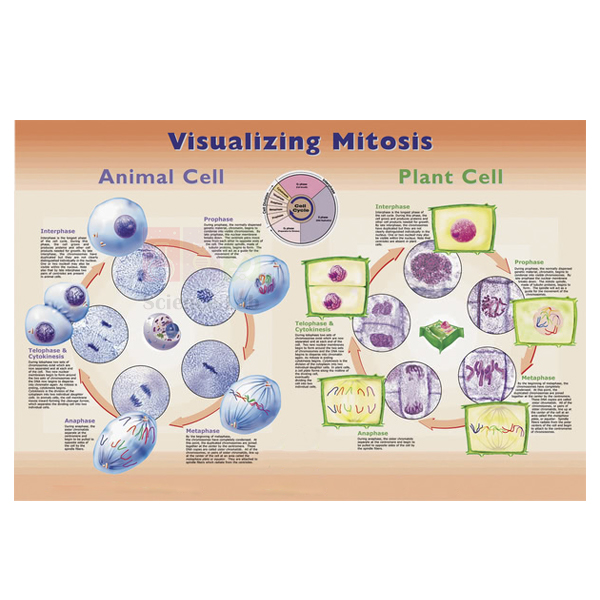
Visualizing Mitosis Poster
Visualizing Mitosis Poster (Product Code: SLE/ECB/18) is a wall poster used to teach the stages of mitosis in school and college biology by setting detailed illustrations of each stage beside close-up photomicrographs of an onion root tip and a whitefish blastula undergoing division. At 890 x 585 mm, it lets students review and compare plant and animal mitosis at a glance. It is supplied by Jain Scientific Equipments Pvt Ltd (ScienceLab), Ambala, India, as school biology lab equipment in India for cell division units.
Specifications
| Parameter | Detail |
|---|---|
| Format | Wall poster |
| Dimensions (w x h) | 890 mm x 585 mm |
| Illustrations | Detailed illustration of each mitotic stage |
| Photomicrographs | Close-ups of an onion root tip (plant) and a whitefish blastula (animal) undergoing mitosis |
| Teaching focus | Reviewing and comparing the steps of plant and animal mitosis |
| Product code | SLE/ECB/18 |
| Supplied by | Jain Scientific Equipments Pvt Ltd (ScienceLab), Ambala, India |
What Is the Visualizing Mitosis Poster Used For?
Onion root tip and whitefish blastula are the two specimens students most often examine when learning to recognise mitosis, because both contain many actively dividing cells. With the poster on the laboratory wall, students can glance up from the microscope and match what they see — condensed chromosomes, a line of chromosomes at the cell equator, chromatids pulled to opposite poles — with the corresponding illustration and photomicrograph.
The pairing of plant and animal images also makes clear the differences teachers want students to explain, such as the absence of centrioles in most plant cells and the way the two cell types complete division. Outside practical sessions, the poster supports quick recaps at the start of a lesson and revision before examinations, and it shares its dimensions with the Visualising Cells Poster so the two hang neatly side by side.
Supplier
ScienceLab is the trade name of Jain Scientific Equipments Private Limited, based in Ambala, Haryana, which supplies the Visualizing Mitosis Poster alongside its cell division models, simulations and prepared slides. Schools and colleges can place the poster on the same quotation as the rest of their biology list, and distributors in India and international markets can order it in quantity. For vendor registration, the supplying company is Jain Scientific Equipments Private Limited (CIN U29309HR2020PTC087597, GSTIN 06AAECJ8618A1ZT). Our company profile gives further details.
Related Cell Biology Equipments
Products that take students from the poster to hands-on work and real specimens:
- Mitosis Simulation Lab Investigation — students model each stage with pop-bead chromosomes, then check against the poster.
- Ts Root Tip Onion — a prepared slide of the plant tissue pictured on the poster.
- Mitosis & Meiosis Set, Microscope Slides — prepared slides for identifying stages under the microscope.
- Animal Mitosis Models — three-dimensional stage models for demonstration.
- Visualising Cells Poster — the matching-size poster on bacterial, animal and plant cell structure.
View all items in Cell Biology Equipments.
Frequently Asked Questions
Which specimens are shown in the photomicrographs on the Visualizing Mitosis Poster?
The Visualizing Mitosis Poster shows close-up photomicrographs of an onion root tip and a whitefish blastula, both photographed while undergoing mitosis, so students see a plant example and an animal example of the same process.
What are the dimensions of the Visualizing Mitosis Poster?
The Visualizing Mitosis Poster measures 890 mm wide by 585 mm high, the same size as the Visualising Cells Poster, so the two can be displayed together as a matching pair on a biology room wall.
Does the Visualizing Mitosis Poster show every stage of mitosis?
Yes. The Visualizing Mitosis Poster features detailed illustrations of each mitotic stage, each with corresponding close-up photomicrographs, so students can review the full sequence and compare plant and animal mitosis.
Is the Visualizing Mitosis Poster laminated?
Our product information for the Visualizing Mitosis Poster does not state a surface finish. If you need a laminated or wipe-clean surface for laboratory use, please ask through our contact page before ordering.
How should the Visualizing Mitosis Poster be displayed?
Hang the Visualizing Mitosis Poster on a dry wall out of direct sunlight, ideally near the microscope benches, and keep it clear of sinks and chemical benches where splashes could mark the print.
Who supplies the Visualizing Mitosis Poster?
The Visualizing Mitosis Poster is supplied by Jain Scientific Equipments Pvt Ltd (ScienceLab), Ambala, India, to schools, colleges and distributors in India and international markets, together with slides and models for the same topic if required.
Can the Visualizing Mitosis Poster be supplied for a tender or several schools?
Yes. Include product code SLE/ECB/18, the number of Visualizing Mitosis Posters and the delivery locations in your enquiry, and the posters can be quoted together with other items on your tender list.
Request a Quote
To order the Visualizing Mitosis Poster, contact us or add it to your enquiry cart together with matching slides and models.
Last updated: 24 September 2026

Visualising Osmosis & Diffusion Investigation
Visualising Osmosis & Diffusion Investigation (Product Code: SLE/ECB/19) is a class investigation used to demonstrate selective permeability with coloured solutions in school biology lessons on the cell membrane. Sized for a class of 40 students, it pairs a model cell with a mixture of special dye solutions, so learners watch the membrane let one dye pass while the other stays inside. It is supplied by Jain Scientific Equipments Pvt Ltd (ScienceLab), Ambala, India, as an osmosis experiment kit for school laboratories.
Key Details
| Parameter | Detail |
|---|---|
| Type | Osmosis and diffusion class investigation |
| Class size | 40 students |
| Working materials | Model cell and a mixture of special dye solutions |
| What students observe | One dye passes through the membrane while the other remains within the cell, producing a colour change |
| Concepts covered | Selective permeability; nutrient uptake and waste discharge; osmosis and diffusion; passive transport of water through a semipermeable membrane; equilibrium |
| Shelf life of solutions | Depends on the chemical, its grade and storage conditions; ask us for the expiry date of the batch you are quoted |
| Product code | SLE/ECB/19 |
| Supplied by | Jain Scientific Equipments Pvt Ltd (ScienceLab), Ambala, India |
What Is the Osmosis & Diffusion Investigation Used For?
The investigation is built around a prediction: once the model cell is placed in the dye mixture, which colour will end up where? As the experiment runs, the membrane lets one dye through and holds the other back, and the colour change inside and outside the cell gives students clear evidence that membranes are selective rather than simply porous. Teachers then use the result to explain how smaller molecules move by diffusion down a concentration gradient, why larger molecules are retained, and how water moves by osmosis through the same semipermeable membrane until equilibrium is approached.
Because it needs no living material, the activity is predictable enough for timed practical lessons and suits younger classes under normal laboratory supervision. It fits well after a simple osmosis demonstration and before work on root hair cells, the small intestine or kidney function, and it gives every group in a biology lab setup the same clear result. Keep the dye solutions capped, label any mixtures prepared in advance and wear gloves if students handle the solutions directly.
Supplier
This investigation is supplied by Jain Scientific Equipments Private Limited, which trades as ScienceLab from Ambala, Haryana, India. The company sources class investigations like this one for schools, colleges and distributors in India and international markets, and can quote it together with the glassware and extension materials a membrane-transport unit needs. More information is on our company profile.
Related Cell Biology Equipments
Use these items to extend the membrane transport topic:
- Osmosis Simulation Activity Model — a quick osmosis demonstration that can be quantified, to run first.
- Visking Tubing, 30m — for a follow-up practical using dialysis-tubing cells made by students.
- Visking Tubing Clips — to close the tubing bags for that extension experiment.
- Glass Beaker Set of 5 — containers for holding the model cell and solutions during the run.
- Visualising Cells Poster — shows the cell membrane in context for the follow-up discussion.
Further investigations are listed in Cell Biology Equipments.
Frequently Asked Questions
How many students is the Visualising Osmosis & Diffusion Investigation designed for?
The Visualising Osmosis & Diffusion Investigation is sized for a class of 40 students. If you teach several sections or are equipping more than one school, give the number of classes when you enquire so the quotation covers every group.
What will students see during the osmosis and diffusion investigation?
In the Visualising Osmosis & Diffusion Investigation, the model cell's membrane lets one dye pass while the other remains within the cell. The resulting colour change is visible evidence of selective permeability and of how a cell absorbs nutrients and discharges wastes.
Does the Visualising Osmosis & Diffusion Investigation cover osmosis as well as diffusion?
Yes. Alongside diffusion of the dyes, the Visualising Osmosis & Diffusion Investigation teaches how osmosis and diffusion maintain equilibrium through the passive transport of water across the cell's semipermeable membrane, linking the demonstration to wider membrane-transport topics.
Are the dye solutions safe for school use?
Treat the dye solutions in the Visualising Osmosis & Diffusion Investigation like any laboratory solution: supervise students, use gloves and eye protection, and avoid ingestion. For safety information on the specific solutions, please contact us before the practical.
What is the shelf life of the dye solutions?
Shelf life depends on the chemical, its grade and storage conditions, so no single expiry period applies to the solutions in the Visualising Osmosis & Diffusion Investigation. Ask us for the expiry date of the batch you are quoted, and store them closed, away from heat and sunlight.
Can replacement solutions be ordered separately?
Refill availability for the Visualising Osmosis & Diffusion Investigation is not stated in the listing. Enquire with the product code SLE/ECB/19 and we will advise what can be supplied as replacement solutions for repeat classes.
Who supplies the Visualising Osmosis & Diffusion Investigation?
The Visualising Osmosis & Diffusion Investigation is supplied by Jain Scientific Equipments Pvt Ltd (ScienceLab), Ambala, Haryana, India, to schools and distributors in India and international markets. Shipping arrangements for the solutions are confirmed with the quotation.
Request a Quote
To request pricing for the Visualising Osmosis & Diffusion Investigation, contact us or add it to your enquiry cart with any related apparatus.
Last updated: 24 September 2026
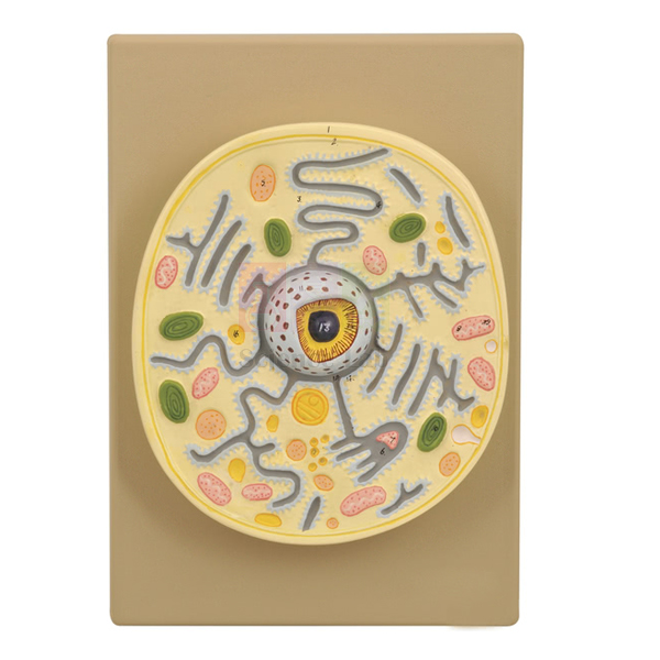
Plant Cell on Base Model
Plant Cell on Base Model (Product Code: SLE/ECB/20) is a base-mounted model of a plant cell's electron microscopic structure, used to teach cell ultrastructure in senior secondary, undergraduate botany and teacher-training classes. A portion of the wall is removed to reveal the ectoplast, endoplast and tonoplast, the vacuoles, nuclear structure, plastids and mitochondria. It is made by Jain Scientific Equipments Pvt Ltd (ScienceLab), an OEM manufacturer, at its own factory in Ambala, India, as an advanced plant anatomy model for institutions in India and abroad.
Key Details
| Parameter | Detail |
|---|---|
| Type | Plant cell model showing electron microscopic structure |
| Presentation | Portion of the cell wall removed to expose the interior |
| Structures shown | Ectoplast, endoplast, tonoplast, vacuoles, nuclear structure, plastids, mitochondria and other cell components |
| Mounting | On a base |
| Product code | SLE/ECB/20 |
| Manufacturer | Jain Scientific Equipments Pvt Ltd (ScienceLab) — OEM |
| Country of origin | India (made in Ambala, Haryana) |
What Is the Plant Cell on Base Model Used For?
The model is most useful once students already know the basic parts of a plant cell and are ready to study membranes in detail. With the wall partly cut away, a teacher can show that the cell contents are bounded by the ectoplast, that the vacuoles are enclosed by their own membrane, the tonoplast, and that the nucleus, plastids and mitochondria sit within the surrounding cytoplasm. This makes it easier to explain plasmolysis, where the protoplast pulls away from the wall, and how substances are stored in or released from the vacuole.
Because it depicts electron microscope detail, the model also helps students interpret published electron micrographs, which can otherwise look like abstract patterns. Colleges often display it beside a conventional plant cell model so that learners see the same cell at two levels of magnification, and the base keeps it steady on a bench or display shelf between lessons.
OEM Manufacturer
ScienceLab is the name used by Jain Scientific Equipments Private Limited, the OEM (original equipment manufacturer) of the Plant Cell on Base Model, which the company makes at its own licensed factory at Saha, Ambala, Haryana, India. Colleges, schools, dealers and importers therefore buy direct from the maker and can return to it for repeat orders or questions about the model — ask us. Buyers who need vendor identifiers for procurement can use the company's GST registration, 06AAECJ8618A1ZT, and its Legal Entity Identifier, 3358004HRD1JIVXKYX49. See the company profile for more.
Related Cell Biology Equipments
Models that complement an ultrastructure lesson on plant cells:
- Plant Cell — a larger translucent model with a multi-layered cell wall and 25 organelles and inclusions.
- Model Plant Cell — a compact model for group work at an introductory level.
- Chloroplast Model — an enlarged plastid for studying its internal membranes.
- Mitochondrion Model — enlarges another organelle shown on this model.
- Plant Cell Plaque — a plaque-format plant cell for comparison and revision.
Browse the full Cell Biology Equipments range.
Frequently Asked Questions
How is the Plant Cell on Base Model different from a standard plant cell model?
The Plant Cell on Base Model shows the electron microscopic structure of the cell, with part of the wall removed, so membranes such as the ectoplast and tonoplast can be studied along with the vacuoles, nuclear structure, plastids and mitochondria.
Is the Plant Cell on Base Model mounted?
Yes. The Plant Cell on Base Model is mounted on a base, which keeps it stable on a demonstration bench or display shelf and lets it stay out for the whole of a practical session without being handled.
What are the dimensions and weight of the Plant Cell on Base Model?
The size and weight of the Plant Cell on Base Model are not given in our current product information. Please ask through our contact page for dimensions and packed weight before ordering, especially for export shipments.
Which level of student is the Plant Cell on Base Model for?
The Plant Cell on Base Model is most useful from senior secondary level upward, where plant cell ultrastructure and membranes are part of the syllabus, and in undergraduate botany and teacher-training courses.
How should the Plant Cell on Base Model be cared for?
Dust the Plant Cell on Base Model with a soft brush or cloth, lift it by the base rather than the cut-away section, and keep it away from heat and direct sunlight so the surface and colours stay in good condition.
Who manufactures the Plant Cell on Base Model?
The Plant Cell on Base Model is manufactured by Jain Scientific Equipments Pvt Ltd (ScienceLab), the OEM, at its own licensed factory at Saha, Ambala, Haryana, India. Institutions and distributors order it and receive quotations directly from the manufacturer.
What should a quotation request for the Plant Cell on Base Model include?
A quotation request for the Plant Cell on Base Model should include product code SLE/ECB/20, the quantity, the delivery location and any tender format you need. Buyers in India and international markets are supplied.
Request a Quote
To get a quotation for the Plant Cell on Base Model, reach our team or add it to the enquiry cart; the quotation comes straight from the manufacturer.
Last updated: 24 September 2026