- Physics Lab Equipment
- Chemistry Lab Equipment
- Biology Lab Equipment
- Anatomical Lab Equipment
- Learning
- Educational Lab Equipment
- Magnets
- Agriculture
- Astronomy
- Earth Science
- Geography Lab Equipment
- Safety Lab Coat
- Laboratory
- Meteorological
- Electricity & Magnetism
- Heat
- School Lab Equipment
- Lab Glassware
- Science Lab Equipment
- Tvet Lab Equipment
- Robotics Kits Supplier and Manufacturer for Schools India
- Math Lab Equipment Manufacturers and Exporters in Ambala
- STEM Kits Manufacturers and Supplier India
- Engineering Lab Equipment
- Hospital Lab Equipment
- Water, Sanitation & Hygiene Supplies
- Cold Chain, Solar & Communication Equipment
- Nutrition
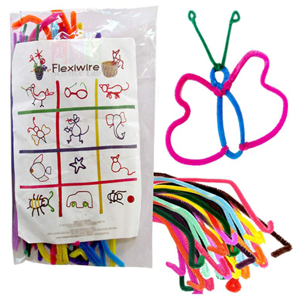
Flexi Wire
Flexi Wire (Product Code: SLE/PP/12) is a bendable craft material made of colourful wires wrapped in yarn, used for twisting and shaping simple models in nursery and kindergarten activity sessions. Children bend it into flowers, animals and many other shapes, which exercises hand-eye co-ordination and small motor skills. It is supplied by Jain Scientific Equipments Pvt Ltd (ScienceLab), Ambala, India, for nurseries adding a craft element to a preschool kit.
Key Details
| Parameter | Detail |
|---|---|
| Construction | Colourful wires with yarn wrapped around them |
| How it is used | Twisted and bent by hand into shapes |
| Example models | Flowers, animals and many other figures |
| Skills practised | Hand-eye co-ordination and small motor skills |
| Learning focus | Creativity and imagination |
| Product code | SLE/PP/12 |
| Supplied by | Jain Scientific Equipments Pvt Ltd (ScienceLab), Ambala, India |
What Is Flexi Wire Used For?
In a pre-primary craft session, each child takes a length of flexi wire and bends, twists and loops it to build a flower, an animal outline or any shape they imagine. The yarn covering makes the wire comfortable to hold, and each bend stays where the child puts it, so finished models can be displayed on a table or used in play afterwards.
Teachers can turn the activity into early learning by asking children to sort pieces by colour, count the petals on a flower or copy a simple shape shown on the board. Gripping and shaping the wire strengthens the small hand muscles children later need for holding a pencil. Supervise young children while they work and check the wire ends before and after each session.
Supplier
Jain Scientific Equipments Private Limited trades as ScienceLab and supplies Flexi Wire alongside the counting, matching and shape activities in its Pre-Primary range. Nurseries, kindergartens, school pre-primary sections, dealers and importers in India and international markets can order it with other early-years aids in one consignment. For vendor registration, the supplying company is listed under GSTIN 06AAECJ8618A1ZT and LEI 3358004HRD1JIVXKYX49. More about the company is on the ScienceLab profile page.
Related Pre-Primary
Pair Flexi Wire with these hands-on activities for a varied fine-motor corner:
- Stringing Beads — another threading task that trains hand-eye co-ordination.
- Tangram — a shape puzzle that builds on the outlines children twist from wire.
- Clay, Modelling, Assorted Color — a second modelling material for creative work.
- Finger Puppets (Set of 5) — use wire flowers and animals as props in puppet stories.
Browse all early-years learning aids in the Pre-Primary category.
Frequently Asked Questions
What is Flexi Wire made of?
Flexi Wire consists of colourful wires with yarn wrapped around them. The yarn covering gives each piece its colour and soft texture, while the wire inside lets children twist and bend it into shapes. Wire gauge and piece length are not listed, so ask us if you need them.
What can children make with Flexi Wire?
Children can twist and bend Flexi Wire into flowers, animals and many other interesting shapes. Teachers often show a simple model to copy first and then let children invent their own designs, which encourages creativity and imagination during pre-primary craft time.
Which skills does Flexi Wire develop?
Flexi Wire develops hand-eye co-ordination and small motor skills, because children must grip, twist and bend the wire accurately to form a shape. It also develops creativity and imagination as they plan and build their own models from the coloured pieces.
How many pieces come in a pack of Flexi Wire?
The number of pieces and the colour mix in a pack of Flexi Wire are not stated in the product listing. Tell us how many children will use it through the contact page, and we will confirm the pack contents in our quotation.
Who supplies Flexi Wire (SLE/PP/12)?
Flexi Wire, product code SLE/PP/12, is supplied by Jain Scientific Equipments Pvt Ltd (ScienceLab) of Ambala, Haryana, India. It can be sent to nurseries, schools, dealers and importers in India and international markets, either alone or with other Pre-Primary learning aids.
Can Flexi Wire be ordered for a whole school?
Yes, Flexi Wire can be quoted in bulk for a whole school, a group of nurseries or a tender. State the product code SLE/PP/12, the quantity required and the delivery location in your enquiry so that we can prepare a single quotation.
Request a Quote
Add Flexi Wire to the enquiry cart, or write to us through the contact page with the quantity and delivery address. We will send a quotation for Flexi Wire and any other pre-primary items you list.
Last updated: 24 September 2026
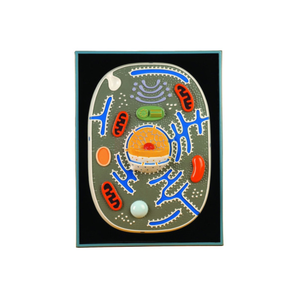
Animal Cell Plaque
Animal Cell Plaque (Product Code: SLE/ECB/06) is a raised-relief teaching plaque used to show the organelles and inclusions of the animal cell in secondary school and college biology lessons. Moulded from heavy vacuum-formed plastic to 20 structures, each named on the accompanying key cards, it measures about 46 x 61 cm yet weighs only 1.2 kg. It is made by Jain Scientific Equipments Pvt Ltd (ScienceLab), an OEM manufacturer, at its own factory in Ambala, India, for schools choosing an animal cell model in India.
Specifications
| Parameter | Detail |
|---|---|
| Type | Relief plaque of the animal cell |
| Construction | Heavy plastic vacuum-formed sheet, moulded in relief |
| Structures moulded | 20 (organelles and cell inclusions) |
| Identification | All structures identified by accompanying key cards |
| Size | 46 x 61 cm (approx.) |
| Weight | 1.2 kg (approx.) |
| Companion item | Plant cell plaque in the same series, moulded to 23 structures |
| Product code | SLE/ECB/06 |
| Manufacturer | Jain Scientific Equipments Pvt Ltd (ScienceLab) — OEM |
| Country of origin | India (made in Ambala, Haryana) |
What Is the Animal Cell Plaque Used For?
Compared with a printed biology chart, the raised relief gives each organelle a shape and depth that students can see from across the room and trace with a finger up close. The series is designed to show every known organelle and inclusion of the cell, so teachers can work through the whole animal cell on one plaque and then ask students to match each of the 20 moulded structures to its name on the key cards.
Displayed beside the plant cell plaque from the same series, it makes the classic comparison lesson straightforward: learners list which structures appear in both cells and which belong only to one. After the lesson, a quick labelling quiz with the key cards set aside shows who has understood the layout of the cell.
What to Specify When Ordering
- Number of Animal Cell Plaques needed, for example one per biology room or one per group bench.
- Whether you also want the plant cell plaque (23 structures) from the same series for comparison lessons.
- Delivery location in India or abroad, and any other cell biology models to include on the same quotation.
OEM Manufacturer
ScienceLab is the trading name of Jain Scientific Equipments Private Limited, the OEM (original equipment manufacturer) behind this plaque. It is produced at the company's own licensed factory at Saha, Ambala, Haryana, India, and sold factory-direct to schools, colleges, dealers and importers. Repeat orders for the animal and plant plaques, and questions about replacement key cards, are handled by the manufacturer itself — ask us. Read more on our company profile.
Related Cell Biology Equipments
Items that build on an animal cell structure lesson:
- Plant Cell Plaque — the companion plaque for plant and animal cell comparison.
- Animal Cell Model — an enlarged three-dimensional animal cell for comparing formats.
- Mitochondrion Model — an enlarged look at one of the organelles on the plaque.
- Microslide Ultra Structure of Animal Cells — photomicrograph images of animal cell ultrastructure.
- Animal Meiosis Plaque — a plaque on reduction division for the next topic.
See all models and plaques in our Cell Biology Equipments range.
Frequently Asked Questions
How many structures are shown on the Animal Cell Plaque?
The Animal Cell Plaque is moulded to 20 structures covering the organelles and inclusions of the animal cell, and every structure is identified on the accompanying key cards. Its companion plant cell plaque in the same series is moulded to 23 structures.
What is the Animal Cell Plaque made of?
The Animal Cell Plaque is formed from heavy plastic sheet that is vacuum-formed to create the raised relief. Moulding the organelles in relief gives them shape and depth, so students can see them from across the room and trace them with a finger up close.
What are the size and weight of the Animal Cell Plaque?
The Animal Cell Plaque measures approximately 46 x 61 cm and weighs about 1.2 kg. One person can lift it easily, so it can be held up in front of a class or carried between biology rooms without help.
How does the Animal Cell Plaque differ from the plant cell plaque?
The Animal Cell Plaque and the plant cell plaque belong to the same relief series. The animal version is moulded to 20 structures and the plant version to 23, so displaying both lets students compare which features the two cell types share.
How should the Animal Cell Plaque be cleaned?
Clean the Animal Cell Plaque with a soft cloth dampened with mild soapy water, and avoid solvents or abrasive pads. Keep it away from heaters and strong sunlight, which can distort formed plastic, and store it flat so the relief is not crushed.
Who manufactures the Animal Cell Plaque?
The Animal Cell Plaque is manufactured by Jain Scientific Equipments Pvt Ltd (ScienceLab), acting as the OEM, at its own licensed factory at Saha, Ambala, Haryana, India. Orders and quotations for the plaque are handled by the manufacturer itself, with no trading intermediary.
How do we request a quotation for several Animal Cell Plaques?
To get a quotation for Animal Cell Plaques, send product code SLE/ECB/06, the number of plaques, whether you also want matching plant cell plaques, and your delivery location through our contact page. Bulk orders for several classrooms are welcome.
Request a Quote
For pricing, contact us or add the Animal Cell Plaque to your enquiry cart; your quotation comes direct from the manufacturer.
Last updated: 24 September 2026
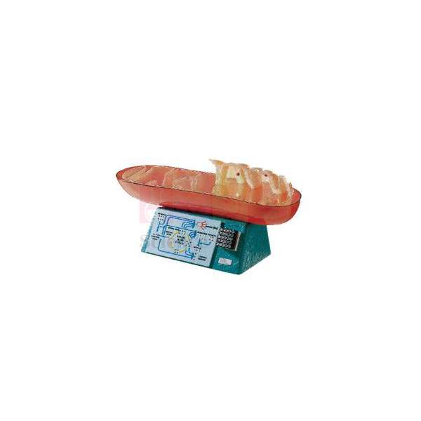
Mitochondrion Model
Mitochondrion Model (Product Code: SLE/ECB/07) is an enlarged organelle model used to teach the structure of the mitochondrion and its role in cellular respiration in senior school and college biology. It magnifies a mitochondrion, normally about 3 microns long, 190,000 times, and its base carries a 1,000,000X membrane relief plus a citric acid cycle schematic. It is made by Jain Scientific Equipments Pvt Ltd (ScienceLab), an OEM manufacturer, at its own factory in Ambala, India, and is a useful piece of cell biology equipment in India.
Specifications
| Parameter | Detail |
|---|---|
| Type | Enlarged mitochondrion model on base |
| Enlargement | 190,000 times (a real mitochondrion is normally 3 microns in length) |
| Structures shown | Two membrane walls, cristae, subunits, matrix and granules |
| Base relief 1 | Cross-section through the membrane and subunits at 1,000,000X |
| Base relief 2 | Simplified schematic of the citric acid cycle |
| Size (approx.) | 58 x 28 x 20 cm (23 in long x 11 in high x 8 in deep) |
| Weight | 4 kg |
| Product code | SLE/ECB/07 |
| Manufacturer | Jain Scientific Equipments Pvt Ltd (ScienceLab) — OEM |
| Country of origin | India (made in Ambala, Haryana) |
What Is the Mitochondrion Model Used For?
In lessons on cellular respiration the model shows why the inner membrane is folded into cristae, giving a large surface for the reactions that release energy, and where the matrix sits in which the citric acid cycle runs. With the cycle moulded on the base, the teacher can move from the organelle to its chemistry without switching to a separate chart, and the 1,000,000X relief brings the membrane subunits into view for more advanced classes.
It also makes a good scale exercise: students multiply the real length of 3 microns by 190,000 and compare the answer with the model's length of about 58 cm. Pairing the model with a respiration practical, such as yeast fermentation, helps students connect what they measure at the bench with the structure that makes it happen.
OEM Manufacturer
The Mitochondrion Model is made by Jain Scientific Equipments Private Limited, trading as ScienceLab, as the OEM (original equipment manufacturer) at the company's own licensed factory at Saha, Ambala, Haryana, India. Colleges, schools, dealers and importers buy it from the manufacturer rather than from a stockist, so questions about the model, repeat orders and spare parts go straight to the people who make it — ask us. Background on the company is on our company profile.
Related Cell Biology Equipments
Models and slides that place the mitochondrion in context:
- Chloroplast Model — the photosynthetic organelle, for comparing the two energy organelles.
- Animal Cell Plaque — shows where mitochondria sit among the other organelles.
- Prokaryotic Cell Model — supports discussion of the bacterial origin of mitochondria.
- Microslide Ultra Structure of Animal Cells — photomicrograph images that include a mitochondrion.
Find further organelle models in our Cell Biology Equipments range.
Frequently Asked Questions
How much has the Mitochondrion Model been enlarged?
The Mitochondrion Model shows a mitochondrion enlarged 190,000 times. A real mitochondrion is normally about 3 microns long, so the model reveals internal detail that no school light microscope can show, at a size the whole class can study.
What is shown on the base of the Mitochondrion Model?
The base of the Mitochondrion Model carries two moulded reliefs: a 1,000,000X cross-section through the membrane and subunits, and a simplified schematic of the citric acid cycle. Together they link the organelle's structure with the chemistry it carries out.
Which internal parts can students identify on the Mitochondrion Model?
On the Mitochondrion Model students can identify the two membrane walls, the cristae, subunits, matrix and granules. Seeing the folded cristae in three dimensions makes it easier to explain why the inner membrane has such a large surface area.
How much space does the Mitochondrion Model need?
The Mitochondrion Model measures approximately 58 x 28 x 20 cm (23 x 11 x 8 in) and weighs 4 kg. It needs a firm bench or cupboard shelf and should be lifted with both hands when moved between rooms.
Is a key card included with the Mitochondrion Model, and what is it made of?
Neither a key card nor the construction material of the Mitochondrion Model is listed in our current product information. Please ask through our contact page and we will confirm both before you order.
Who manufactures the Mitochondrion Model?
The Mitochondrion Model is manufactured by Jain Scientific Equipments Pvt Ltd (ScienceLab), the OEM, at its own licensed factory at Saha, Ambala, Haryana, India. Institutions and distributors deal directly with the manufacturer for quotations and repeat orders of the model.
Can the Mitochondrion Model be ordered in quantity or for export?
Yes. The Mitochondrion Model is supplied to colleges, schools and distributors in India and international markets. Include product code SLE/ECB/07, the quantity and the delivery destination in your enquiry so that the quotation covers packing and shipping.
Request a Quote
For pricing, contact us or add the Mitochondrion Model to your enquiry cart. Quotations are issued directly by the manufacturer.
Last updated: 24 September 2026
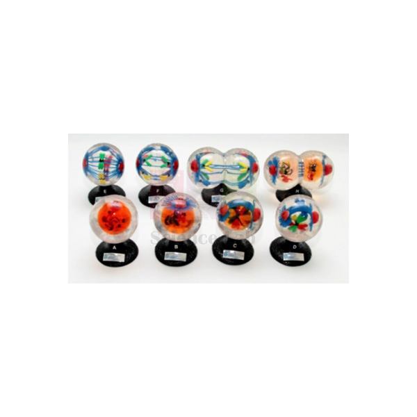
Animal Mitosis Models
Animal Mitosis Models (Product Code: SLE/ECB/08) is a set of 8 sequential models used to show how the chromosomes and mitotic apparatus of an animal cell divide, from interphase to late telophase, in school and college biology. Each stage is embedded in a strong, crystal-clear plastic globe with colour-coded double-thread chromosomes, so students can study it from any side. The set is made by Jain Scientific Equipments Pvt Ltd (ScienceLab), an OEM manufacturer, at its own factory in Ambala, India, among its biology teaching aids for India and abroad.
Specifications
| Parameter | Detail |
|---|---|
| Type | Set of 8 sequential animal cell mitosis models |
| Construction | Organelles and cell inclusions embedded in strong, crystal-clear plastic globes |
| Structures shown | Nucleus, nucleolus, double-thread chromosomes (each in a different colour), centrioles, centromeres (kinetochores), aster and spindle fibres |
| Phases in the set | Interphase, Early Prophase, Late Prophase, Mesophase, Metaphase, Anaphase, Early Telophase, Late Telophase |
| Size, models 1 to 6 | 5-1/4 in high, 3-1/4 in diameter (approx.) |
| Size, models 7 and 8 | 6 in wide x 3-1/4 in deep x 5-1/4 in high |
| Weight of set | 5.8 kg (approx.) |
| Product code | SLE/ECB/08 |
| Manufacturer | Jain Scientific Equipments Pvt Ltd (ScienceLab) — OEM |
| Country of origin | India (made in Ambala, Haryana) |
What Are the Animal Mitosis Models Used For?
Lined up on the bench in order, the eight globes let students follow a single cell cycle step by step: chromosomes condensing in prophase, gathering at the centre in metaphase, separating in anaphase and forming two new nuclei in telophase. Because each double-thread chromosome is a different colour, learners can track one chromosome through every stage and see for themselves that each daughter cell receives an identical set.
A useful classroom activity is to shuffle the models and ask groups to put them back in sequence, explaining the change between each pair. The set then leads naturally into prepared microscope slides, where students search real dividing cells for the same stages they have just handled. Models 7 and 8 are wider than the others, so allow a little extra bench space for them.
OEM Manufacturer
Jain Scientific Equipments Private Limited, known by its trading name ScienceLab, is the OEM (original equipment manufacturer) of the Animal Mitosis Models and makes the set at its own licensed factory at Saha, Ambala, Haryana, India. Buying from the maker means one point of contact for the quotation, for adding sets later as more sections start the topic, and for enquiries about a replacement globe — ask us. Our company profile has the full background.
Related Cell Biology Equipments
Resources that support a complete cell division unit:
- Animal Meiosis Plaque — illustrates reduction division for comparison with mitosis.
- Mitosis Simulation Lab Investigation — a pop-bead student activity on the same process.
- Visualizing Mitosis Poster — a wall reference for the stages.
- Mitosis & Meiosis Set, Microscope Slides — prepared slides for finding the stages in real cells.
Browse our complete Cell Biology Equipments range.
Frequently Asked Questions
How many models are in the Animal Mitosis Models set, and which phases do they show?
The Animal Mitosis Models set contains 8 models showing Interphase, Early Prophase, Late Prophase, Mesophase, Metaphase, Anaphase, Early Telophase and Late Telophase. Placed in order, they take students through one complete division of an animal cell.
What can students see inside each globe of the Animal Mitosis Models?
Each globe of the Animal Mitosis Models contains the nucleus, nucleolus, double-thread chromosomes, centrioles, centromeres (kinetochores), aster and spindle fibres, embedded in strong, crystal-clear plastic so the structures are visible from every side.
Why are the chromosomes in the Animal Mitosis Models different colours?
In the Animal Mitosis Models each double-thread chromosome has its own colour. That makes it easy to follow an individual chromosome from one stage to the next and to show that both daughter nuclei end up with the same chromosome set.
What are the sizes and total weight of the Animal Mitosis Models?
In the Animal Mitosis Models, models 1 to 6 are about 5-1/4 in high with a 3-1/4 in diameter, and models 7 and 8 measure 6 in wide, 3-1/4 in deep and 5-1/4 in high. The whole set weighs about 5.8 kg.
How should the clear globes of the Animal Mitosis Models be cared for?
Clean the Animal Mitosis Models with a soft, lint-free cloth only, and avoid abrasive cleaners and solvents that could scratch or cloud the plastic. Store each model so the globes do not knock against one another.
Is a teacher's key or storage case included with the Animal Mitosis Models?
Neither a teacher's key nor a storage case is listed in our current product information for the Animal Mitosis Models. Please ask through our contact page to confirm exactly what is supplied with the set.
Who manufactures the Animal Mitosis Models?
The Animal Mitosis Models are manufactured by Jain Scientific Equipments Pvt Ltd (ScienceLab), the OEM, at its own licensed factory at Saha, Ambala, Haryana, India. Schools, colleges, dealers and importers order the set directly from the manufacturer, which also prepares their quotation.
Request a Quote
Tell us the number of sets and your delivery location through our contact page, or add the Animal Mitosis Models to your enquiry cart. The quotation comes direct from the manufacturer.
Last updated: 24 September 2026
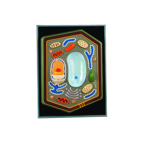
Plant Cell Plaque
Plant Cell Plaque (Product Code: SLE/ECB/09) is a raised-relief display of the plant cell used to teach its organelles and inclusions in secondary school and college botany. Twenty-three structures are moulded on a heavy vacuum-formed plastic sheet of 18 x 24 in (46 x 61 cm), and every one is identified on the accompanying key cards. As a plant cell model for India's biology classrooms, it is made by Jain Scientific Equipments Pvt Ltd (ScienceLab), an OEM manufacturer, at its own factory in Ambala, India.
Specifications
| Parameter | Detail |
|---|---|
| Type | Relief plaque of the plant cell |
| Structures moulded | 23 (organelles and cell inclusions) |
| Construction | Heavy plastic vacuum-formed sheet |
| Identification | All structures identified by accompanying key cards |
| Size | 46 x 61 cm (18 x 24 in) |
| Weight | 1.2 kg |
| Companion item | Animal cell plaque in the same series, moulded to 20 structures |
| Product code | SLE/ECB/09 |
| Manufacturer | Jain Scientific Equipments Pvt Ltd (ScienceLab) — OEM |
| Country of origin | India (made in Ambala, Haryana) |
What Is the Plant Cell Plaque Used For?
The plaque gives the whole class a shared, three-dimensional reference while the teacher explains the parts of a plant cell and what each one does, from the cell wall and chloroplasts to the vacuole and nucleus. Students then work in pairs with the key cards, pointing to each raised structure and naming it, which turns identification into an active exercise instead of copying a diagram.
When the animal cell plaque from the same series is shown alongside, students notice that the plant version carries more moulded structures and can use the key cards to work out which features are found only in plant cells. The pair covers the plant-versus-animal cell comparison found in school biology syllabuses, and both plaques are light enough to move between classrooms.
What to Specify When Ordering
- How many Plant Cell Plaques you need across your botany or biology rooms.
- Whether the matching animal cell plaque (20 structures) should be quoted as a pair for comparison teaching.
- The delivery address and any vendor documents your purchase department requires.
OEM Manufacturer
ScienceLab is the brand of Jain Scientific Equipments Private Limited, which is the OEM (original equipment manufacturer) of the Plant Cell Plaque rather than a reseller of it. Institutions, dealers and importers receive the plaque factory-direct, and any request for extra key cards or a matching set later on is answered by the maker — just ask us. The company is registered under CIN U29309HR2020PTC087597 and makes its products at its licensed factory at 449, HSIIDC Industrial Area, Saha, Ambala, Haryana (Factory Licence AMB-ONLINE-CHD-J-414). Read more on our company profile.
Related Cell Biology Equipments
Items that pair well with the plant cell plaque:
- Animal Cell Plaque — the matching animal cell version for comparison lessons.
- Plant Cell Model — an enlarged model of a typical plant cell with all types of organelle.
- Chloroplast Model — a close-up of the organelle that makes plant cells photosynthetic.
- Plant Cell Study Kit — slides, stains and specimens for moving from the plaque to real plant cells.
- Hands-On Cell Model Act-Plant — a foam plant cell model with removable parts for younger classes.
The complete selection is in our Cell Biology Equipments range.
Frequently Asked Questions
How many structures does the Plant Cell Plaque show?
The Plant Cell Plaque is moulded to 23 structures, the organelles and inclusions of the plant cell, and each one is identified on the key cards that accompany it. That is three more than the animal cell plaque from the same series.
What size is the Plant Cell Plaque in inches and centimetres?
The Plant Cell Plaque measures 18 x 24 in, which is 46 x 61 cm, and it weighs 1.2 kg. The size suits front-of-class display, while the low weight lets a teacher move it easily between rooms.
Is a matching animal cell plaque available for the Plant Cell Plaque?
Yes. The Plant Cell Plaque has a companion Animal Cell Plaque in the same relief series, moulded to 20 structures. Ordering both lets students compare plant and animal cells side by side using the two sets of key cards.
Can students handle the Plant Cell Plaque?
Yes, with care. The Plant Cell Plaque weighs only 1.2 kg, but the raised areas of the vacuum-formed sheet should not be pressed hard or stacked under heavy items; students should hold it by the edges.
Who manufactures the Plant Cell Plaque?
The Plant Cell Plaque is manufactured by Jain Scientific Equipments Pvt Ltd (ScienceLab), the OEM, at its own licensed factory at Saha, Ambala, Haryana, India. Schools, colleges and distributors buy it directly from the manufacturer, not through a trading company.
Can we get vendor registration details for the Plant Cell Plaque purchase file?
Yes. The company registration and factory licence numbers for the maker of the Plant Cell Plaque are given in the manufacturer section above, and any further documents your institution needs can be requested through our contact page.
Request a Quote
For pricing, contact us or add the Plant Cell Plaque to your enquiry cart; quotations are prepared directly by the manufacturer.
Last updated: 24 September 2026
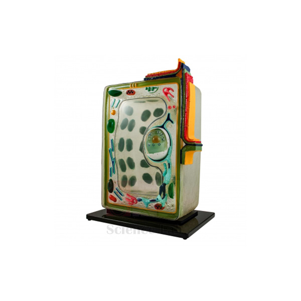
Plant Cell
Plant Cell (Product Code: SLE/ECB/10) is a large, translucent demonstration model used to teach the 25 organelles and cell inclusions of a plant cell in school, college and teacher-training biology. Its fibreglass-reinforced plastic body gives the appearance of living matter, and the cell wall is divided into distinct layers from the middle lamella to a warted tertiary wall. It is made by Jain Scientific Equipments Pvt Ltd (ScienceLab), an OEM manufacturer, at its own factory in Ambala, India, as a centrepiece of botany lab equipment in India.
Specifications
| Parameter | Detail |
|---|---|
| Organelles and inclusions shown | 25 |
| Body material | Translucent, fibreglass-reinforced plastic that gives the appearance of living matter |
| Detail parts | Moulded plastic, so colours do not chip, crack, peel or fade |
| Key structures | Endoplasmic reticulum, cross-sectioned chloroplast with distinct grana and grana discs, large central vacuole, mitochondria and ribosomes, along with the other known organelles and inclusions |
| Cell wall layers | Middle lamella, primary wall, three secondary walls, warted tertiary wall and plasma membrane |
| Size (approx.) | 16 in high x 11-1/2 in wide x 8 in deep (41 x 29 x 20 cm) |
| Weight (approx.) | 4 kg |
| Product code | SLE/ECB/10 |
| Manufacturer | Jain Scientific Equipments Pvt Ltd (ScienceLab) — OEM |
| Country of origin | India (made in Ambala, Haryana) |
What Is the Translucent Plant Cell Model Used For?
Teachers usually work from the outside in: first the middle lamella that joins neighbouring cells, then the primary wall, the three secondary walls and the warted tertiary wall, and finally the plasma membrane. From there the lesson moves to the cross-sectioned chloroplast, where the grana and grana discs are easy to point out, and on to the large central vacuole, mitochondria and endoplasmic reticulum. Because the body is translucent, internal organelles stay visible from several angles when small groups gather round for labelling or oral questioning.
The model is most effective when paired with microscope practicals on onion epidermis or leaf peels, so students connect the enlarged structures with what they actually see under the lens. It also serves as a revision aid before practical examinations. At around 4 kg it should rest on a stable bench or shelf rather than be passed hand to hand across a room.
OEM Manufacturer
The Plant Cell model comes from Jain Scientific Equipments Private Limited, the company behind the ScienceLab name, which is its OEM (original equipment manufacturer) and makes it at its own licensed factory in Saha, Ambala, Haryana, India. Because there is no trading layer between maker and buyer, schools, colleges, dealers and importers get their quotation, repeat supply and after-sales questions handled by the manufacturer — ask us about anything you need. Background on the company is on our company profile.
Related Cell Biology Equipments
These items support the same lessons on plant cell structure and comparison with other cell types:
- Chloroplast Model — a dedicated, enlarged view of the organelle shown here in cross-section.
- Mitochondrion Model — lets students study the respiration organelle in more depth after the whole-cell overview.
- Animal Cell Plaque — for side-by-side comparison of plant and animal cell features such as the wall and vacuole.
- Plant Cell Plaque — a plaque-format plant cell reference for a second bench or display.
- Visualising Cells Poster — a laminated wall poster comparing bacterial, animal and plant cells.
See the full range of Cell Biology Equipments for more models, plaques and classroom investigations.
Frequently Asked Questions
What does the Plant Cell model show?
The Plant Cell model shows 25 organelles and cell inclusions, including the endoplasmic reticulum, a cross-sectioned chloroplast with grana and grana discs, the large central vacuole, mitochondria and ribosomes, and a cell wall divided into middle lamella, primary, secondary and tertiary layers and plasma membrane.
How large and heavy is the Plant Cell model?
The Plant Cell model measures approximately 41 x 29 x 20 cm (16 x 11-1/2 x 8 in) and weighs about 4 kg. That makes it a teacher's demonstration piece for the front bench rather than an item for every student group.
Will the colours of the Plant Cell model wear off with classroom use?
The detail parts of the Plant Cell model are moulded plastic, so the colours do not chip, crack, peel or fade. Normal care still applies: lift the model with both hands and keep it away from direct heat.
How should the Plant Cell model be cleaned?
Wipe the Plant Cell model with a soft cloth, slightly dampened with mild soapy water if needed. Avoid solvents and abrasive cleaners, which can dull the translucent fibreglass-reinforced surface, and dry it before returning it to the shelf.
Is a labelling key supplied with the Plant Cell model?
Our product information for the Plant Cell model does not mention a key or guide. Please ask through our contact page to confirm what documentation is sent with the model before you order.
Who manufactures the Plant Cell model?
The Plant Cell model (SLE/ECB/10) is manufactured by Jain Scientific Equipments Pvt Ltd (ScienceLab), its OEM, at the company's own licensed factory at Saha, Ambala, Haryana, India, and is supplied factory-direct to institutions and distributors.
What should an institutional quotation request for the Plant Cell model include?
A quotation request for the Plant Cell model should mention product code SLE/ECB/10, the quantity, the delivery location and any tender or purchase-order format you must follow. Orders from India and international markets, including bulk orders, are welcome.
Request a Quote
To order the Plant Cell model or ask about quantities, contact our team or add it to your enquiry cart with other items from your lab list. You will receive a quotation directly from the manufacturer.
Last updated: 24 September 2026
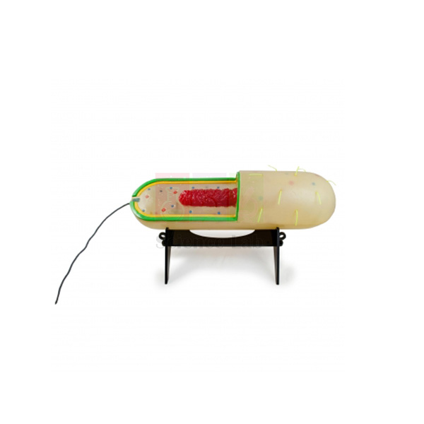
Prokaryotic Cell Model
Prokaryotic Cell Model (Product Code: SLE/ECB/11) is an enlarged bacterial-type cell model used to teach prokaryotic cell structure in school biology, college and microbiology courses. Shown at 280,000X and partly cut away, it displays pili and flagella outside and chromosomal DNA, plasmids and ribosomes inside, and the cell lifts off its stand, with key cards supplied. It is made by Jain Scientific Equipments Pvt Ltd (ScienceLab), an OEM manufacturer, at its own factory in Ambala, India, for colleges assembling microbiology lab equipment in India.
Specifications
| Parameter | Detail |
|---|---|
| Magnification | Enlarged 280,000X |
| Presentation | External cellular structures shown; partially cut away to reveal internal structures |
| Structures illustrated | Pili, flagella, chromosomal DNA, an inclusion body, plasmids, ribosomes, the cell envelope and the plasma membrane |
| Mounting | Removable from its stand |
| Supplied with | Key cards |
| Size (approx.) | 22 in long x 6-1/2 in (56 x 17 x 17 cm) |
| Weight (approx.) | 2.6 kg |
| Product code | SLE/ECB/11 |
| Manufacturer | Jain Scientific Equipments Pvt Ltd (ScienceLab) — OEM |
| Country of origin | India (made in Ambala, Haryana) |
What Is the Prokaryotic Cell Model Used For?
The model is most useful in the lesson that contrasts prokaryotes with eukaryotes. With the cutaway facing the class, the teacher can show that the chromosomal DNA lies free in the cytoplasm rather than inside a nucleus, point out the small plasmids and the ribosomes, and then turn the model to discuss the cell envelope, the pili used for attachment and the flagella used for movement. Lifting the cell off its stand makes it easy to pass to a group, and the key cards let students check their own identification of each part.
In microbiology courses it gives context before students look at stained bacterial smears, where individual structures are far too small to see. Foundation courses in nursing, pharmacy and life sciences use it to build a clear picture of bacterial anatomy that supports later topics such as antibiotic action and gene transfer through plasmids.
OEM Manufacturer
Jain Scientific Equipments Private Limited operates under the ScienceLab name and is the OEM (original equipment manufacturer) of the Prokaryotic Cell Model, which it makes at its own licensed factory at Saha, Ambala, Haryana, India. Departments, distributors and importers source the model factory-direct, so the same manufacturer handles their quotation, follow-up orders and requests for replacement key cards or a stand — ask us. Read more on the ScienceLab company profile.
Related Cell Biology Equipments
Pair the model with these items to build a complete lesson on cell types:
- Plant Cell — a detailed eukaryotic model for direct comparison with the prokaryotic layout.
- Visualising Cells Poster — a wall reference showing how bacterial, animal and plant cells differ.
- Bacteria slide Set of 12 — prepared slides of real bacteria, including cocci, bacilli and spirilla.
- Amoeba Model — a single-celled eukaryote that contrasts well with a prokaryote.
- Mitochondrion Model — supports discussion of how mitochondria resemble bacteria.
More models and investigations are listed under Cell Biology Equipments.
Frequently Asked Questions
Which structures can students identify on the Prokaryotic Cell Model?
On the Prokaryotic Cell Model students can identify pili, flagella, chromosomal DNA, an inclusion body, plasmids, ribosomes, the cell envelope and the plasma membrane. The partial cutaway shows the internal parts, while the outer surface displays the external structures.
What scale and size is the Prokaryotic Cell Model?
The Prokaryotic Cell Model represents the cell enlarged 280,000X. Its overall size is about 56 x 17 x 17 cm (22 in long) and it weighs roughly 2.6 kg, so it suits a demonstration bench or a group table.
Can the Prokaryotic Cell Model be taken off its stand?
Yes. The Prokaryotic Cell Model is removable from its stand, which makes close inspection and group work easier. Return it to the stand for storage so the flagella and pili are not bent or knocked.
What are the key cards with the Prokaryotic Cell Model for?
The key cards supplied with the Prokaryotic Cell Model identify the illustrated structures, so students can use the model for self-checking and revision without a separate textbook diagram, and teachers can run quick labelling quizzes.
How do I keep the Prokaryotic Cell Model in good condition?
Dust the Prokaryotic Cell Model with a soft brush or dry cloth and store it away from direct sunlight and heat. Do not use solvents or abrasive cleaners, and lift it by the stand or body rather than by the flagella.
Who manufactures the Prokaryotic Cell Model?
The Prokaryotic Cell Model is manufactured by Jain Scientific Equipments Pvt Ltd (ScienceLab), acting as the OEM, at its own licensed factory at Saha, Ambala, Haryana, India. Buyers in India and international markets deal directly with the manufacturer.
Can several Prokaryotic Cell Models be supplied for a department or distributor?
Yes. Send product code SLE/ECB/11 with the quantity of Prokaryotic Cell Models and the destination, and we will quote for delivery within India or to international markets. Other cell models can be added to the same quotation.
Request a Quote
For pricing and availability, send us an enquiry or place the Prokaryotic Cell Model in your enquiry cart with the rest of your requirements; the manufacturer replies with the quotation directly.
Last updated: 24 September 2026
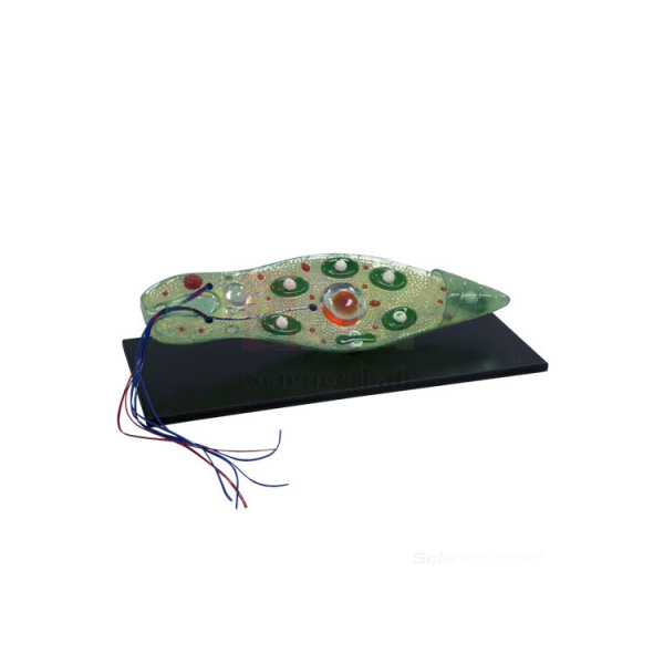
Euglena Model
Euglena Model (Product Code: SLE/ECB/12) is an enlarged, translucent fibreglass model of Euglena used to teach protist structure in school zoology, botany and college biology. It shows the features seen in living and stained preparations, from the flagellum and stigma to the pyrenoid, contractile vacuole and endosome, on a base of 7 x 19-1/2 in. It is made by Jain Scientific Equipments Pvt Ltd (ScienceLab), an OEM manufacturer, at its own factory in Ambala, India, and suits any biology lab setup for schools in India studying protists.
Specifications
| Parameter | Detail |
|---|---|
| Material | Translucent fibreglass |
| Structures shown | Flagellum, stigma, blepharoplast, contractile vacuole, chloroplast, pyrenoid, rhizoplast, nucleus and endosome |
| Basis of detail | Features visible in living and stained preparations |
| Base size | 7 in x 19-1/2 in |
| Overall size (approx.) | 44 x 15 x 15 cm |
| Weight (approx.) | 2.4 kg |
| Product code | SLE/ECB/12 |
| Manufacturer | Jain Scientific Equipments Pvt Ltd (ScienceLab) — OEM |
| Country of origin | India (made in Ambala, Haryana) |
What Is the Euglena Model Used For?
A good way to use the model is as a bridge between a pond-water practical and the theory lesson. Students first try to spot live Euglena under the microscope, where they may see only a green, spindle-shaped cell swimming with a whip-like flagellum; the enlarged model then shows the stigma that senses light, the blepharoplast and rhizoplast at the base of the flagellum, the chloroplasts and pyrenoid involved in photosynthesis, and the contractile vacuole that removes excess water.
Because Euglena combines plant-like chloroplasts with animal-like movement, the model also opens a discussion on why some organisms do not fit neatly into one kingdom. The translucent body keeps internal features such as the nucleus and endosome visible without a cut section, and many departments keep it beside an amoeba model so both protists can be compared in one session.
OEM Manufacturer
This model is made by Jain Scientific Equipments Private Limited, which trades as ScienceLab and is the OEM (original equipment manufacturer) of the Euglena Model, at its own licensed factory at Saha, Ambala, Haryana, India. Schools, colleges, dealers and importers order it straight from the manufacturer, not from a trader, and can come back to the same team for repeat orders or questions about the model — ask us. See our company profile for more information.
Related Cell Biology Equipments
Useful additions for a protist lesson built around this model:
- Amoeba Model — contrasts pseudopod movement with the flagellum-driven swimming of Euglena.
- Chloroplast Model — a closer look at the photosynthetic organelle that makes Euglena green.
- Student Monocular Microscope — for observing live Euglena in pond-water samples.
- Microscope Slides Depression — cavity slides that hold a drop of culture so the cells can swim freely.
- Prokaryotic Cell Model — compares a flagellated prokaryote with this flagellated eukaryote.
Explore other cell and protist models in Cell Biology Equipments.
Frequently Asked Questions
Which parts of Euglena does the Euglena Model represent?
The Euglena Model represents the flagellum, stigma, blepharoplast, contractile vacuole, chloroplast, pyrenoid, rhizoplast, nucleus and endosome — the features that are visible in living and stained preparations of the organism under the microscope.
What are the dimensions of the Euglena Model?
The Euglena Model measures about 44 x 15 x 15 cm overall, stands on a base of 7 x 19-1/2 in and weighs roughly 2.4 kg, so it can sit on a demonstration bench for a whole lesson.
Why is the Euglena Model translucent?
The Euglena Model is made of translucent fibreglass so that students can see internal structures such as the nucleus and pyrenoid through the body of the cell, much as they would through a thin living cell under the microscope.
Does the Euglena Model replace microscope work?
No. The Euglena Model is meant to support microscope work: students learn what to look for before examining live or stained specimens, and they can check their observations against the enlarged structures afterwards.
Are labels or a guide included with the Euglena Model?
Our product information for the Euglena Model does not specify labelling material. If you need a key for student use, please ask through our contact page what accompanies the model before ordering.
Who manufactures the Euglena Model?
The Euglena Model is manufactured by Jain Scientific Equipments Pvt Ltd (ScienceLab), the OEM, at its own licensed factory at Saha, Ambala, Haryana, India. Institutions and distributors receive the model and its quotation directly from the manufacturer.
Is the Euglena Model supplied outside India?
Yes. The Euglena Model is supplied to institutions and distributors in India and international markets. Include product code SLE/ECB/12, the quantity and the destination in your enquiry so that the quotation covers delivery.
Request a Quote
To request a quotation for the Euglena Model, write to us or add it to the enquiry cart and submit your full list in one go. Quotes come direct from the manufacturer.
Last updated: 24 September 2026